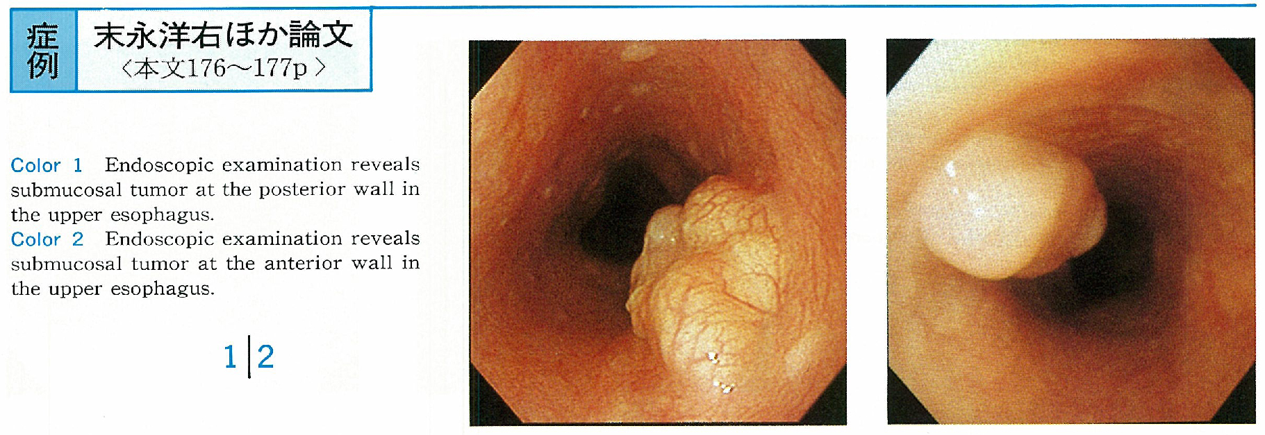
In 2 patients with no symptom, esophageal tumors were detected by screening endoscopy. The tumors were located at the upper esophagus and covered with normal esophageal mucosa. Endoscopic ultrasonography used by a small sized probe (20MHz) revealed that the tumors was connected with the lamina muscularis mucosa. And margin was clear, internal echo was homogeneous. Therefore, the tumors were diagnosed leiomyoma originated from the lamina muscularis mucosa and had indication for resection via endoscopy. These tumors were removed using sliding tube.
Microscopic examination revealed that they were leiomyoma derived from the lamina muscularis mucosa and resected completely. So the diagnosis of location and origin of the tumor was accurate. Endoscopic ultrasonography using a small sized probe is useful for selection of endoscopic treatments in esophageal submucosal tumors.