消化器内視鏡の進歩:Progress of Digestive Endoscopy
Online ISSN : 2189-0021
Print ISSN : 0389-9403
49 巻
選択された号の論文の73件中1~50を表示しています
掲載論文カラー写真集
臨床研究
  • 豊島 明, 伊藤 正秀, 角田 誠之, 馬越 正通, 桜井 四郎, 湯川 雅枝
    1996 年 49 巻 p. 86-89
    発行日: 1996/12/02
    公開日: 2015/03/20
    ジャーナル フリー
     胃内への逆流胆汁酸と胃粘膜障害の関連について,様々の研究が行われてきた。われわれは逆流胆汁酸を分画に分け,それぞれの胆汁酸濃度とその背景胃粘膜との関連を病理組織学的に観察し検討を行ってきた。今回は胃内への逆流胆汁酸濃度が胃内pHに影響を与えるとの考えより,逆流胆汁酸分画とHelicobacter pylori,さらに胃粘膜内微量元素濃度に関して検討した。53例の消化性潰瘍の患者と8例の慢性胃炎の患者を対象として,逆流胆汁酸分画を高速液体クロマトグラフィー法により,微量元素(Cu,Fe,Zn)をPIXE法により各々測定した。胃内逆流胆汁酸中ケノデオキシコール酸(TCDC)濃度の多い群にHpの生息の少ない結果を得た。よってTCDCがHpの胃内での至適環境に影響を与えている可能性があると考えられた。微量元素Zn濃度はHp生息例において低かった。これはZnのもつHpウレアーゼ活性阻害作用,増殖抑制作用,活性酸素消去作用などでZnが消費されたためと考えられた。またFe濃度も低い傾向が認められ,胃粘膜萎縮との関与が考えられた。次にTCDCと微量元素の関係は,TCDC高濃度群の方にZnの濃度が高いことがわかった。理由の1つにTCDC濃度が高くなるに従って,胃内pHが変化し,Hp菌数の減少が生じ,粘膜内の活性酸素の生産量が減り,Znの消費量が減ったためと考えられた。
  • 松田 摩也, 木田 光広, 菅野 聡, 近藤 一英, 山田 至人, 坂口 哲章, 野登 誠, 西元寺 克禮
    1996 年 49 巻 p. 90-95
    発行日: 1996/12/02
    公開日: 2015/03/20
    ジャーナル フリー
     上部消化管粘膜下腫瘍(SMT)の診断に超音波内視鏡(EUS)は有効な手段として広く普及しつつある。今回われわれの施設のEUSによるSMTの鑑別診断について再検討した。対象は1992年12月から現在までにEUSにより診断した上部消化管SMT症例のうち,組織学的診断が得られた29例で,内訳は平滑筋腫12例,平滑筋肉腫5例,カルチノイド3例,ブルンネル腺腫2例,迷入膵2例,顆粒細胞腫2例,リンパ管腫2例,神経鞘腫1例,EUS診断と組織診断を比較検討した。われわれは1992年に筋原性腫瘍の良悪性の鑑別にmalignant scoring systemを提唱した。①大きさ3cm以上,②結節状,③5mm以上の潰瘍を有する,④内部エコー不均一,⑤無エコー域を有する。以上のうち3つ以上認められれば悪性が疑われる。この診断法で今回の検討で得られた筋原性悪性腫瘍の正診率は感度は100%,特異度は73.6%と高かった。その他の疾患の特徴をあげれば,カルチノイド : 主存在層は第2-3層由来で,やや低エコーから低エコーで均一,迷入膵 : 主存在層は第3層が多く一部は4層まで達し,等からやや低エコーで,顆粒状内部エコーパターン,管腔構造が認められる,顆粒細胞腫 : 主存在層は第2-3層で低エコーで,全体としてカルチノイドに類似する,神経鞘腫 : 主存在層は第4層で,やや低エコーから低エコー,平滑筋腫に類似する。全体の正診率は81.5%であった。EUSは上部消化管のSMTの診断に有用と思われた。malignant scoring systemは,筋原性腫瘍の良悪性の鑑別に有効であった。
症例
  • 竹森 政樹, 岡本 真紀代, 箭頭 正徳, 上野 義隆, 仲村 洋, 鈴木 紘一, 高橋 隆一
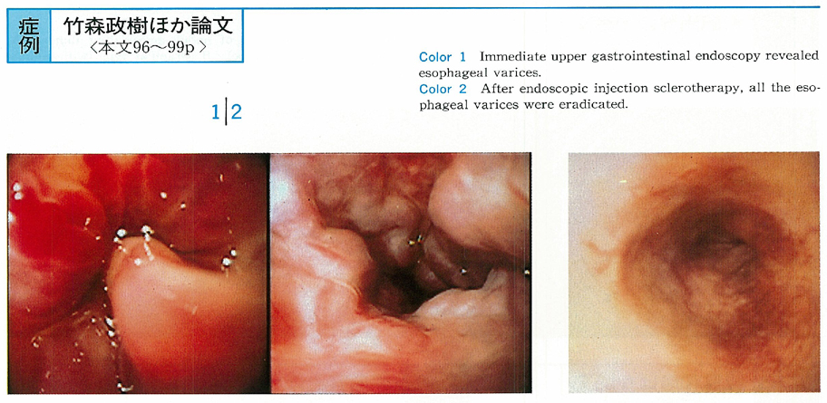
    1996 年 49 巻 p. 96-99
    発行日: 1996/12/02
    公開日: 2015/03/20
    ジャーナル フリー
     原発性骨髄線維症より門脈圧亢進症をきたし,食道静脈瘤硬化療法を繰り返した1例を報告する。症例は46歳,女性。昭和49年血小板減少を指摘され,脾機能亢進として脾摘除を施行し,この際原発性骨髄線維症と診断された。平成7年10月吐血にて入院。食道静脈瘤よりの出血と診断し,内視鏡的治療を繰り返した。門脈圧亢進の原因として明らかな肝硬変の所見なく,原発性骨髄線維症との関連を考え,腹部CT,腹部血管造影を行い,門脈本幹に血栓像を認めたが,門脈血流は十分保たれており,肝内門脈の血流障害が考えられた。原発性骨髄線維症と門脈圧亢進症との合併を考える上で示唆に富む症例と考え,文献的考察を加え報告する。
  • 谷 穣, 三治 哲哉, 緑川 昌子, 半田 豊, 森田 重文, 大野 博之, 吉田 肇, 鶴井 光治, 三坂 亮一, 川口 実, 斉藤 利彦 ...
    1996 年 49 巻 p. 100-103
    発行日: 1996/12/02
    公開日: 2015/03/20
    ジャーナル フリー
     症例は51歳男性。1989年6月健診の食道透視にて異常を指摘され,上部消化管内視鏡検査および超音波内視鏡検査を施行し,食道粘膜下腫瘍(平滑筋腫)と診断された。以後経過観察していたが,1995年2月EUS・CTにて腫瘍の増大傾向が認められ,腫瘍核出目的で入院した。術前の内視鏡所見では門歯より38-43cmの右側半周に粘膜下腫瘍様の隆起性病変を認めた。MRIではT1強調で高信号,T2強調で特徴的な高信号を認め,粘液性の嚢胞が疑われ,食道嚢胞と術前診断し,左開胸開腹食道嚢胞核出術を行った。摘出された腫瘍は大きさ40×35×10mmで,内容物は粘稠性の高い粘液であった。組織学的には嚢胞壁の外側には2層の発達した平滑筋層がみられ,嚢胞壁の内面上皮は繊毛を有する1層の円柱上皮細胞により構成されていた。また組織内に軟骨組織を欠いており,食道重複嚢胞(duplication cyst)と診断した。
  • 竹内 浩紀, 浜田 節雄, 松木 盛行, 石崎 嘉宏, 大久保 学, 岡村 維摩, 水口 博之, 村上 三郎, 里見 昭, 石田 清, 平山 ...
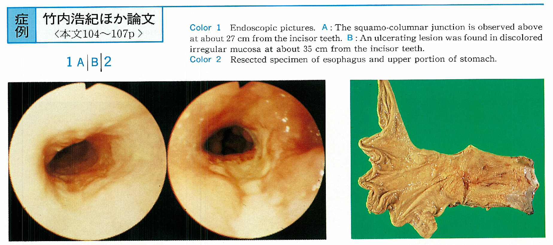
    1996 年 49 巻 p. 104-107
    発行日: 1996/12/02
    公開日: 2015/03/20
    ジャーナル フリー
     われわれは,Barrett食道に発生した比較的珍しい表層拡大型食道粘膜腺癌を経験したので報告する。症例は64歳,男性。胸痛を主訴に平成7年8月近医を受診し,精査の結果Barrett食道腺癌と診断され,当科に紹介された。内視鏡所見では切歯列より27cmの部位から胃噴門部までルゴール染色不染のBarrett上皮が連続し,約35cmの部位には0-Ⅲ様病変2個と粘膜粗糙な局面を認めた。上部消化管造影検査ではⅠm領域に10×3mmの潰瘍を伴う狭窄病変とひだ集中を伴う7×2mmの潰瘍性病変,および食道裂孔ヘルニアを認めた。Barrett食道腺癌の診断で右開胸開腹,食道亜全摘D2郭清を行った。切除標本の肉眼型分類は0-Ⅱb+Ⅲ,全周性で大きさ9×5cm,病理組織学的検査ではBarrett食道腺癌,m,n(-),ly0,v0であった。
  • 田中 洋一, 真船 健一, 関根 毅, 山本 邦男, 二ツ木 浩一
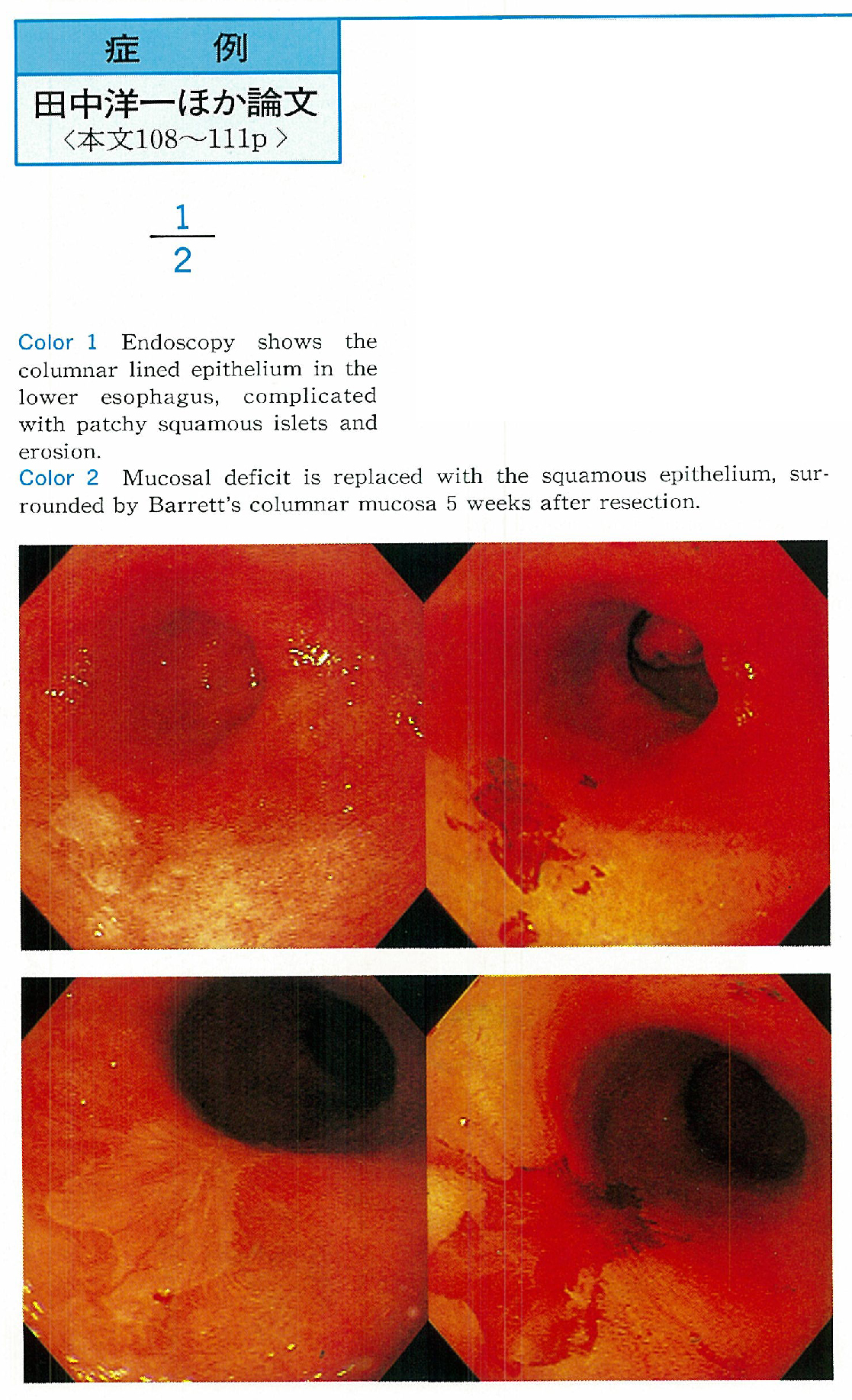
    1996 年 49 巻 p. 108-111
    発行日: 1996/12/02
    公開日: 2015/03/20
    ジャーナル フリー
     症例は70歳男性。1994年に食道潰瘍に対する保存的治療歴あり。1995年3月心窩部不快感を主訴に内視鏡検査を受け,食道腺癌と診断され,当院受診となった。約13cm長の全周型円柱上皮を有するBarrett食道で,腺癌を合併していた。同年8月4日,内視鏡的に腺癌部分を含むBarrett上皮の粘膜切除術を行った。切除標本の組織像は特殊円柱上皮型Barrett上皮で,特有の粘膜筋板の二重化を認めた。標本中央に4.5×2mm大の微小上皮内腺癌(tub1)を認めた。制酸治療を継続したところ,粘膜欠損部に扁平上皮の再生を認めた。Barrett腺癌粘膜切除治療後の扁平上皮再生として,最初の症例報告と思われる。Barrett上皮の起源および合併腺癌の予防・治療上に示唆を与える興味ある症例として報告した。
  • 古河 哲哉, 小金井 裕之, 香山 秀之, 山村 晴男, 山本 和夫, 池内 孝夫
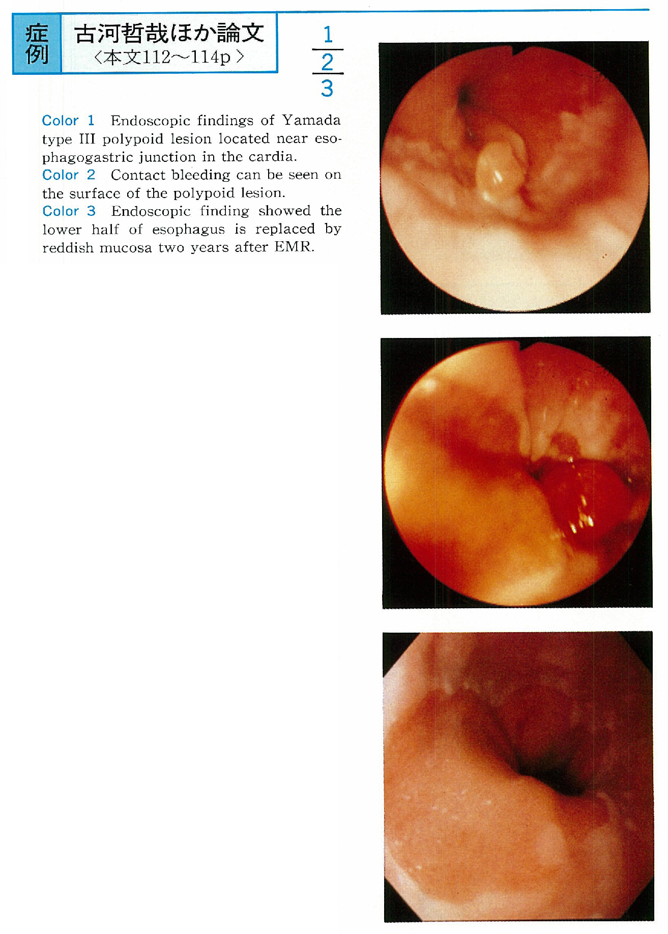
    1996 年 49 巻 p. 112-114
    発行日: 1996/12/02
    公開日: 2015/03/20
    ジャーナル フリー
     症例は50歳男性。1993年11月右季肋部痛で近医受診。近医での内視鏡検査の結果,胃癌が疑われた。当科で再検査を行い,食道胃粘膜接合部付近の噴門に白色の山田Ⅲ型のポリープ様の小隆起が認められた。易出血性で,内視鏡検査中に出血がみられた。生検の結果はGroup Ⅳで,内視鏡的胃粘膜切除術施行。腫瘍径は8×6.5mm,上皮が欠損し,病理組織学的には強い炎症細胞浸潤を伴った毛細血管の拡張と増生のみられる肉芽組織で,pyogenic glanulomaと診断された。pyogenic glanulomaは,皮膚や粘膜にみられる易出血性腫瘤で,消化管では口腔粘膜や舌に比較的多いが,胃粘膜での発生は本邦で1例のみ報告されている。本症例の場合,EMR施行後約2年間経過しているが,再発は認められていない。
  • 松本 宙明, 門馬 久美子, 小澤 広, 加藤 久人, 荒川 丈夫, 榊 信廣, 中村 二郎, 小池 盛雄, 藤谷 幹浩, 池上 雅博
    1996 年 49 巻 p. 115-117
    発行日: 1996/12/02
    公開日: 2015/03/20
    ジャーナル フリー
     生検にて胃顆粒細胞腫と診断し,内視鏡的粘膜切除術にて治療した1例を経験したので報告する。症例は39歳男性。検診の内視鏡検査にて胃体下部大彎に,直径約7mm大,山田Ⅰ型で表面が正常上皮で被われた,やや黄白色調の胃粘膜下腫瘍が指摘された。生検組織の免疫染色などから顆粒細胞腫と診断され,内視鏡的粘膜切除術を施行した。切除標本では,6×5×2mmの灰白色の充実性腫瘤が粘膜下層を中心に存在し,一部粘膜筋板を超え粘膜固有層まで侵入していた。粘膜下層の神経にそって腫瘍細胞の進展が確認されたが,切除断端,深部断端ともに腫瘍細胞を認めなかった。本症の本邦報告例10例の検討では,8例が胃体部に存在し,大きさ10mm程度で,内視鏡的に色調は淡黄色-黄白色調であった。本症を疑診された場合には,生検で診断しえなかった場合でも,全生検目的に内視鏡的粘膜切除術を施行すべきと考えた。
  • 杉山 みどり, 多田 修治, 小山 浩徳, 池田 和隆, 中村 太造, 上野 直嗣, 江上 のり子, 原口 修, 須古 博信, 安藤 正幸, ...
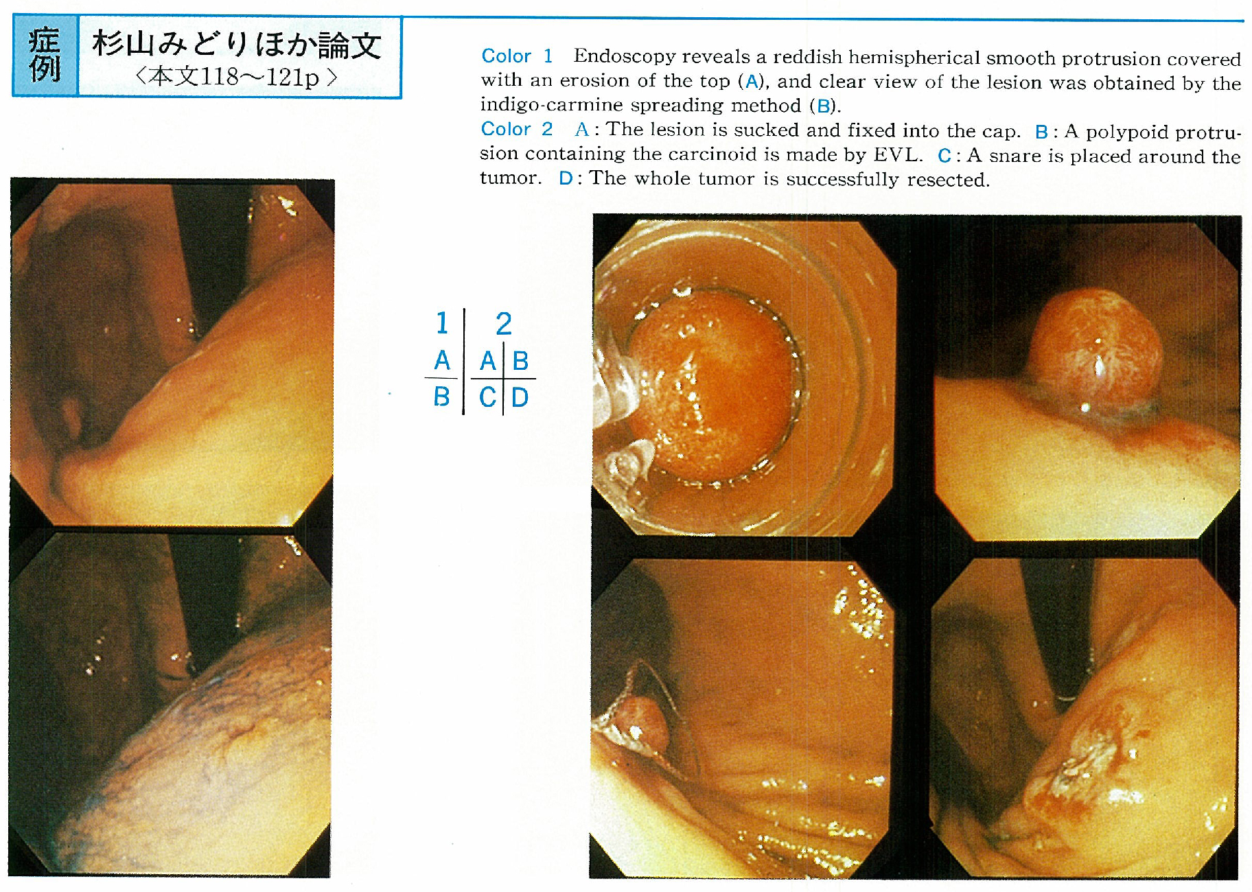
    1996 年 49 巻 p. 118-121
    発行日: 1996/12/02
    公開日: 2015/03/20
    ジャーナル フリー
     患者は44歳,男性。胃集団検診にて胃角の変形を指摘され,胃内視鏡検査にて胃体上部前壁に径約5mmの中心陥凹をもつ発赤した微小隆起が認められた。生検にてカルチノイドと診断されたが,超音波内視鏡検査において病変は粘膜固有層と粘膜下層上部に限局していたため,内視鏡的静脈瘤結紮術(endoscopic variceal ligation ; EVL)を用いて内視鏡的摘除を行った。切除標本の組織学的検索において,カルチノイド腫瘍は粘膜下層上部にとどまり,断端は水平・垂直方向ともに陰性で,切除1年2カ月後再発の徴候はない。内視鏡的治療の根治性を示唆する症例と思われ,今後のカルチノイド腫瘍の治療手段の1つとなりうると考え報告した。
  • 金井 明彦, 森 淑美, 金田 繁樹, 木幡 義彰, 中島 昌人, 清水 直樹, 渡辺 浩一, 片山 麻子, 田口 夕美子, 白鳥 泰正, ...
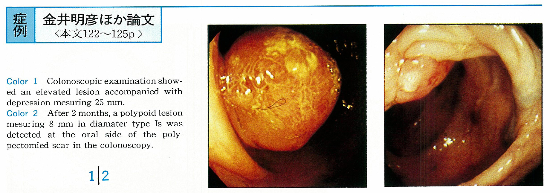
    1996 年 49 巻 p. 122-125
    発行日: 1996/12/02
    公開日: 2015/03/20
    ジャーナル フリー
     症例は47歳,女性。平成5年3月,検診で便潜血反応陽性を指摘された。大腸X線検査では横行結腸に有茎性ポリープを,大腸内視鏡検査で同部位に25mm大の表面に凹凸のある発赤調ポリープを認め,スネアポリペクトミーを施行した。病理組織像では深達度smの高分化型腺癌であった。2カ月後の大腸内視鏡検査では,ポリペクトミー後の瘢痕部口側に8mm大の無茎性ポリープを認めた。ポリペクトミーした病理組織像は深達度mの印環細胞癌であった。横行結腸部分切除術を施行したが,切除標本には癌細胞の残存はなかった。両病変の関連を考慮し連続切片を作成したが,初回病変は切除断端に高分化型腺癌が確認され,分割切除された可能性が示唆された。また2カ月後の病変は印環細胞癌で,連続切片のいずれにも両組織の混在はなかった。今回われわれは,特異な経過で発見された大腸早期印環細胞癌症例を経験したので報告した。
  • 中溝 裕雅, 浜田 慶城, 芹澤 宏, 渡辺 憲明, 熊谷 直樹, 土本 寛二, 山田 好則, 三浦 総一郎, 日比 紀文, 石井 裕正
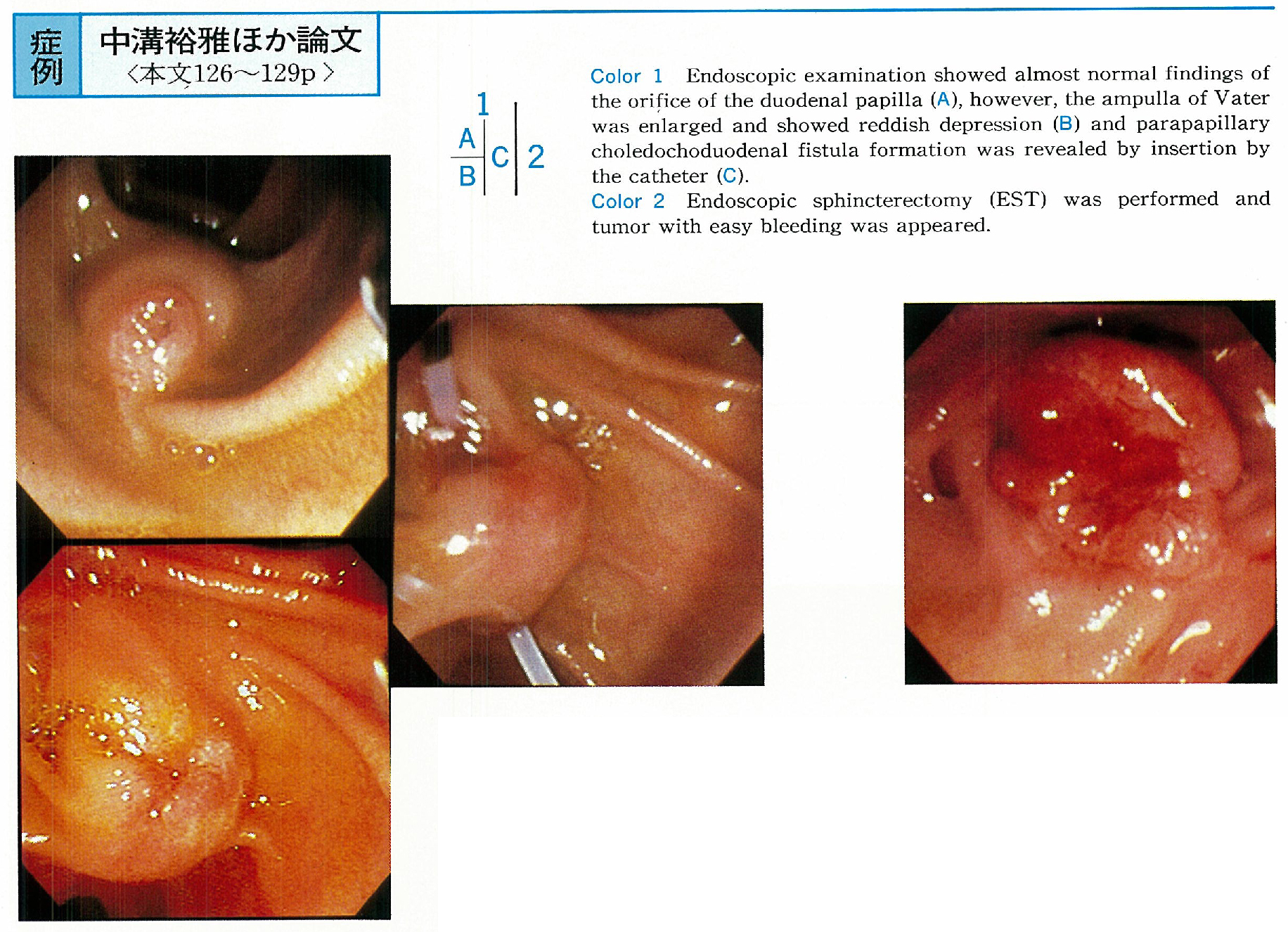
    1996 年 49 巻 p. 126-129
    発行日: 1996/12/02
    公開日: 2015/03/20
    ジャーナル フリー
     症例は77歳,女性。既往に胆石があり,近医に通院していた。平成7年12月11日より発熱,黄疸を認め,血液検査にて胆道系優位の肝機能障害がみられ,12月16日当院に紹介入院となった。腹部エコー,腹部CT検査より胆石を伴う急性胆囊炎が疑われ,抗生剤投与にて10日後には解熱,血液検査も改善したが,総胆管,肝内胆管の拡張が持続し,腫瘍マーカーも高値であったため,内視鏡的逆行性膵胆管造影を施行した。Vater乳頭膨大部の腫大,傍乳頭総胆管十二指腸瘻の形成が確認され,瘻孔部からの造影で総胆管の拡張,陰影欠損がみられ,腫瘍性病変が疑われた。内視鏡的乳頭括約筋切開術(EST)を施行し,腫瘍を露出させ,同部の生検より中等度分化型腺癌と診断した。傍乳頭総胆管十二指腸瘻を合併するVater乳頭部癌はまれであり,ESTを利用した組織生検の有用性を示した貴重な症例と考えられた。
  • 長田 達郎, 橋詰 清江, 山口 開, 小林 裕太郎, 鈴木 啓弘, 奥瀬 千晃, 高橋 泰人, 巴 雅威, 遠山 裕樹, 林 毅, 吉田 ...
    1996 年 49 巻 p. 130-132
    発行日: 1996/12/02
    公開日: 2015/03/20
    ジャーナル フリー
     症例は54歳女性。既往歴に胆囊内結石および胆囊腺筋症あり。平成7年8月人間ドックの腹部超音波検査で進行胆囊癌を指摘され,当科に紹介入院となった。腹部CT検査で肝床部,膵頭部および十二指腸への直接浸潤を認めた。ERCPでは,①胆囊体部から底部の内腔の狭小化,②胆囊管のVater乳頭近傍での低位付着,③総肝管とは独立した肝右葉後区域からVater乳頭まで続く副肝管をもつ重複胆管,④主膵管が副肝管で合流する膵管型の膵胆管合流異常,以上4点の異常所見を認めた。進行胆囊癌の診断のもと,当院外科において肝三区域切除兼尾状葉切除,膵頭十二指腸切除が行われた。重複胆管は非常にまれな疾患であり,本邦において胆道系悪性腫瘍を合併した報告はない。本例は重複胆管に胆囊癌が発症した興味深い症例と考え報告した。
臨床研究
  • 神沢 輝実, 荒川 丈夫, 江川 直人, 榊 信廣, 石渡 淳一
    1996 年 49 巻 p. 133-135
    発行日: 1996/12/02
    公開日: 2015/03/20
    ジャーナル フリー
     症例は49歳,女性。主訴は食後の胃部不快感と背部痛。上部消化管造影検査では十二指腸下行脚上部に圧排狭窄像を,腹部CT scanでは膵頭部に近接して,十二指腸下行脚の右側にも膵実質像を認めた。主乳頭からの内視鏡的逆行性膵胆管造影では,十二指腸をとり囲む環状膵管のみを認め,胆管はこの部分より下方で狭小化を呈した。副乳頭からの造影では拡張のない背側膵管が尾部まで造影され,2本の下頭枝を認めたが,環状膵との交通はなかった。膵管非癒合を伴った輪状膵(吉岡3型)と診断した。患者は内服治療で症状が改善し退院した。吉岡3型の輪状膵はまれであり,その成因として,右側腹側膵原基の過剰発現により形成された環状膵管が背側膵管と癒合しなかったのか,左側腹側膵原基の遺残と右側腹側膵原基の低形成によるものの,2つの可能性が考えられた。
内視鏡の器械と技術
  • 石原 学, 森 一博, 鳥居 尚隆, 高橋 敬二, 織田 良夫, 長山 徹, 古谷 亮, 吉谷 和男, 尾崎 元信, 西野 執, 大塚 幸雄
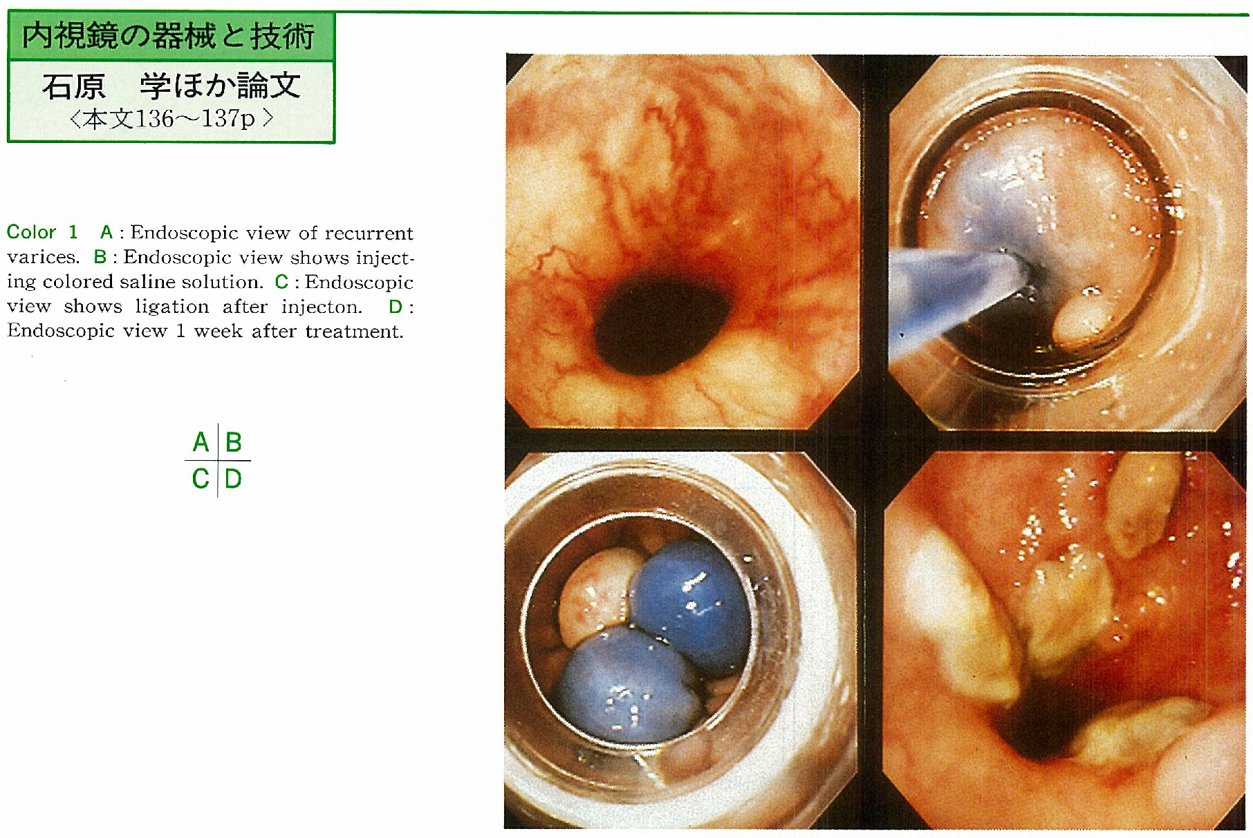
    1996 年 49 巻 p. 136-137
    発行日: 1996/12/02
    公開日: 2015/03/20
    ジャーナル フリー
    Endoscopic variceal ligation (EVL) is now common treatment of esophageal varices for the reason of safety and easy handling. But in the case of recurrent varices on hard fibrotic tissue EVL is not easy because endoscopic aspiration is difficult. We devised ligation method using paravariceal saline injection.
    Twelve cases of recurrent esophageal varices were treated by this method. All cases were performed disappearance of varices. EVL after saline injection is useful method for the reason for easy aspiration and ligation due to increasing submucosal volume. This method also has merits no use of sclerosant and no special apparatus.
  • 渋谷 進, 足立 信也, 河島 孝彦, 青柳 啓之, 飯田 浩行, 五本木 武士, 井坂 直秀, 大塚 雅昭, 深尾 立, 中原 朗, 福富 ...
    1996 年 49 巻 p. 138-139
    発行日: 1996/12/02
    公開日: 2015/03/20
    ジャーナル フリー
    In recent years, endoscopic variceal ligation (EVL) has been performed in the treatment of esophageal varices. It has been reported that esophageal perforation due to the over tube is a serious complication. Larger differentiation between diameters of endoscope and over tube make the insertion of the tube more painful. Therefore, we designed double over tube that has conventional over tube (outer tube) and thin one (inner tube) .
    We performed EVL in 13 cases of the primary treatment and 9 cases of the retreatment after the first EVL using conventional over tube for esophagogastric varix. As a result, insertion of double one is easier than that of conventional one in all the cases.
    The retreatment using the double over tube is more convenient for all the cases than the first treatment. No serious complications due to double over tube was encountered in all cases. Therefore, it was thought that our technique of a double over tube is easier to perform and more convenient for the patients.
  • 今泉 弘, 大井田 正人, 菅野 聡, 田辺 聡, 木田 光広, 小泉 和三郎, 勝又 伴栄, 西元寺 克禮
    1996 年 49 巻 p. 140-141
    発行日: 1996/12/02
    公開日: 2015/03/20
    ジャーナル フリー
    A prototype gastro-videoendoscope (GIF-XQ230AY) has been tested in 87 cases for routine upper gastrointestinal examination. The bending section of the tip of this scope was improved.
    The capacity of the observation was compared with prototype and two conventional scopes (GIF-XQ200 and XQ230) . The capacity of the prototype scope was higher than that of GIF-XQ200 significantly in angular region, upper and middle body, lesser curvature of cardiac region and duodenal bulb. And the capacity of this scope was higher than that of GIF-XQ230 in angular region, upper body and bulb.
    Improved bending section of a prototype scope was considered to be useful for upper gastrointestinal examination, especially the capacity of the observation with this scope was superior to the conventional videoendoscope.
  • 山崎 震一, 山崎 健二, 宮崎 高明
    1996 年 49 巻 p. 142-143
    発行日: 1996/12/02
    公開日: 2015/03/20
    ジャーナル フリー
    The short splinting device has been recognized as an effective instrument, for facilitating deeper insertion of the colonoscope and for taking back specimens repeatedly. However, there was a fear of the short splinting device slipping into the rectum because of the lubricant. So, we devised a sword guard for preventing this type of accident.
    It is made of urethane or silicone. The latter is more superior than the former because of its flexibility. The size is 80mm in outer diameter, 25mm in inner diameter, 5mm (3.5mm silicone) in thickness and 2mm in width of the groove. The sword guard can be taped to the patient or tied with string using the four holes. It is also possible to carry out the procedure without involving an assistant holding the sliding tube using this sword guard.
  • 中島 俊一, 古谷 正伸, 市川 一仁, 浮田 雄生, 松村 修志, 石井 俊也, 掛村 忠義, 石塚 俊一郎, 吉田 光宏, 高田 洋孝, ...
    1996 年 49 巻 p. 144-145
    発行日: 1996/12/02
    公開日: 2015/03/20
    ジャーナル フリー
    Development of the ultrasonic probe produced application to the gastrointestinal diseases easily. But in the cases with colonic lesions in the proximal part, the length of the probe sometimes became insufficient. According to the wide application of the probe to the colon disease, elongated probe was developed to avoid the difficulty of manipulation.
    Twenty seven cases of colorectal flat lesion were examined using long ultrasonic probe (XUM-3T, Olympus Co Ltd) and 31 cases were examined using ordinary probe (UM-3R) from October 1995 to May 1996. XUM-3T (20MHz) was elongated about 200mm from ordinary probe (UM-3R) . Controlability and quality of picture were studied using these probes.
    Controlability in the proximal part of colon, long type probe was used in all cases (100%) but cases with UM-3R was disturbed in a few cases (70%) . Visualization rate was 92.6% with XUM-3T and 87.1% with UM-3R in the whole colon. Especially in the proximal part of colon, it was 71.4% with XUM-3T and 80% with UM-3R, respectively.
    In summary, a long ultrasonic probe was useful when the lesion was located in the proximal part of the colon without evidence of diminishing quality of ultrasonogram.
  • 三上 哲弥, 河井 博明, 野津 史彦, 坂本 仁, 梅田 知幸, 秋田 泰, 吉川 望海, 三田村 圭二, 藤森 基次, 千葉 俊哉, 平 ...
    1996 年 49 巻 p. 146-147
    発行日: 1996/12/02
    公開日: 2015/03/20
    ジャーナル フリー
    We evaluated the usefulness of endoscopic papillary dilation (EPD) for treatment of patients with choledocholithiasis. Bleeding tendency, Vater's papilla in duodenal diverticulum, and poor general condition were risk factor of treatment with endoscopic sphincterotomy in 4 patients. A function of Vater's papilla had to be preserved in a young patient.
    Therefore, EPD was performed and all of 5 patients were successfully treated without serious complications. The maximal size of stones treated with EPD was 10 mm in diameter. Atrophy and fibrous changes were observed histologically in the musclar layer of Vater's papilla, but its fundamental structure was conserved in a patient died from normal pressure hydrocephalus 2 months after EPD. We considered that EPD was a safe and usefull therapy for choledocholithiasis.
臨床研究
  • 田村 茂生, 山田 俊彦, 市川 武, 阿部 毅彦
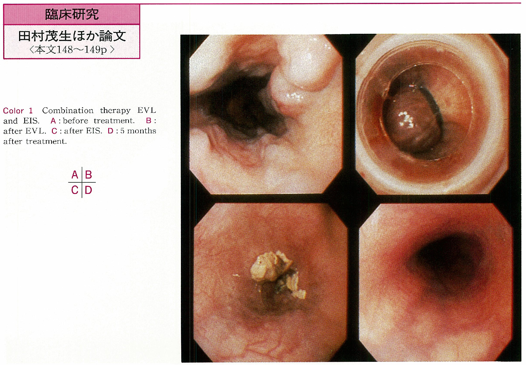
    1996 年 49 巻 p. 148-149
    発行日: 1996/12/02
    公開日: 2015/03/20
    ジャーナル フリー
    We analyzed the effects of combination therapy for esophageal varices, endoscopic variceal ligation (EVL) and endoscopic injection sclerotherapy (EIS) . Subjects were 7 cases treated by the combination therapy. And compared subjects which were treated by EVL only were 27 cases. All cases were positive of red color (RC) sign and therapeutic goal were negative of RC sign. Effective cases were negative of RC sign at one month after those treatments.
    Seven cases (100%) were effective after the combination therapy and 21 cases (77.8%) were after EVL only. As for the treated form of varices, 7 cases (100%) were F1 or F0 after combination therapy, but 14 cases (66.7%) were after EVL only. The relapse rate of these effective cases after combination therapy was 20.0% at 6 months, and after EVL only was 29.9% at 6 months, 56.2% at 12 months.
    These results suggest that combination therapy have no significant statistical difference in relation to efficacy and relapse rate at 6 months. But we consider that the combination therapy is more eradicated therapy than EVL only and further study is necessary for comparing the relapse rate. And we think that intravariceal EIS is effective to eradicate gastric varices.
  • 松久 威史, 井上 泰夫, 羽山 享宏, 山田 宣孝
    1996 年 49 巻 p. 150-151
    発行日: 1996/12/02
    公開日: 2015/03/20
    ジャーナル フリー
    A relation between serum pepsinogen I/II (PG I/II) ratio in gastroduodenal diseases and gastric mucosa observed with endoscope, Helicobacter pylori (H. pylori) infection, histological activity in biopsied specimen were studied.
    1) PG I/II ratio in H. pylori positive cases was significantly higher than that of negative cases. It was same in patients with gastric ulcer and control group which has no region except atrophic gastritis (each p<0.001) .
    2) According to Oshima's classification of gastric mucosa, which was classified into 5 groups by appearance of vascular pattern in lesser curvature of lower body, PG I/II ratio in H. pylori infected cases decreased with increase of atrophic grade.
    3) Histological activity by the Sydney system and PG I/II ratio were examined. It was higher in (-, ±) group, which has no activity, than in (+) , (⧺) , (⧻) groups, which have activity. H. pylori positive rate was low in (-, ±) group. On the other hand, it was high in (+) , (⧺) , (⧻) groups (each 100.0%, 100.0%, 88.2%) .
    4) We got a closely reration between PG I/II ratio and histological activity accompanied with H. pylori infection.
  • 坊 英樹, 伊藤 正秀, 豊島 明, 角田 誠之, 小山 雅章, 田中 賢助, 陸川 秀智, 堀口 実, 河西 徹, 福田 達弥, 吉森 勝 ...
    1996 年 49 巻 p. 152-153
    発行日: 1996/12/02
    公開日: 2015/03/20
    ジャーナル フリー
    We investigated the relationship between neutrophil infiltration and trace minerals with Helicobacter pylori. The subject of investigation are 10 cases which reacted to the Hp proved by positive 4 cases by negative. We studied the number of neutrophilia in gastric lamina propria mucosae per an unit area and quantity of zinc and copper in same mucosa.
    Hp positive gastric mucosa showed more marked neutrophil infiltration compared to Hp negative mucosa (p<0.05) . On the other hand, Hp positive gastric mucosa was more likely to be associated with low concentration of zinc and copper than Hp negative mucosa (p<0.05) . The abovementioned data disclosed the fact that the degree of neutrophil infiltration altered in reverse proportion to the amount of trace minerals in Hp positive gastric mucosa (r zn=0.65133, r cu=0.67195) .
    We confirmed the correlation between the neutrophil infiltration and the quantity of zinc and copper, in gastric mucosa infiltrated with Helicobacter pylori.
  • 小山 博, 成木 行彦, 西野 執, 北條 裕, 三浦 富宏, 近藤 栄作, 中谷 尚登, 長山 徹, 進藤 彦二, 石原 学, 岩崎 格, ...
    1996 年 49 巻 p. 154-155
    発行日: 1996/12/02
    公開日: 2015/03/20
    ジャーナル フリー
    We evaluated 13C-urea breath test (UBT) in a diagnosis of Helicobacter pylori (HP) after treatment with lansoprazole and clarithromycin. Fifty four patients inclused 24 peptic ulcer and 54 chronic gastritis, treated for 14 days with 30mg of lansoprazole and 600mg of clarithromycin in a day. Antral, lower body and upper body biopsy samples were taken for culture and histology. We evaluated 54 chronic gastritis with Sydney classification.
    Eradication was defined as the absence of HP in all speciments. HP was eradicated by treatment in 55.5% (30/54) . Severity of the side effects did not occurred. It was possible that low grade erosive gastritis were eradicated rather than severe grade erosive gastritis. Eradication of HP with lansoprazole and clarithromycin provided a safe and effective, and 13C-UBT examination were useful in observing the eradication of HP.
  • 小山 博, 成木 行彦, 西野 執, 有木 寿史, 北條 裕, 中谷 尚登, 長山 徹, 山田 秀一, 進藤 彦二, 岩崎 格, 瓜田 純久 ...
    1996 年 49 巻 p. 156-157
    発行日: 1996/12/02
    公開日: 2015/03/20
    ジャーナル フリー
    We evaluated 13C-urea breath test (UBT) in a diagnosis of Helicobacter pylori (HP) after treatment with lansoprazole and clarithromycin (CAM) or amoxicillin (AMPC) . Seventy three patients inclused 35 gastric ulcer, 15 duodenal ulcer and 73 chronic gastritis, treated for 14 days with 30mg of lansoprazole, 600mg of CAM or 2000mg of AMPC in a day. Antral, lower body and upper body biopsy samples were taken for culture and histology. We evaluated 73 chronic gastritis with Sydney classification.
    Eradication was defined as the absence of HP in all speciments. HP was eradicated by treatment in 63.0% (46/73) , by CAM/lansoprazole in 84.3% (16/19) and by AMPC/lansoprazole in 55.5% (30/54) . Severity of the side effects did not occurred. It was valid that AMPC/lansoprazole were eradicated rather than CAM/lansoprazole. Eradication of HP with lansoprazole and CAM or AMPC provided a safe and effective, and 13C-UBT examination were useful in observing the eradication of HP.
  • 角谷 宏, 糸井 隆夫, 武井 和夫, 荻原 正示, 真田 淳, 二木 修司, 武田 一弥, 三輪 一彦, 真神 易, 篠原 靖, 堀部 俊 ...
    1996 年 49 巻 p. 158-159
    発行日: 1996/12/02
    公開日: 2015/03/20
    ジャーナル フリー
    Gastric varices are frequently associated with inferior phrenic vein as an accessory shunt vessel (ASV) in addition to main shunt. How to control the blood flow of ASV is a matter of importance for transvenous obliteration therapy. Cases of successfully eradicated solitary gastric varices by controlling the blood flow of ASV using selfretaining catheter method (transvenous obliteration of porto-systemic shunt, TOPS) or coil embolization method are presented.
    In order to control the blood flow of ASV, ethanolamine oleate iopamidol (EO) was administrated through selfretaining catheter depend on varicearography findings and could be infused every day as occasion demands.
    We also performed coil embolization to stop the blood flow of ASV. Coil embolization was examined before ordinary transvenous obliteration therapy. Our results suggest that solitary gastric varices including usual difficult cases can be easily eradicated by these methods.
  • 中島 俊之, 西谷 篤史, 柿沼 徹, 岩堀 泰基, 吉岡 久, 矢部 諭, 大西 久仁彦, 藤原 研司, 中島 美智子, 林 浩司, 折茂 ...
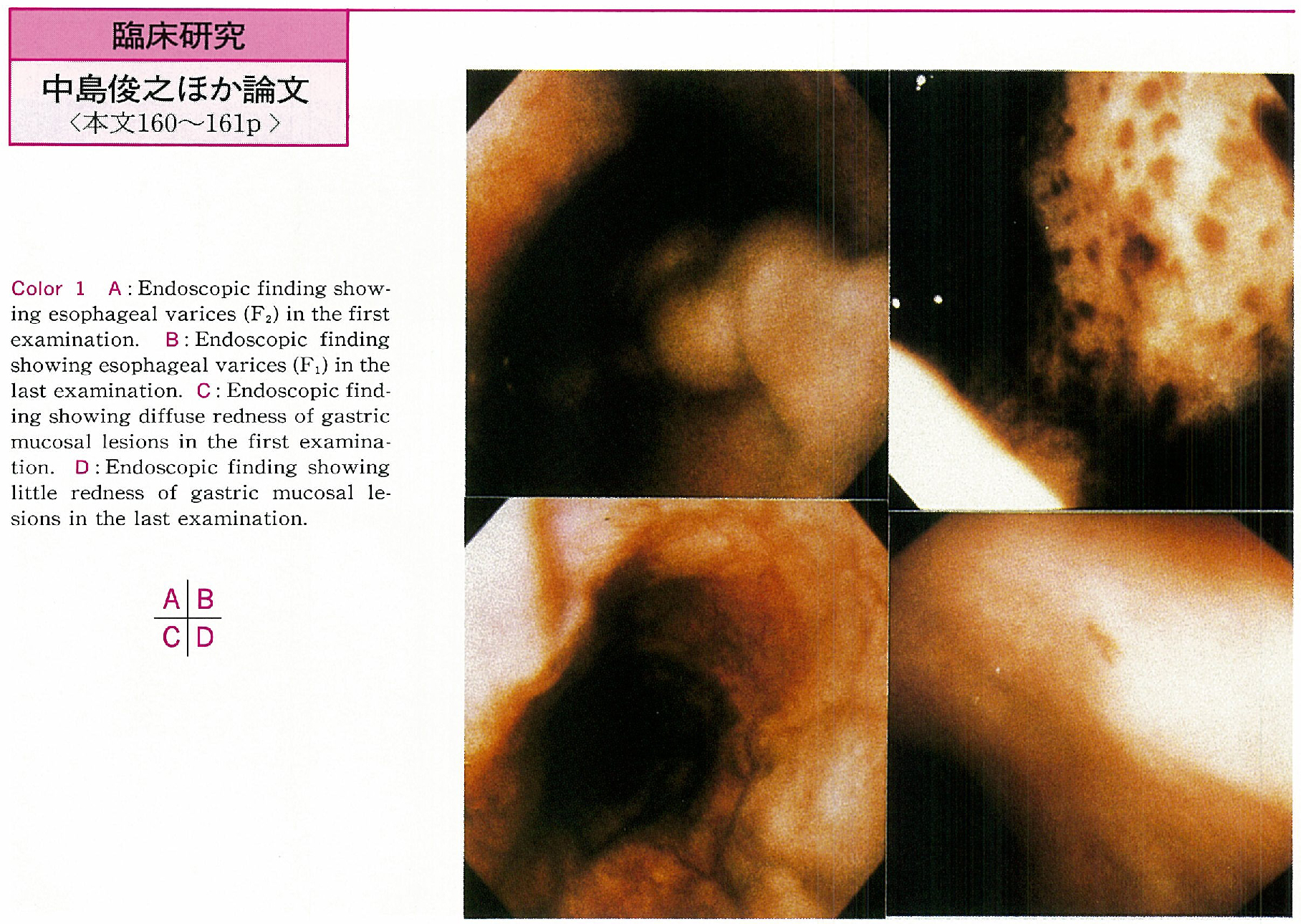
    1996 年 49 巻 p. 160-161
    発行日: 1996/12/02
    公開日: 2015/03/20
    ジャーナル フリー
    We evaluated the association between endoscopic findings of the upper digestive tract and portal hemodynamics. The subjects were 42 patients with liver cirrhosis complicated by esophageal varices in whom the clinical course were observed for 5 years. They were classified into the group who developed a paraumbilical vein during the course and that who did not. Etiology, child classification, male-female ratio and average of the age before observation was similar between two groups.
    Changes in endoscopic findings of the upper digestive tract during the course were slighter in the group II than the group I. Significant differance was observed in changes the mucosal lesions in the group I, the group I was significantly increased (p<0.05) , but the group II was not increased. Development of the major porto-systemic shunts without gastroesophageal varices was increased the incidence of endoscopic findings of the upper digestive tract caused by portal hypertension.
  • 小山 雅章, 伊藤 正秀, 角田 誠之, 田中 賢助, 馬越 正通
    1996 年 49 巻 p. 162-163
    発行日: 1996/12/02
    公開日: 2015/03/20
    ジャーナル フリー
    Forty-eight patients with functional dyspepsia who underwent endoscopy in the division during the past year were studied. Endoscopy was performed during the period on 4,153 patients with epigastrium complaints. Forty-eight patients (14 males, 34 females) were diagnosed functional dyspepsia based on the AGA definition.
    In this study we evaluated its background. The patient's ages ranged from 20-65 years, with the majority in their 20s in both sexes. The disorder developed most predominantly in males 40 years of age or younger, but throughout a wide range of ages in the female. Most of the patients complained of the symptoms mainly in January and April, suggesting that this disease is associated with social causes. No clear relationship between this condision and alcohol and/or tabacco was observed. The understanding of its background is essential to the therapy of functional dyspepsia.
  • 井上 詠, 矢島 知治, 中澤 敦, 久松 理一, 岡本 晋, 高石 官均, 上野 義隆, 細田 泰雄, 石井 裕正, 岩男 泰, 渡辺 守 ...
    1996 年 49 巻 p. 164-165
    発行日: 1996/12/02
    公開日: 2015/03/20
    ジャーナル フリー
    The details of mucosal surface in normal and inflamed lesions were investigated by magnifying colonoscope. Normal architecture of area colonicae (round or diamond-shaped) was easily visualized in colonic mucosa of healthy controls by the magnifying colonoscopy with dye-spraying contrast technique.
    Even in remission stage of ulcerative colitis, the abnormalities of area colonicae such as irregularity, redness or disordered arrangement, associated with or without microerosions, were demonstrated by magnifying colonoscopy. Relapsing rates in patients without abnormalities by magnifying colonoscopy were lower than in those with abnormal findings.
    In Crohn's disease, the magnifying colonoscopic observation with dye-spraying, revealed small erosions in the rectal mucosa with normal appearance. Two kinds of erosions, microerosions surrounded by almost normal architecture of the gland pits and small elevations associated with central erosions, were demonstrated by the close observation with high magnification.
    The present study suggests that the precise observation of the colonic minute lesions with magnifying colonoscopy will be helpful to elucidate the pathophysiology of the earlier lesions in inflammatory bowel disease.
  • 角田 誠之, 伊藤 正秀, 福田 達也, 小山 雅章, 佐藤 薫隆, 馬越 正通, 櫻井 四郎, 湯川 雅枝
    1996 年 49 巻 p. 166-167
    発行日: 1996/12/02
    公開日: 2015/03/20
    ジャーナル フリー
    Essintial elements deficiency or heavy metal poisoning were observed clinically. Physiological functions of trace elements in organism have not been known well. The purpose of this study was to verify the physiological validity of using trace elements in colonic mucosa as a mesure of the cancer stage. Therefore, we analyzed various trace elemens in colonic mucosa. Fifteen colonic adenomas were chosen as subjects of the study. Ten cases of normal colonic mucosa were used as the control group.
    The biopsy of colonic mucosa was performed with well informed concent using an electric endoscope EVIS-200HI (Olympus Co) . The sample was lyophilized at -80℃ immediatery. The feature of PIXE is its ability to analyze trace elements quantitatively without decomposing samples. Pd thin layer was deposited onto the sample on carbon adhesive tape uising metal ion sputtering equipment. The proton energy was 2.3MeV, 10nA. The beam spot area was 2×3mm, and the irradiation time was approximatery 10 minutes. Proton bombardment was carried out at National Institute of Radiological Sciences.
    Calcium, zinc, copper, iron, were determined quantitativerly. Among trace elements, Zn, and Cu and among non-trace elements Ca, and Fe were highly significant among three groups (significant at the p<0.1% level) . The concentration of Zn, Cu and Fe were higher in the adenoma group than in control group.
  • 吉本 一哉, 酒井 義浩, 古谷 正伸, 浮田 雄生, 市川 一仁, 松村 修志, 中島 俊一, 石井 俊也, 広瀬 安紀, 小林 博之, ...
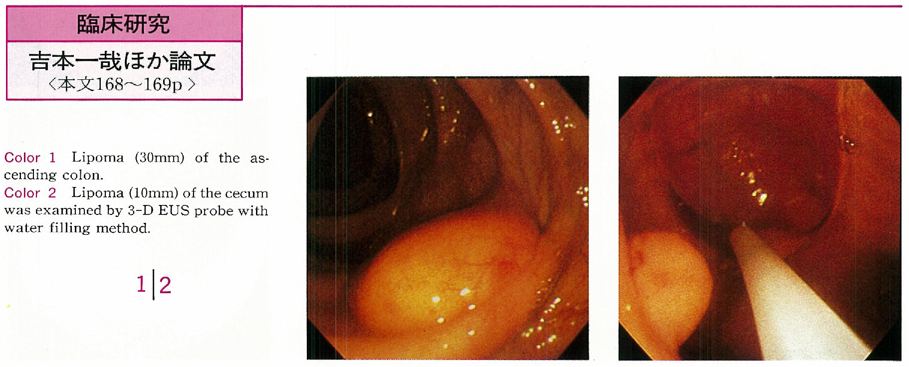
    1996 年 49 巻 p. 168-169
    発行日: 1996/12/02
    公開日: 2015/03/20
    ジャーナル フリー
    Endoscopic ultrasonography (EUS) was widely performed according to the routine application of the ultrasonic probe. 3-dimentional endoscopic ultrasonography (3-D EUS) system was developed using ordinary ultrasonic probe (UM-3R, UM-3T) by Olympus Co. From 1995 July to March 1996, 4 cases of lipoma of the colon were examined using 3-D EUS to evaluate the usefulness and the visualization.
    Four cases of lipoma were located in the right side colon. Tumor size were ranged from 10mm to 30mm and noted yellowish surface with cushion-sign endoscopically. EUS image were able to visualize in all cases. Ordinary radial image and linear image were also useful to diagose the lipoma in all cases. Reconstructed 3-D EUS image of the large lipoma provide much information for the diagnosis. But image of the samll lipoma was not so different from radial or linear image. Linear image sometimes seemed to be quite useful in the cases of the lesion on the fold or unvisible lesion.
    3-D EUS image thought to be given diagnosable information by internal echo findings also three dimentional tumor recognition in many cases. 3-D EUS will be advanced much more in near future and will be given much information for the endoscopic diagnosis.
  • 中村 尚志, 大倉 康男, 細井 董三, 塩田 滋朗, 郷 佳克, 知念 信昭, 益満 博, 荻原 英夫, 東 馨, 山田 耕三, 中井 呈 ...
    1996 年 49 巻 p. 170-171
    発行日: 1996/12/02
    公開日: 2015/03/20
    ジャーナル フリー
    Indication of endoscopic treatment to Type-I colon cancer is still controversial. In order to diagnose the cancerous depth invasion precisely, 65 lesions of Type-I were analysed in terms of endoscopic appearance.
    1) Type-I sm cancers were split into two subdivisions : typical group (30 lesions) and modified group (35) .
    2) Both groups significantly differed in the cancerous depth invasion (p<0.01, chisquare test) , resulting in the fact that modified group reaches much deeper than typical group (Table 1) . In typical group, sm1 80%, sm2 20%. In modified group, sm1 2.9%, sm2 and sm3 97.1%. Furthermore, modified group comprised erosive-ulcerative, nodular, and mixed type. With erosive-ulcerative, all 18 lesions proved sm2 or deeper. With nodular, 9 sm2 but one sm1. With mixed, all 7 lesions proved sm2 or deeper.
    3) Pertaining to the group and differentiation, group of typical and modified was significantly associated with the incidence of well/moderately differentiated cancer (p<0.01) . Typical group was linked with well differentiated cancer (73.3%) , modified group linked with moderately differentiated cancer (82.9%) .
    4) With vessel invasion, ly (+) was significantly higher in modified group (p<0.01) , while also so was v (+) in modified group. Ly (+) by 20% in typical group while in modified, ly (+) 80%. V (+) by 3.3% in typical while in modified, v (+) by 31.4%.
    To sum up, it was concluded that it is of the first importance to deal with Type-I sm cancer depending on the subdivision of typical group and modified group in order to discriminate sm1 from sm2 and deeper. This subdivision is useful to decide on a proper treatment between endoscopic removal and surgical operation with the precise preoperative diagnosis over cancerous depth invasion.
  • 笹木 淳司, 野崎 貞彦, 小山 洋, 尾関 豊, 松原 長樹
    1996 年 49 巻 p. 172-173
    発行日: 1996/12/02
    公開日: 2015/03/20
    ジャーナル フリー
    Twenty cases of rectal carcinoid were reviewed, which were treated with endoscopic resections at our hospital from October 1989 to April 1996. The mean age of patients was 55 years (range 27-80) . The ratio of men to women was 0.54 : 1 (=7 : 13) . The size of the tumors were less than 5mm in diameter in 6 cases and 5-8mm in 14 cases. The locations of the lesions were the upper rectum (Ra) in 4 cases and the lower rectum (Rb) in 16 cases. The depth of the tumors were submucosal layer in all cases.
    For the initial treatments snare polypectomies were done in 9 cases, endoscopoic mucosal resections (EMR) in 4 cases and hot biopsies in 7 cases. In 8 cases (40%) the lesions were completely resected without recurrences. But in 12 cases cut-end were positive with tumors. By follow-up study, no residual lesions were seen in 8 cases (40%) because of burning effects. But in 2 cases residual lesions were recognized histopathologically. Both cases were treated with hot biopsies initially. In the other 2 cases no follow-up study has been done yet.
    The rate of cut-end positive cases was high (6/7=86%) by hot biopsy technique, and there was the risk of residual lesions. So we recommend snare polypectomy or EMR for the initial endoscopic treatment of rectal carcinoids.
症例
  • 佐藤 治夫, 神津 照雄, 磯野 可一
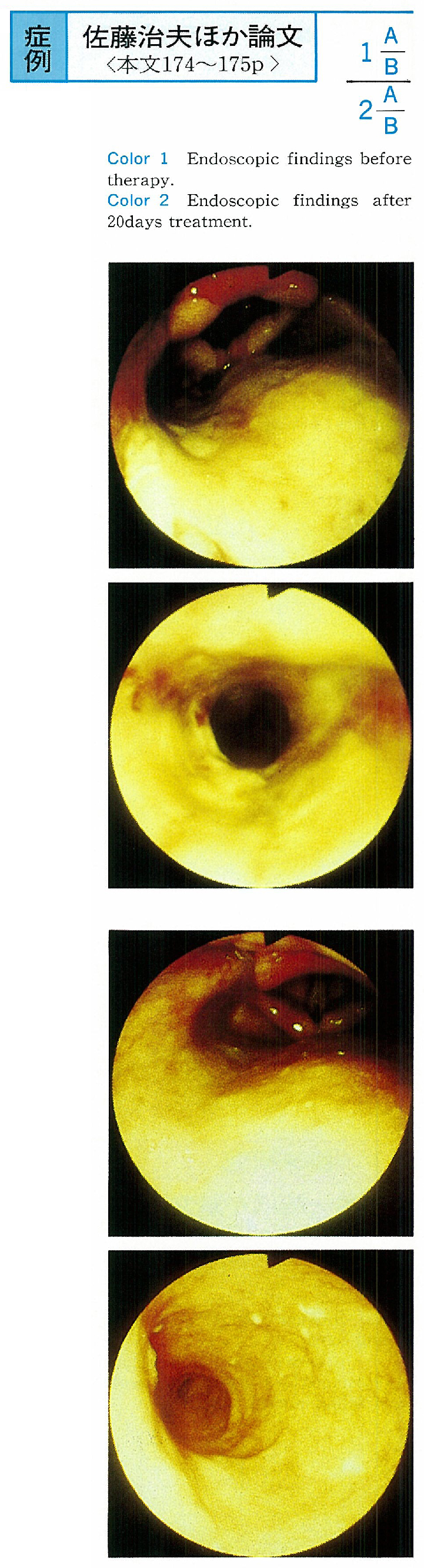
    1996 年 49 巻 p. 174-175
    発行日: 1996/12/02
    公開日: 2015/03/20
    ジャーナル フリー
    A case of 82-year-old woman who had esophagus lesion and bullous pemphigoid was reported. Hematoemesis was occured in her pemphigoid therapy. Endoscopic examination was done for upper gastrointestinal hemorrhage. The endoscopic findings of upper esophagus were covered with dick whitish exudate, in lower esophagus esophagitis, friable mucosa and no bullous lesions was seen. After her bullous pemphigoid therapy, her esophagus lesion was disappeared.
  • 末永 洋右, 村田 洋子, 鈴木 茂, 吉田 一成, 中村 英美, 井手 博子, 高崎 健, 藤井 昭義
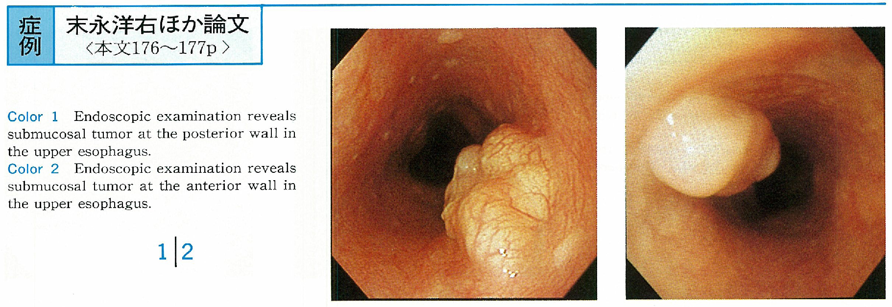
    1996 年 49 巻 p. 176-177
    発行日: 1996/12/02
    公開日: 2015/03/20
    ジャーナル フリー
    In 2 patients with no symptom, esophageal tumors were detected by screening endoscopy. The tumors were located at the upper esophagus and covered with normal esophageal mucosa. Endoscopic ultrasonography used by a small sized probe (20MHz) revealed that the tumors was connected with the lamina muscularis mucosa. And margin was clear, internal echo was homogeneous. Therefore, the tumors were diagnosed leiomyoma originated from the lamina muscularis mucosa and had indication for resection via endoscopy. These tumors were removed using sliding tube.
    Microscopic examination revealed that they were leiomyoma derived from the lamina muscularis mucosa and resected completely. So the diagnosis of location and origin of the tumor was accurate. Endoscopic ultrasonography using a small sized probe is useful for selection of endoscopic treatments in esophageal submucosal tumors.
  • 立松 秀樹, 町村 貴郎, 郭 宗宏, 篠原 央, 米川 甫, 古川 俊治, 栗原 直人, 長谷川 健
    1996 年 49 巻 p. 178-179
    発行日: 1996/12/02
    公開日: 2015/03/20
    ジャーナル フリー
    Benign tumors of the esophagus are few. Even among these tumors, suquamous cell of the esophagus of the esophagus is rare. With the recent progress in the diagnostic technology of the upper gastrointestinal endoscopy, the number of case reports about such tumors is gradually increasing, these cases are of clinical interest to understand the problems related to these diseases such as the etiologic factors and malignant change. In this report, meticulous endoscopic examination was carried out for the patient.
    This patient was a 60-year-old male. Barium meal examination showed a projecting lesion in the upper part of the esophagus. Upper gastrointestinal endoscopy revealed a mulberry-like tumor in the same location, with a longitudinal diameter of 10mm. Endoscopic mucosal resection (EMR) was carried out, and the lesion was diagnosed as squamous cell papilloma of the esophagus. A qualitative diagnosis of squamous cell papilloma of the esophagus can be made by through endoscopic observation on the basis of its characteristic morphological features. EMR is believed to be useful in the diagnosis and treatment of this tumor.
  • 竹内 裕也, 小澤 壯治, 安藤 暢敏, 石 志紘, 中村 榮一, 小柳 和夫, 林 浩二, 熊井 浩一郎, 北島 政樹
    1996 年 49 巻 p. 180-181
    発行日: 1996/12/02
    公開日: 2015/03/20
    ジャーナル フリー
    A 51-year-old man who underwent esophagectomy for intrathoracic esophageal cancer and reconstruction of gastric tube in January 1994 and had postoperative radiotherapy, was admitted to Keio University Hospital with a complaint of hematemesis and tarry stool in November 1995.
    Endoscopic examination showed a easily bled multiple erosions in the gastric tube. He was treated with proton pump inhibitor (PPI) and H2 receptor antagonist (H2RA) , but failed. Endoscopic laser-Doppler flowmetry revealed that mucosal tissue blood flow of the erosive lesion was elevated. The 24-hour pH monitoring showed pH was low in the gastric tube. The plasma gastrin level was just below the upper limit. The erosions were significantly improved and healed by the treatment with secretin in addition to PPI and H2RA.
    The above results suggest that it is important to perform not only endoscopy, but also mucosal tissue blood flow measuring, pH monitoring and the plasma gastrin level examination.
  • 渡辺 昌則, 伊藤 正秀, 原 一郎, 馬越 正通
    1996 年 49 巻 p. 182-183
    発行日: 1996/12/02
    公開日: 2015/03/20
    ジャーナル フリー
    A successful endoscopic treatment for the postoperative esophageal stricture is reported. The postoperative esophageal stricture secondary to a major leakage of anastomosis occurred after subtotal esophagectomy. The axial aberration in the esophageal stricture brought a difficulty to conventional endoscopic dilatation. Accordingly, we inserted endoscope from the mouth to the gastrostomy, and performed endoscopic retrograde dilatation, by using the Savary-Gilliard dilator. An approach from the dual directions made it possible to modify the axial aberration, therefore the guide wire was safely inserted aiming at the oral endoscopic fiber light. This approach from the dual directions enabled a secure endoscopic retrograde dilatation, that lead to the improvement of the passage and the suture insufficiency.
    The endoscopic retrograde dilatation can be applied not only to the postoperative stricture but also to the malignant stricture. Such endoscopic treatment will become increasingly useful for improving the quality of life, and offers significant benefits to the patients with terminal esophageal cancer.
  • 今枝 博之, 岩男 泰, 都築 義和, 宮口 信吾, 永田 篤文, 大久保 明子, 竹内 一郎, 亀谷 麒與隆, 小林 研介
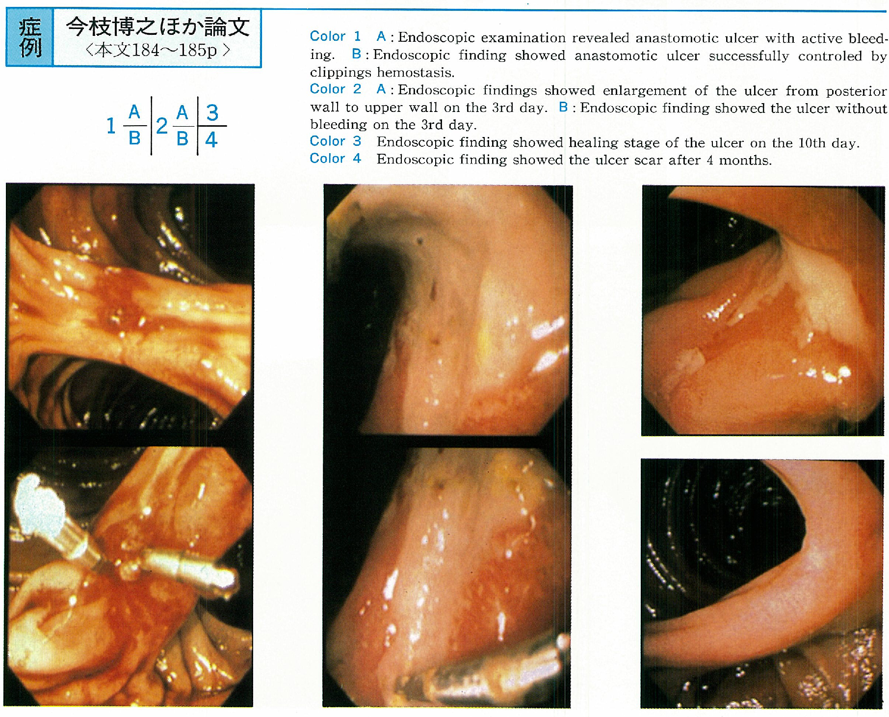
    1996 年 49 巻 p. 184-185
    発行日: 1996/12/02
    公開日: 2015/03/20
    ジャーナル フリー
    We report a patient with enlargement of anastomotic ulcer after clippings hemostasis. A 40-year-old man was admitted to our hospital complaining of hematemesis on July 30, 1994. He was received distal partial gastrectomy for bleeding from duodenal ulcer at age 38.
    Emergency endoscopic examination revealed an anastomotic ulcer with active bleeding in site of the jejunum. The bleeding was successfully controled by endoscopic clippings hemostasis. Endoscopic examination revealed enlargement of the ulcer from posterior wall to upper wall on the 3rd day.
    A case with bleeding gastrointestinal ulcer which was enlarged after clippings hemostasis has never been reported in Japan. This was the first case with bleeding gastrointestinal ulcer which was enlarged after clippings hemostasis. It is suggested that clippings hemostasis may disturb the supply of blood to the submucosal layer as same as in pure ethanol injection.
  • 大久保 明子, 今枝 博之, 都築 義和, 宮口 信吾, 永田 篤文, 竹内 一郎, 亀谷 麒與隆, 岩男 泰
    1996 年 49 巻 p. 186-187
    発行日: 1996/12/02
    公開日: 2015/03/20
    ジャーナル フリー
    A 73-year-old female was admitted to our hospital because of melena. Endoscopic findings showed an angiodysplasia with hematin, about 5mm in diameter on the greater curvature of gastric body. The lesion was ligated endoscopically using O-ring. It changed to necrotic tissue with O-ring after 4 days and ulcer scar after about 2 months.
    A 68-year-old male was admitted to our hospital because of hematoemesis. Endoscopic findings showed an angiodysplasia, about 6mm in diameter on the posterior wall of gastric body. The lesion was ligated endoscopically using O-ring. It changed to necrotic tissue with O-ring after 2days and small ulcer after a month.
    A case of hemorrhagic gastric angiodysplasia successfully controlled by endoscopic ligation has been rarely reported with only 3 cases in Japan, but this procedure is useful and safe for the treatment of hemorrhagic gastric angiodysplasia among several endoscopic procedures.
  • 山岡 佐世, 國分 茂博, 松本 偉男, 浅野 朗, 西元寺 克禮, 遠藤 高, 磯辺 義憲
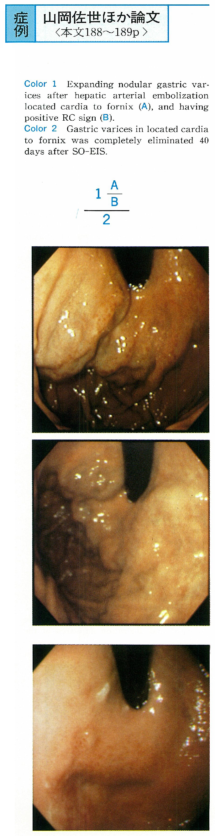
    1996 年 49 巻 p. 188-189
    発行日: 1996/12/02
    公開日: 2015/03/20
    ジャーナル フリー
    A 70-year-old male patient accompanying with liver cirrhosis complicated hepatoma was having solitary gastric varices enlarged after transcatheter hepatic arterial embolization. His endoscopic findings showed that varices were nodulary enlarged shape and having positive red color sign located from cardia to fornix. And also angiographic study showed gastrorenal shunt.
    He was tried to BRTO. But we could not visualze varices on BRTV by using 40 ml contrast medium. We thought the method of BRTO was not indicated because of poor obliteration for gastric varices of this case. We performed SO-EIS. BRTV just after SO-EIS showed filling defect meant thrombosed gastric varices.
    In this case endoscopic findings post 40 days since SO-EIS performed showed solitary gastric varices was completely eliminated. We thought SO-EIS had usefull for solitary gastric varices in cases where BRTO was not indicated.
  • 栗原 直人, 久保田 哲朗, 篠原 央, 米川 甫, 北洞 哲冶, 町村 貴郎, 古川 俊治, 郭 宗宏, 西堀 英樹, 立松 秀樹, 大谷 ...
    1996 年 49 巻 p. 190-191
    発行日: 1996/12/02
    公開日: 2015/03/20
    ジャーナル フリー
    A 45-year-old man with ulcer in the residual stomach was treated with anti-Helicobacter pylori (HP) therapy. After the patients had been treated intermittently because of his gastrointestinal ulcer for 6 years, his duodenal ulcer was perforated and he was treated with wide resection of the stomach followed by Billroth I reconstruction on November 1994. He visited our hospital on July 1995 because of epigastralgia, and the endoscopic examination revealed the ulceration in his residual stomach at H1 stage.
    He was diagnosed as positive HP-infection by rapid urease test, pathological finding, and bacterial culture using by selected Schirow's medium. After the patient was treated with H2 receptor anagonist (famotidine) for 4 weeks, and the ulceration improved at S1 stage, lansoprazole, amoxicillin and clarithromycin were applied for the eradication of HP. The eradication of HP infection was confirmed by 13C urea breath test, as well as rapid urease test, pathology and culture before and after the treatment. Ulceration in the residual stomach improved to S2 stage after eradication of HP with a recovery of gastrointestinal symptom of the patient.
  • 山口 研成, 加藤 雅士, 松岡 正記, 吉田 行哉, 早川 和雄, 福地 創太郎, 藤井 達也, 箱崎 幸也, 大庭 健一, 和田 了, ...
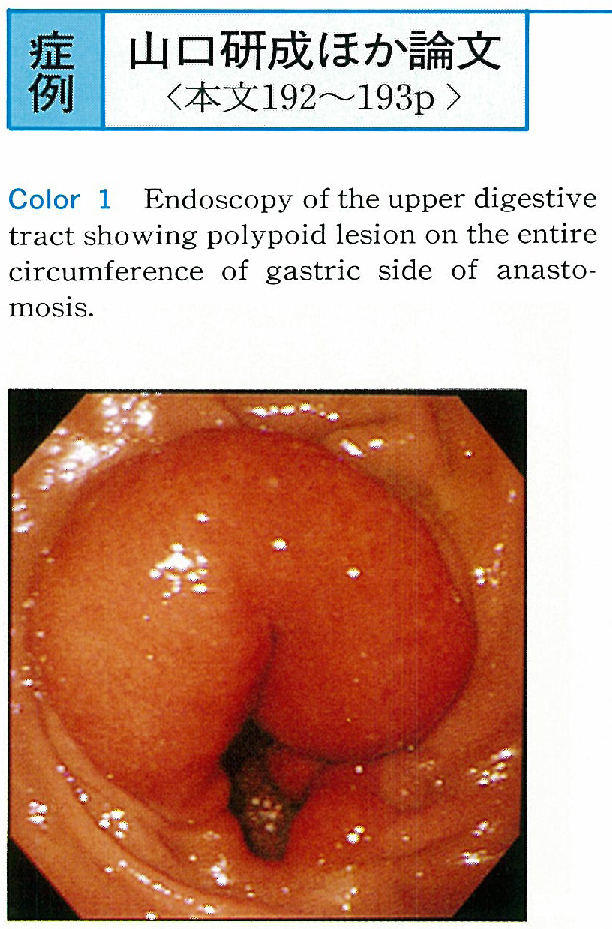
    1996 年 49 巻 p. 192-193
    発行日: 1996/12/02
    公開日: 2015/03/20
    ジャーナル フリー
    The patient was a 54-year-old male who had undergone distant partial gastrectomy with reconstruction by the Billroth II procedure 10 years earlier. Endoscopic examination of the upper digestive tract performed due to discomfort during swallowing showed a red tumorous lesion with a smooth surface on the entire circumference of the anastomosis site. Biopsy specimen revealed cystic dilatation and hyperplasia of the glands without atypia. Based on these findings, a diagnosis of gastritis cystica polyposa was made.
    Endoscopic ultrasonography showed high echoic tumor image in the submucosal layer and an aechoic-low echo multiple small cystic image within, which was covered with nearly normal first and second layers. Compared with histological image, these findings of endoscopic ultrasonography appeared to be specific to gastritis cystica polyposa.
  • 奥田 桂子, 毛利 勝昭, 真坂 彰, 田中 守義
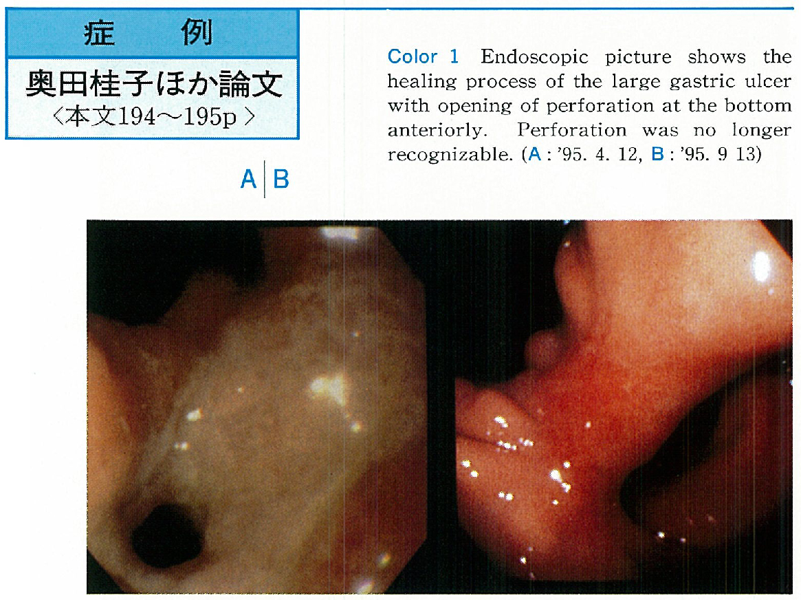
    1996 年 49 巻 p. 194-195
    発行日: 1996/12/02
    公開日: 2015/03/20
    ジャーナル フリー
    A 71-year-old woman suddenly vomited on March 21, 1995, and became giddy. The chest X-ray suggested cardiac failure and she entered the cardiac unit of this hospital. On admission, there was a marked anemia, and upper GI endoscopy on March 23 disclosed a large amount of coffee grounds and a large ulcer at the angulus ; she was transferred to the GI unit. Her general condition was poor, and was treated conservatively with fasting, intravenous hyperalimentation and an H2-blocker.
    On the 7th hospital day, endoscopy revealed an opening of perforation at the bottom of the ulcer anteriorly. Plain abdominal film did not show free air and fluoroscopy demonstrated a large pseudocyst communicating with the stomach. Abdominal CT scan showed some pleural fluid, and a cystic lesion near but separate from the stomach. There was no ascites.
    She was followed with continuous conservative treatment. With time, the ulcer became smaller and scarred, and the perforation no longer recognizable. The pseudocyst seen on the earlier barium examination progressively shrank, and eventually disappeared. This has been a rare experience of giant perforation with formation of a pseudocyst which was successfully cured by only conservative treatment.
  • 佐伯 伊知郎, 竹下 公矢, 谷 雅夫, 本田 徹, 神戸 文雄, 斎藤 直也, 遠藤 光夫
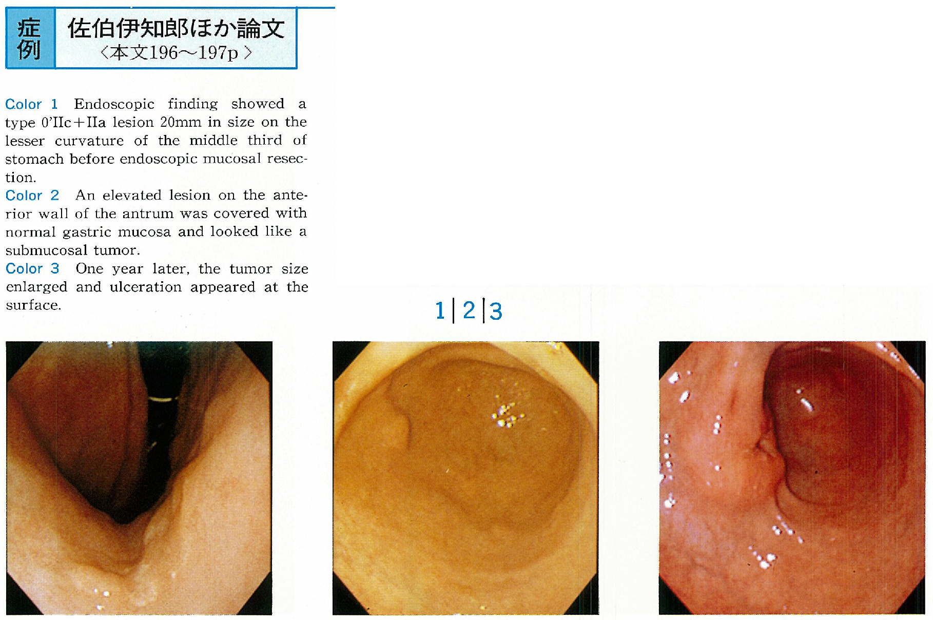
    1996 年 49 巻 p. 196-197
    発行日: 1996/12/02
    公開日: 2015/03/20
    ジャーナル フリー
    A 76-year-old male patient who had undergone endoscopic mucosal resection for a type 0́IIc+IIa lesion on the lesser curvature of the lower gastric body in February 1993, was pointed out an elevated submucosal-tumor-like lesion on the anterior wall of the antrum in October 1994.
    Follow up study in November 1995 showed that the tumor size enlarged and the ulceration appeared at the surface. The biopsy specimen revealed adenocarcinoma and gastrectomy was carried out. Histologically, the tumor was well differentiated adenocarcinoma and invaded to the proper muscle layer with lymph node metastasis.
    The clinical course was suggestive that it was the residual cancer after endoscopic resection. But the distance between the edge of the ulcer scar and the center of tumor was 15mm, so we finaly diagnosed that the tumor was a metachronous multiple cancer.
  • 伊東 祐一, 高木 和俊, 冨田 利夫, 佐久間 敦, 伊藤 生二, 小暮 洋暉
    1996 年 49 巻 p. 198-199
    発行日: 1996/12/02
    公開日: 2015/03/20
    ジャーナル フリー
    We report a rare case of extraluminal giant leiomyosarcoma of the stomach. A 60-year-old male was admitted with a palpable fist-sized tumor in the upper abdomen. Endoscopy showed a smooth elevated lesion in the posterior wall of the middle body of the stomach, but mucous membrane showed no other lesions. By endoscopic ultrasonography, the tumor was demonstrated as a heterogeneous lesion including partly anechoic area. Angiogram revealed a tumor in the middle body of the stomach similar to that in CT scanning and MRI.
    Wedge resection of the stomach was performed for the tumor. The resected specimen was a giant leiomyosarcoma with a pedicle. It measured 14.5×12.0×10.0cm and weighed 950g. The cross section showed a solid tumor with white capsule including several parts of necrosis inside. Histologically, the tumor consisted of scattered spindle-shaped cells.
  • 日野 昌力, 藤崎 順子, 秋庭 宏紀, 千葉井 基泰, 奥脇 秀一郎, 新井 弥生, 荒川 広志, 小泉 大樹, 金 哲宇, 田中 文彦, ...
    1996 年 49 巻 p. 200-201
    発行日: 1996/12/02
    公開日: 2015/03/20
    ジャーナル フリー
    Case 1 : A 44-year-old man. Endoscopic examination showed a submucosal tumor 30mm in size, in the lesser curvature of the lower body of the stomach. Endoscopic ultrasonography (EUS) showed a hypoechoic lesion and a heterogenous internal echo. We resected the mucosa covering the tumor by the EMR-L method and removed the specimen from the exposed tumor. Pathological study showed heterotopic pancreatic tissue.
    Case 2 : A 64-year-old woman who was found to have a submucosal tumor 30 mm in size in the lower body of the stomach. We removed the spcimen from the exposed tumor by EMR-L. Pathological findings showed heterotopic pancreatic tissue.
    We could definitely diagnose both hetrotopic pancreatic tissues by biopsy after EMR-L and have followed up the cases. This biopsy technique is useful for defferential diagnosis and to determine how to manage submucosal tumors.
  • 関根 正幸, 卜部 元道, 溝渕 昇, 三島 吾朗, 天野 高行, 榊原 宣
    1996 年 49 巻 p. 202-203
    発行日: 1996/12/02
    公開日: 2015/03/20
    ジャーナル フリー
    Endoscopic polypectomy for duodenal bulb tumor has not been performed aggressively, because of the polypectomic technique in the narrow lumen being difficult, and having a high risk. We report two cases of duodenal bulb tumor removed safely by the endoscopic polypectomy.
    Case 1 : A 44-year-old woman, who had a duodenal submucosal tumor located at the anterior wall of the duodenal bulb pointed out 4 years ago. Recently, the tumor grew up to 2.5cm in size, was elastic soft, and the pedunculated type. The tumor easily prolapsed into the stomach. The CT scanning revealed that the tumor was localized within the duodenal wall. An endoscopic polypectomy was performed in the stomach with a two-channeled endoscope. The histological examination revealed a Brunnerioma.
    Case 2 : A 39-year-old man, who had a duodenal polyp located at the anterior wall of the doudenal bulb pointed out 10 years ago. Recently, the tumor grew up to 1.5cm in size, was elastic soft, and the semipedunculated type. An endoscopic ultrasonography revealed the main location of the tumor was in the mucosal layer. An endoscopic polypectomy was performed in the doudenal bulb. The histological examination revealed a hyperplasia of Brunner's gland.
  • 丸田 和夫, 日高 道生, 清水 慎介, 中田 健一, 小林 智子, 五頭 三秀, 井川 守仁, 須藤 一郎, 杉浦 弘和, 堀向 文憲, ...
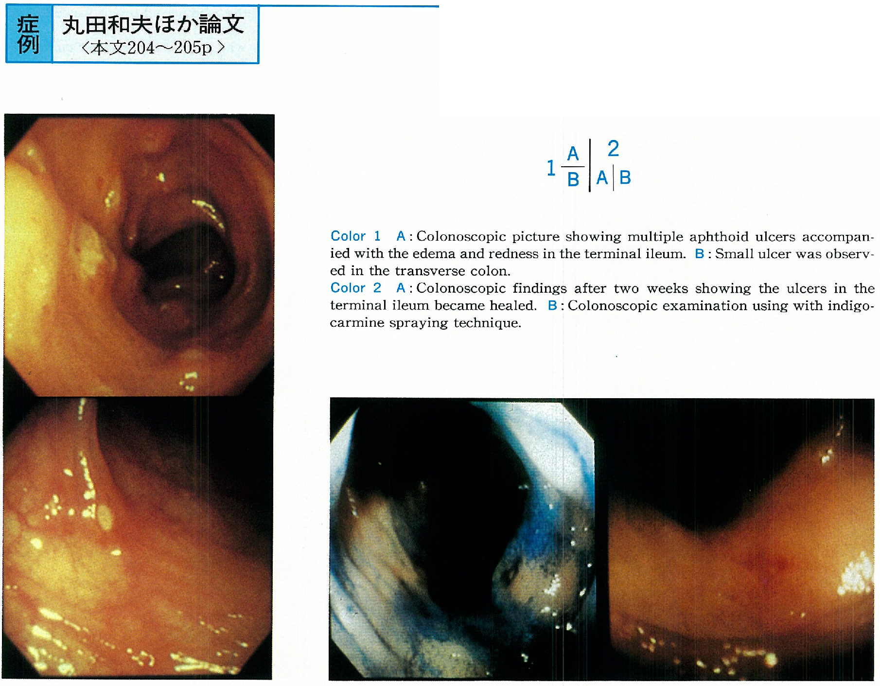
    1996 年 49 巻 p. 204-205
    発行日: 1996/12/02
    公開日: 2015/03/20
    ジャーナル フリー
    A 54-years-old female visited our hospital for bloody stool. She had a history of having taken diclofenac soudium as suppositories 10 days and tablet for 3 days for her lumbago.
    Colonoscopic finding showed multiple aphthoid ulcers accompanied with the edema and redness in the terminal ileum and revealed some small ulcers in the transverse colon. Another colonic mucosa showed normal vascular pattern and no inflammatory findings. Microscopic view of the biopsy specimen obtained from the margin of ulcer in the terminal ileum revealed erosin and slight infiltration of lymphocyte.
    We diagnosed NSAIDs induced enterocolitis, and we ordered her to stop taking NSAIDs. After two weeks, the ulcers in the terminal ileum became healed colonoscopically. This kind of case were rare and the literatures regarding the similar cases were also reviewed.
  • 伊佐山 浩通, 加藤 善久, 辻野 武, 皆川 伸幸, 山田 尚士, 関谷 祐之, 中田 良, 吉次 通泰, 庵 政志, 永岡 栄, 喜島 ...
    1996 年 49 巻 p. 206-207
    発行日: 1996/12/02
    公開日: 2015/03/20
    ジャーナル フリー
    A 56-year-old male visited our outpatient clinic for the examination for rectal polyp pointed out by barium enema. A granular superficial spreading tumor of the cecum was disclosed by colonoscopy. Biopsied specimen from the tumor showed a carcinoma in tubulovillous adenoma. The tumor resected revealed carcinoma in tubulovillous adenoma with severe atypia.
    Histologically the carcinoma and tubulovillous adenoma with severe atypia are indistinguishable on HE stain. Therefore, we stained the tumor immunohistochemically, with Ki-67 and p53 antibodies, to differentiate objectively carcinoma from adenoma with severe atypia. Many Ki-67 positive cells in both tissues reveal high grade proliferation. This suggests whole tissue is carcinoma. On the other hand, neither was stained by p53 although the immunohistochemistry with the gene is useful for the detection of cancer. Thus we could not objectively decide whether the whole is cancer or the cancer exists in part. Tumors stained by p53 have a tendency to invade more deeply.
    Generally speaking, a granular superficial spreading tumor has a tendency to grow rather horizontally than vertically in the colon mucosa. The immunohistochemistry in this case as mentioned above supports this.
feedback
Top