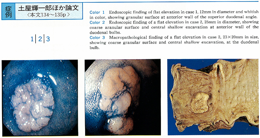
Case 1 : A 58-year-old female was first seen in our hospital because of abnormality found by gastric checkup study. Endoscopic examination revealed a flat elevation, 12mm in diameter, and whitish in color, at the duodenal bulb. Endoscopic mucosal resection (EMR) of the carcinoma was carried out. Histological examination showed well differentiated adenocarcinoma proliferating within the mucosal layer.
Case 2 : Endoscopic examination of a 67-year-old female was performed because of anemia revealing a flat elevation, 25mm in diameter, at the duodenal bulb. Subtotal gastrectomy and duodenectomy were carried out for curative resection of carcinoma. Histological examination showed well differentiated adenocarcinoma with submucosal invasion at the excavation. Histological staging was represented as follows : p0, h0, n0, sm, Stage I. Carcinoma cells permeated in the lymphatic vessels and small veins (ly1, v1) .