
52 巻
選択された号の論文の63件中1~50を表示しています
掲載論文カラー写真集
-
1998 年 52 巻 p. 1-22
発行日: 1998年
公開日: 2015/01/22
PDF形式でダウンロード (28985K)
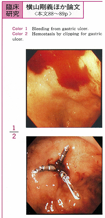
臨床研究
-
1998 年 52 巻 p. 57-61
発行日: 1998/10/20
公開日: 2015/01/22
PDF形式でダウンロード (573K) -
1998 年 52 巻 p. 62-65
発行日: 1998/10/20
公開日: 2015/01/22
 PDF形式でダウンロード (425K)
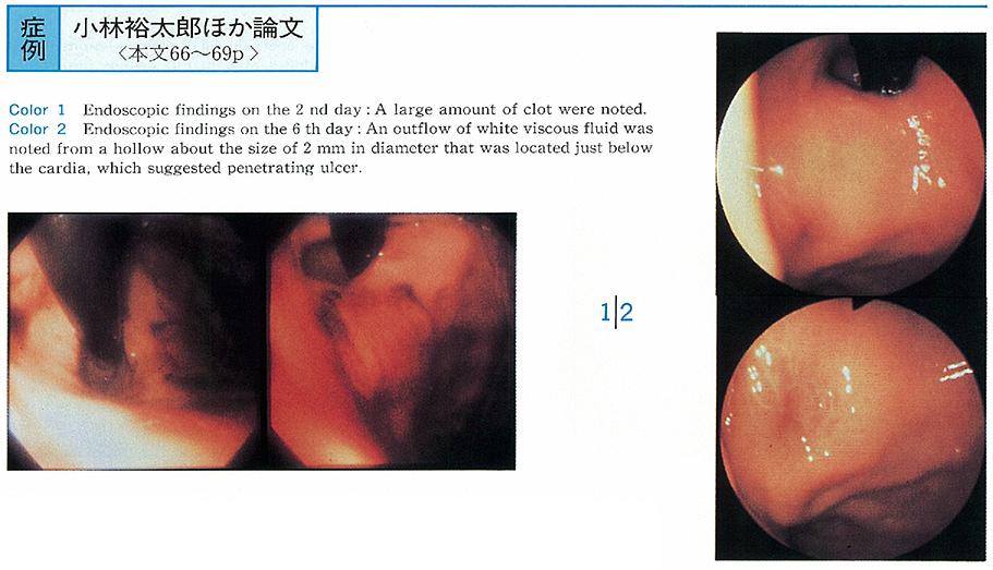
症例
-
1998 年 52 巻 p. 66-69
発行日: 1998/10/20
公開日: 2015/01/22
 PDF形式でダウンロード (931K)
 -
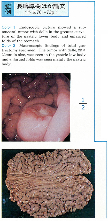
1998 年 52 巻 p. 70-73
発行日: 1998/10/20
公開日: 2015/01/22
 PDF形式でダウンロード (1178K)
 -
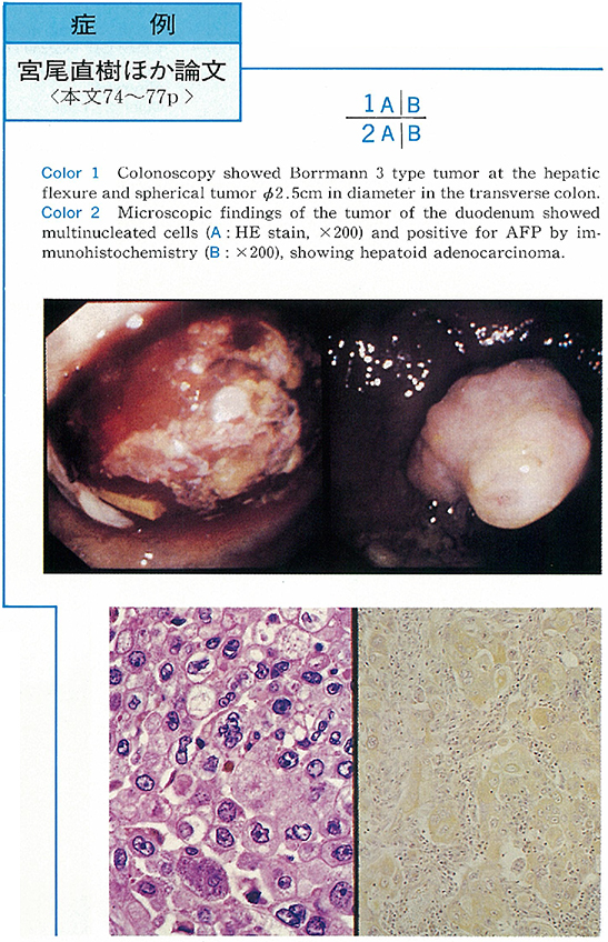
1998 年 52 巻 p. 74-77
発行日: 1998/10/20
公開日: 2015/01/22
 PDF形式でダウンロード (1409K)
 -
1998 年 52 巻 p. 78-81
発行日: 1998/10/20
公開日: 2015/01/22
 PDF形式でダウンロード (1101K)
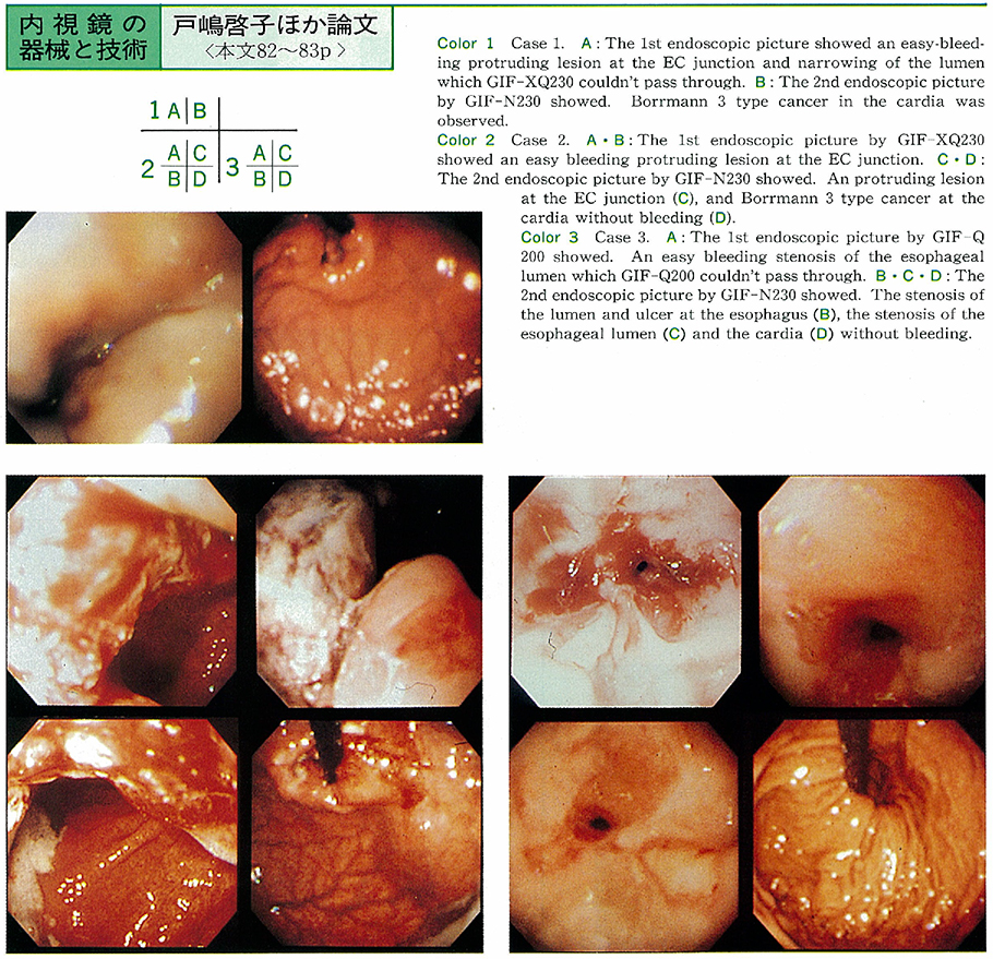
内視鏡の器械と技術
-
1998 年 52 巻 p. 82-83
発行日: 1998/10/20
公開日: 2015/01/22
 PDF形式でダウンロード (501K)
 -
1998 年 52 巻 p. 84-85
発行日: 1998/10/20
公開日: 2015/01/22
PDF形式でダウンロード (175K) -
1998 年 52 巻 p. 86-87
発行日: 1998/10/20
公開日: 2015/01/22
 PDF形式でダウンロード (663K)
臨床研究
-
1998 年 52 巻 p. 88-89
発行日: 1998/10/20
公開日: 2015/01/22
 PDF形式でダウンロード (239K)
 -
1998 年 52 巻 p. 90-91
発行日: 1998/10/20
公開日: 2015/01/22
PDF形式でダウンロード (396K) -
1998 年 52 巻 p. 92-93
発行日: 1998/10/20
公開日: 2015/01/22
PDF形式でダウンロード (290K) -
1998 年 52 巻 p. 94-95
発行日: 1998/10/20
公開日: 2015/01/22
PDF形式でダウンロード (246K)
症例
-
1998 年 52 巻 p. 96-97
発行日: 1998/10/20
公開日: 2015/01/22
 PDF形式でダウンロード (239K)
 -
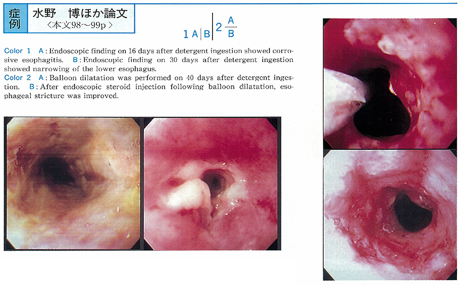
1998 年 52 巻 p. 98-99
発行日: 1998/10/20
公開日: 2015/01/22
 PDF形式でダウンロード (544K)
 -
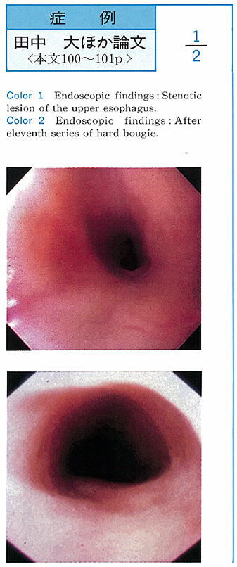
1998 年 52 巻 p. 100-101
発行日: 1998/10/20
公開日: 2015/01/22
 PDF形式でダウンロード (1195K)
 -
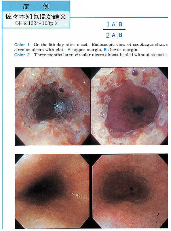
1998 年 52 巻 p. 102-103
発行日: 1998/10/20
公開日: 2015/01/22
 PDF形式でダウンロード (469K)
 -
1998 年 52 巻 p. 104-105
発行日: 1998/10/20
公開日: 2015/01/22
 PDF形式でダウンロード (533K)
 -
1998 年 52 巻 p. 106-107
発行日: 1998/10/20
公開日: 2015/01/22
 PDF形式でダウンロード (958K)
 -
1998 年 52 巻 p. 108-109
発行日: 1998/10/20
公開日: 2015/01/22
 PDF形式でダウンロード (1231K)
 -
1998 年 52 巻 p. 110-111
発行日: 1998/10/20
公開日: 2015/01/22
 PDF形式でダウンロード (782K)
 -
1998 年 52 巻 p. 112-113
発行日: 1998/10/20
公開日: 2015/01/22
 PDF形式でダウンロード (1063K)
 -
1998 年 52 巻 p. 114-115
発行日: 1998/10/20
公開日: 2015/01/22
 PDF形式でダウンロード (252K)
 -
1998 年 52 巻 p. 116-117
発行日: 1998/10/20
公開日: 2015/01/22
 PDF形式でダウンロード (627K)
 -
1998 年 52 巻 p. 118-119
発行日: 1998/10/20
公開日: 2015/01/22
 PDF形式でダウンロード (457K)
 -
1998 年 52 巻 p. 120-121
発行日: 1998/10/20
公開日: 2015/01/22
 PDF形式でダウンロード (445K)
 -
1998 年 52 巻 p. 122-123
発行日: 1998/10/20
公開日: 2015/01/22
 PDF形式でダウンロード (954K)
 -
1998 年 52 巻 p. 124-125
発行日: 1998/10/20
公開日: 2015/01/22
 PDF形式でダウンロード (500K)
 -
1998 年 52 巻 p. 126-127
発行日: 1998/10/20
公開日: 2015/01/22
 PDF形式でダウンロード (679K)
 -
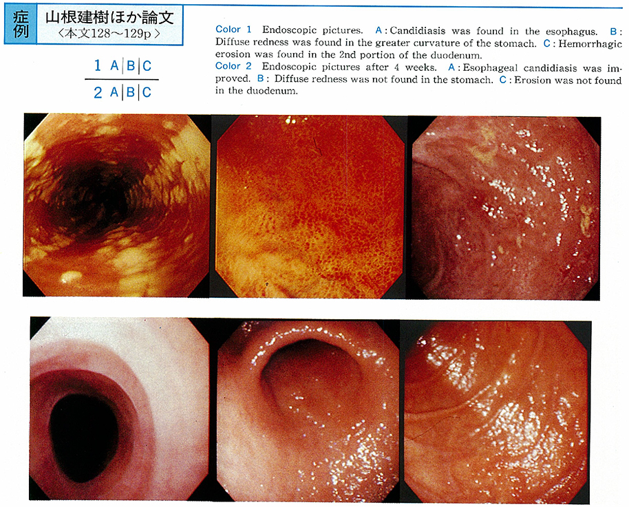
1998 年 52 巻 p. 128-129
発行日: 1998/10/20
公開日: 2015/01/22
 PDF形式でダウンロード (642K)
 -
1998 年 52 巻 p. 130-131
発行日: 1998/10/20
公開日: 2015/01/22
 PDF形式でダウンロード (466K)
 -
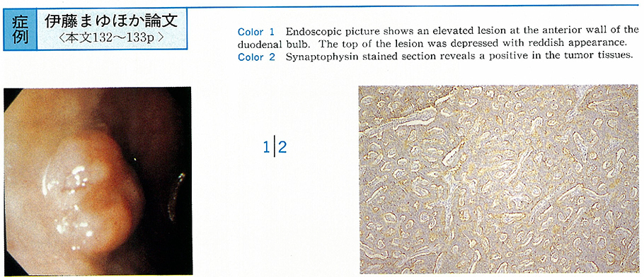
1998 年 52 巻 p. 132-133
発行日: 1998/10/20
公開日: 2015/01/22
 PDF形式でダウンロード (844K)
 -
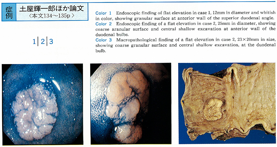
1998 年 52 巻 p. 134-135
発行日: 1998/10/20
公開日: 2015/01/22
 PDF形式でダウンロード (1077K)
 -
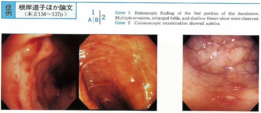
1998 年 52 巻 p. 136-137
発行日: 1998/10/20
公開日: 2015/01/22
 PDF形式でダウンロード (579K)
 -
1998 年 52 巻 p. 138-139
発行日: 1998/10/20
公開日: 2015/01/22
 PDF形式でダウンロード (591K)
 -
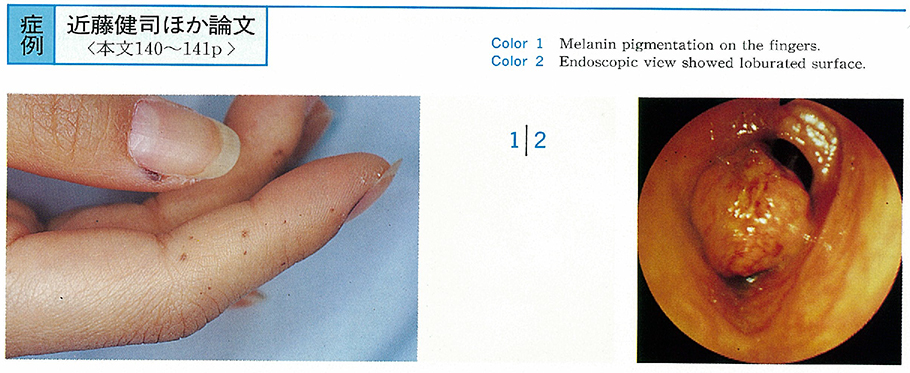
1998 年 52 巻 p. 140-141
発行日: 1998/10/20
公開日: 2015/01/22
 PDF形式でダウンロード (1364K)
 -
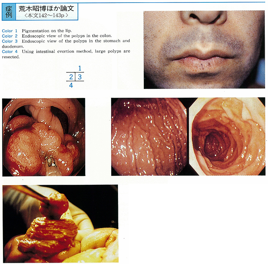
1998 年 52 巻 p. 142-143
発行日: 1998/10/20
公開日: 2015/01/22
 PDF形式でダウンロード (754K)
 -
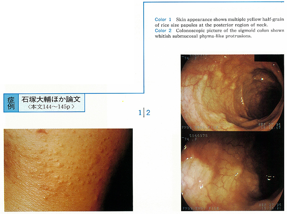
1998 年 52 巻 p. 144-145
発行日: 1998/10/20
公開日: 2015/01/22
 PDF形式でダウンロード (718K)
 -
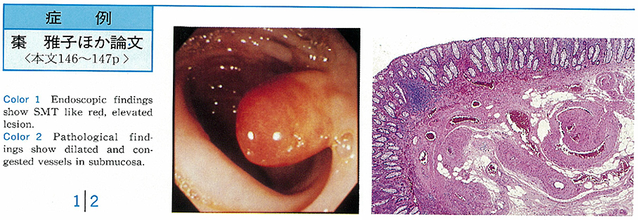
1998 年 52 巻 p. 146-147
発行日: 1998/10/20
公開日: 2015/01/22
 PDF形式でダウンロード (525K)
 -
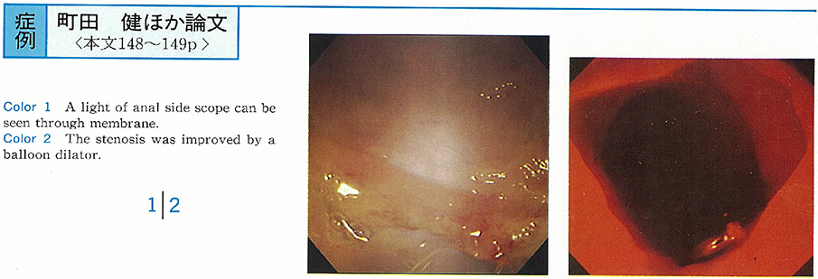
1998 年 52 巻 p. 148-149
発行日: 1998/10/20
公開日: 2015/01/22
 PDF形式でダウンロード (732K)
 -
1998 年 52 巻 p. 150-151
発行日: 1998/10/20
公開日: 2015/01/22
 PDF形式でダウンロード (636K)
 -
1998 年 52 巻 p. 152-153
発行日: 1998/10/20
公開日: 2015/01/22
 PDF形式でダウンロード (189K)
 -
1998 年 52 巻 p. 154-155
発行日: 1998/10/20
公開日: 2015/01/22
 PDF形式でダウンロード (483K)
 -
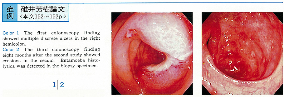
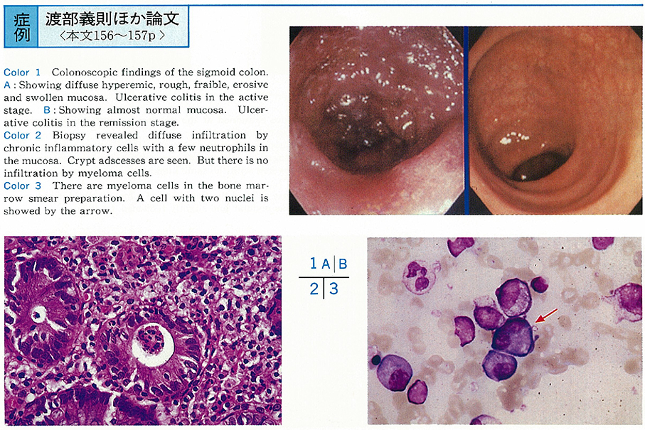
1998 年 52 巻 p. 156-157
発行日: 1998/10/20
公開日: 2015/01/22
 PDF形式でダウンロード (665K)
 -
1998 年 52 巻 p. 158-159
発行日: 1998/10/20
公開日: 2015/01/22
 PDF形式でダウンロード (762K)
 -
1998 年 52 巻 p. 160-161
発行日: 1998/10/20
公開日: 2015/01/22
 PDF形式でダウンロード (230K)
 -
1998 年 52 巻 p. 162-163
発行日: 1998/10/20
公開日: 2015/01/22
 PDF形式でダウンロード (1152K)
 -
1998 年 52 巻 p. 164-165
発行日: 1998/10/20
公開日: 2015/01/22
 PDF形式でダウンロード (982K)
 -
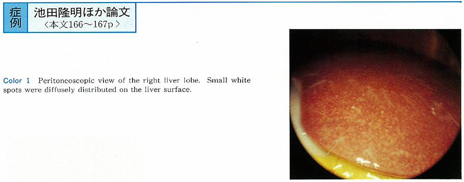
1998 年 52 巻 p. 166-167
発行日: 1998/10/20
公開日: 2015/01/22
 PDF形式でダウンロード (545K)