抄録
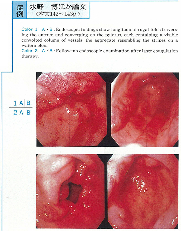
A 75-year-old man in whom hypertension was diagnosed in 1987 developed a mild anemia for the first time in December 1996. He complained of passage of tarry stool since March 1997. Endoscopic examination was said to reveal chronic hemorrhagic gastritis and he treated with oral iron and famotidine 40mg twice a day. But, recurrent tarry stool did not improved. Because of anemia and hypoproteinemia, he was admitted to our hospital in August 1997. Endoscopic examination revealed longitudinal red stripes and diffuse erythematous spots indicating dilated vessels in the gastric antrum. Endoscopic mucosal resection specimen demonstrated dilated vessels in the proper gastric mucosal layer, leading to a diagnosis of gastric antral vascular ectasia (GAVE) .
Because he refused surgical and endoscopic treatment at first, he received medical treatment again. But he had no improvement. In November 1997, laser treatment was started by his agreement. Photo coagulation was carried out using Nd-YAG laser. The power setting for photo coagulation averaged 39W and the pulses were delivered at a duration of 0.5 seconds. After three sessions, the dilated vessels in the antrum were almost obliterated and his anemia and hypoproteinemia improved subsequently. There were no complications as a result of laser treatment. Laser photo coagulation is safe and effective treatment for GAVE and should be considered as first line therapy.