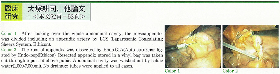
54 cases of acute appendicitis were operated laparoscopically for past 3 years (Three cases were converted to open exploration.) . Patients were divided into three groups according to postoperative pathological findings of appendix. (Group A : catarrhal, Group B : phlegmonous, Group C : gangrenous) . In each groups, operation time, postoperative period of bed rest and NPO (nothing per os) , pyretic term after operation, length of hospital stay and postoperative complications were analyzed to estimate the quality of laparoscopic appendectomy. Three ports were inserted on the abdomen ; under umblication, right upper abdomen (3 or 5 mm) and above left pubic (12 mm in a diameter) . After looking over the whole abdominal cavity, the mesoappendix was divided including an appendix artery by LCS (Laparosonic Coagulating Sheers System, Ethicon) and then, the root of appendix was dissected by Endo-GIA (Auto suture) or ligated by Endo-loop (Ethicon) . Resected appendix stored in a vinyl bag was taken out through a port of above pubic. Abdominal cavity was washed out by saline water (1,000-7,000 ml) . No drainage tubes were applied to all cases.
In Group C, period of bed rest was longer than Group A. No significant difference was detected in another classification. There were no remarkable complications as wound infection or abdominal abscess after surgery except paralytic ileus in two patients. These results demonstrate laparoscopic surgery is appropriate, and should be standard method for acute appendicitis.