International Journal of Surgical Wound Care
Online ISSN : 2435-2128
Volume 4, Issue 4
Displaying 1-8 of 8 articles from this issue
Original Articles
  • Rie Uehara, Akiyoshi Kubo, Yasuyuki Oyatsu, Saeko Yui, Shunichiro Ego, ...
    2023 Volume 4 Issue 4 Pages 128-132
    Published: December 01, 2023
    Released on J-STAGE: December 01, 2023
    JOURNAL FREE ACCESS
    Background: Infected urachal anomalies are rare, and there have been few reports on their causative bacteria. This study aimed to understand the clinical features and determine the bacteriological profile of patients with infected urachal anomalies at our hospital.
    Methods: This retrospective study included 55 patients with infected urachal anomalies managed at Saku Central Hospital Advanced Care Center in Japan for 10 years. We reviewed hospital records and collected information about diagnoses, age, sex, symptoms, type classification, treatments, pathological specimens, and bacteriological examinations.
    Results: The mean patient age was 29 years (range: 0–93 years), with 37 males and 18 females. The most common symptoms were umbilical pain or discharge (76%), followed by abdominal pain (18%) and urinary symptoms (5%). The most common anatomical classification of the urachus was external sinus in 46 patients (84%). Surgery was performed in 42 patients (76%), and 13 (24%) were treated conservatively. Bacteriological examinations were performed in 46 patients. Bacterial growth was observed in 44 samples (96%), and 97 microorganisms were isolated. Bacteriological examinations revealed mixed infections caused by anaerobic bacteria. Among the isolated bacteria, the most commonly cultured were Prevotella spp. (9%), followed by Bacteroides spp. (8%), Staphylococcus spp. (6%), Streptococcus spp. (6%), and Corynebacterium spp. (6%).
    Conclusions: This is the first study to report the bacteriological profiles of patients with infected urachal anomalies. When patients present with an infected urachal anomaly, the initial management should include broad-spectrum antibiotic therapy targeting anaerobes, and anaerobic culture during bacteriological examination is essential.
    Drawings illustrating the classifications of the types of urachal anomalies: patent urachus, urachal cyst, external urachal sinus, and urachal diverticulum. Fullsize Image
    B = bladder.
    Download PDF (1157K)
  • A Pilot Study
    Atsuhiko Iwao, Akihito Higashi, Yuki Moriuchi, Shoko Ashizuka, Noriko ...
    2023 Volume 4 Issue 4 Pages 133-138
    Published: December 01, 2023
    Released on J-STAGE: December 01, 2023
    JOURNAL FREE ACCESS
    Introduction: Wound odors are caused by bacteria and necrotic tissue, which severely degrade a patient’s quality of life. However, the management of wound odors is difficult for physicians. Dialkylcarbamoyl chloride-coated dressings can reduce bacterial load in wounds through hydrophobic interactions. In this study, we evaluated whether dialkylcarbamoyl chloride-coated dressings reduced wound odor by reducing bacterial load.
    Materials and methods: The target population was composed of patients with wounds and those who met the criteria for the use of dialkylcarbamoyl chloride-coated dressings. The dressing and gauze were changed every 24 hours. Before and 7 days after the use of the dialkylcarbamoyl chloride-coated dressing, gauze soaked in exudate was collected, then the odor was subjectively rated using a six-stage odor intensity scale, and objectively assessed using a handheld odor meter. A swab culture test of the wounds was performed simultaneously and the bacterial loads were compared.
    Results: Dialkylcarbamoyl chloride-coated dressing use tended to reduce wound odor as evaluated by the six-stage odor intensity scale (2.0 ± 1.6 vs. 3.7 ± 1.2, P = 0.005) and the handheld odor meter (334.7 ± 409.4 vs. 754.8 ± 352.0, P = 0.003). However, there was no difference in bacterial load (7.7 × 10⁶ ± 1.2 × 10⁷ vs. 2.8 × 10⁷ ± 6.0 × 10⁷, P = 0.21).
    Conclusion: This study demonstrated that dialkylcarbamoyl chloride-coated dressings reduce wound odor in subjective and objective assessments. These findings may provide a new option for managing odorous wounds.
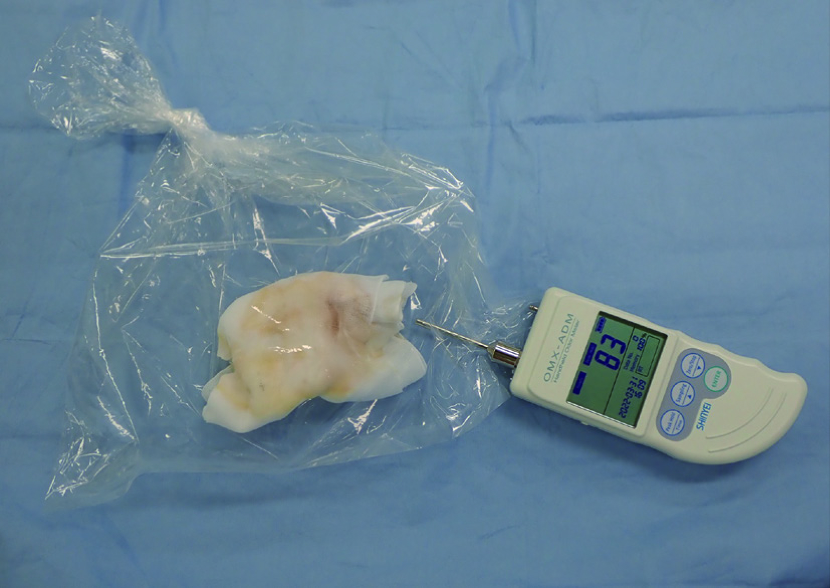
    Gauze soaked in exudate was packed in a polyethylene bag, then the odor in the bag was evaluated using OXM-ADM. Fullsize Image
    Download PDF (1117K)
Case Reports
  • A Case Report
    Kento Takaya, Rieko Shimizu, Kazuo Kishi
    2023 Volume 4 Issue 4 Pages 139-141
    Published: December 01, 2023
    Released on J-STAGE: December 01, 2023
    JOURNAL FREE ACCESS
    Pentamidine is one of the oldest drugs currently used to treat Pneumocystis carinii pneumonia. Reports of complications from administration include skin side effects, such as skin necrosis, sterile abscesses, and ulceration at the injection site when administered intramuscularly, and urticaria formation near the site of injection when administered intravenously. We herein report an unintended subcutaneous injection of pentamidine in the abdomen of a patient with asthma instead of omalizumab, which resulted in soft tissue depressions and adipose tissue necrosis secondary to dermatitis without skin ulceration. The dermatitis improved with the use of a topical steroid ointment, but the depressions persisted; therefore, scar resection and liposuction were performed to improve the morphology of the abdomen.
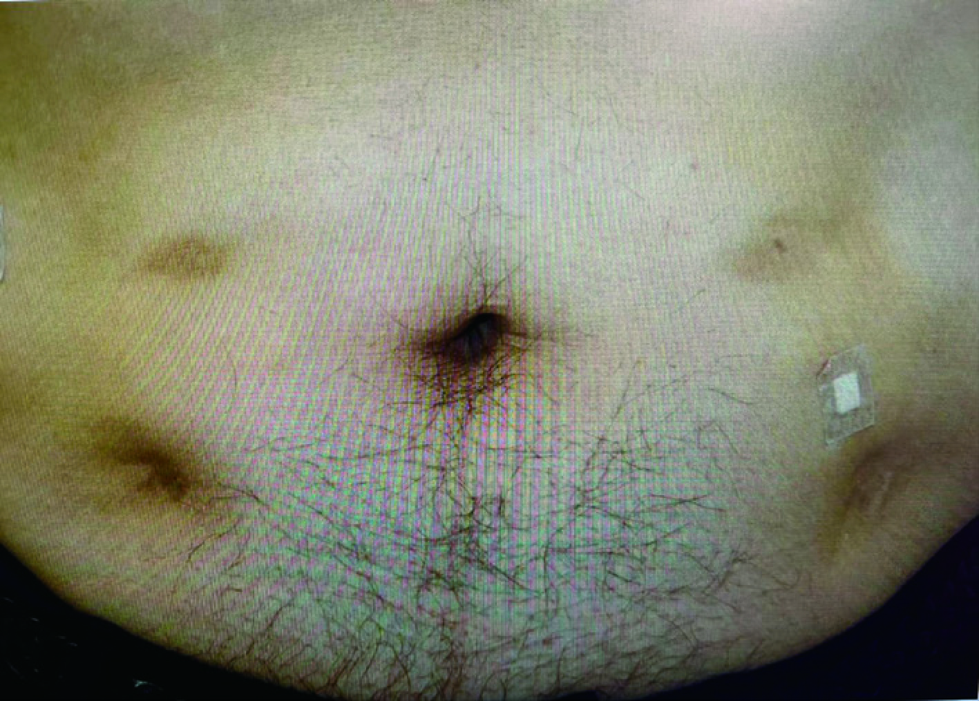
    Sixty days after the subcutaneous administration of pentamidine, prominent skin depressions at a visually discernible level and significant hyperpigmentation were evident. Fullsize Image
    Download PDF (1218K)
  • Tomomi Uchida, Karin Ikeda, Takashi Yamaki
    2023 Volume 4 Issue 4 Pages 142-145
    Published: December 01, 2023
    Released on J-STAGE: December 01, 2023
    JOURNAL FREE ACCESS
    Although there are numerous reports on fungal infections in other implanted medical devices, fungal infections in silicone gel breast implants are considered extremely uncommon. We herein report a rare case in which Candida albicans infection after breast implant replacement resulted in implant removal. The patient presented with symptoms of infection 35 days after breast implant replacement. The patient’s symptoms were mild, persistent, and unresponsive to antibiotics. Culture tests of fluid aspirate showed Candida albicans, which was initially suspected to be contaminated and left untreated. The recognition of fungal infections in augmented breasts is challenging, as they are rarely encountered but should always be considered as a possibility.
    Appearance of skin post implant replacement. Fullsize Image
    Right: Post implant replacement day 15: no symptoms. Middle: Day 35 when she first presented symptoms. Left: Day 84 when redness of the skin gradually worsened.
    Download PDF (1200K)
  • Airi Yamashita, Erika Guilpain Kusajima, Susumu Honda, Chu Kimura
    2023 Volume 4 Issue 4 Pages 146-149
    Published: December 01, 2023
    Released on J-STAGE: December 01, 2023
    JOURNAL FREE ACCESS
    Ehlers-Danlos syndrome is an umbrella term that includes a heterogeneous group of inherited connective tissue disorders. Classical Ehlers-Danlos syndrome is characterized by skin fragility, easy bruising, skin hyperextensibility, and joint hypermobility. Patients with this condition often develop wound dehiscence; however, limited information is available on wound care in these patients. This report describes the case of a 12-month-old boy who sustained a deep laceration on his right knee when he fell out of bed. Noting that the dermis was thinner and more fragile than normal, we performed suturing carefully with tension in two layers and waited for longer sutures. The wound took 4 weeks to heal but did so without unsightly scarring. However, the patient continued to present with recurrent injuries. Therefore, we performed a detailed examination, including molecular testing, and finally diagnosed the patient with classical Ehlers-Danlos syndrome. We identified several specific considerations that are important for wound management in classical Ehlers-Danlos syndrome.
    Clinical photographs showing the laceration to the right knee. Fullsize Image
    (a) Photograph taken on the day of injury showing a 6-cm laceration with exposed subcutaneous tissue. (b) Photograph taken on day 8 after suturing. (c) Photograph taken on day 15 after removal of all sutures showing poor adhesion at some of the wound edges. (d) Photograph taken 2 months after suturing showing that the wound has healed without scarring.
    Download PDF (1173K)
  • Mami Shoji, Hiroaki Kuwahara, Rei Ogawa, Satoshi Akaishi
    2023 Volume 4 Issue 4 Pages 150-154
    Published: December 01, 2023
    Released on J-STAGE: December 01, 2023
    JOURNAL FREE ACCESS
    Congenital antithrombin III deficiency is an inherited disorder associated with a tendency toward thrombosis. As laboratory findings are not always correlated with the clinical severity, there is no standardized approach for the treatment of leg ulcers, which are often associated with disease or perioperative anticoagulation management. We herein report the case of a patient with congenital antithrombin III deficiency with a stasis skin ulcer on the leg that was refractory to conservative treatment due to repeated episodes of cellulitis, who underwent segmental skin grafting and treatment with a combination of heparin and congenital antithrombin. The patient had an uncomplicated and satisfactory outcome, with no graft loss due to thrombosis or perioperative bleeding. Perioperative anticoagulation management of leg ulcers due to congenital antithrombin III deficiency has not been reported previously. We believe that appropriate perioperative management with anticoagulants is necessary for patients with chronic leg ulcers, considering the high rate of comorbidity of this disease. The strict management of this case resulted in successful treatment; however, the extent to which management is required requires further investigation.
    Warfarin, AT3, and heparin administration and changes in APTT, AT3, and PT-INR over time: AT3 activity increased after its administration, with the target level reached on day 3, and remained constant thereafter. Fullsize Image
    Warfarin was resumed after complete withdrawal of bed rest following skin tie-off. On the seventh day of resumption, after the PT-INR reached > 2, both AT3 and heparin were discontinued. AT3: antithrombin III, APTT: activated partial thromboplastin time, PT-INR: prothrombin timeinternational normal ratio
    Download PDF (1726K)
  • A Case Report
    Toshifumi Yamashiro, Izumi Yasutake, Ryuichi Azuma
    2023 Volume 4 Issue 4 Pages 155-159
    Published: December 01, 2023
    Released on J-STAGE: December 01, 2023
    JOURNAL FREE ACCESS
    Traumatic lumbar hernia is a rare condition, and there is no consensus regarding its treatment. Although direct closure or mesh reconstruction is preferred for the treatment of traumatic abdominal wall hernias, we successfully repaired a traumatic lumbar hernia using a fascia lata autograft in an acute setting. A 55-year-old man experienced traumatic inferior lumbar hernia after being caught in heavy machinery. Emergency laparotomy was performed to rule out intra-abdominal organ injury and simultaneous repair of the lumbar hernia was planned. Owing to the high degree of tissue contusion around the hernia, hernia repair was performed using a fascia lata autograft rather than an artificial mesh, given the concerns surrounding secondary adverse events. The patient did not experience any major postoperative complications or recurrence. This is the first report to discuss the use of a fascia lata autograft as a repair material in a patient with acute traumatic lumbar hernia.
    Intraoperative photographs. Fullsize Image
    (a) A trial laparotomy was performed through a midline abdominal incision, and several serosal injuries were repaired. No obvious intestinal injury was observed. (b) An incision was added directly above the hernia. (c) The hernial portal was identified. The size of the hernia portal was 5 × 4 cm, and a contusion of the surrounding musculature was observed. (d) A 7 × 8 cm fascia lata autograft was harvested from the opposite thigh. (e) The hernia portal was repaired using a fascia lata autograft. (f) Images were taken immediately after wound closure. In comparison to the extent shown in the photo (b) taken at the beginning of the surgery, the tissue damage around the hernia portal was more advanced.
    Download PDF (2036K)
  • Riho Takayanagi, Noriko Aramaki-Hattori, Masanori Inoue, Jitsuro Tsuka ...
    2023 Volume 4 Issue 4 Pages 160-164
    Published: December 01, 2023
    Released on J-STAGE: December 01, 2023
    JOURNAL FREE ACCESS
    Arteriovenous malformations commonly cause ulcers, but less emphasis has been placed on the fact that venous hypertension in the leg is responsible. We report the case of a 25-year-old man with a venous leg ulcer caused by an arteriovenous malformation, who presented with a painful left dorsal foot ulcer that had not healed for over a month. Contrast-enhanced computed tomography showed an arteriovenous malformation with a feeder from the three branches of the lower leg on the medial side of the ulcer. After three rounds of embolization and wound management, although arteriovenous malformations remained, the shunt volume was reduced, and the ulcer became epithelialized. It is important to distinguish arteriovenous malformations from venous leg ulcer-like lesions in young patients. We recommend performing multidisciplinary treatment, including embolization, at a specialized institution at an early stage.
    The condition at the time of the patient’s first visit to our hospital. Fullsize Image
    A: A painful 2-cm ulcer is seen on the dorsum of the foot. B: Contrast-enhanced CT images at the initial presentation showed dilated vessels fed by PTA and ATA in the left endocondyle, suggesting arteriovenous malformation. CT: computed tomography, PTA: posterior tibial artery, ATA: anterior tibial artery
    Download PDF (1641K)
feedback
Top