Images in Cardiovascular Medicine
Radiation-Induced Myocardial Injury Detected on Computed Tomography Myocardial Perfusion Imaging
2019 年 83 巻 12 号 p. 2571-

詳細
2019 年 83 巻 12 号 p. 2571-
A 69-year-old woman was referred to hospital due to exertional dyspnea. She had history of partial mastectomy followed by hormone therapy and radiation therapy for left breast cancer 3 years earlier. Radiation therapy was delivered through tangential photon beams with a total dose of 50 Gy/25 fr using opposing bilateral portal irradiation for residual breast cancer. On admission, laboratory tests showed elevated troponin I of 19.3 ng/mL (normal, <15.6 ng/mL) and brain natriuretic peptide of 49.3 pg/mL (normal, <18.4 pg/mL). Transthoracic echocardiography showed preserved left ventricular (LV) ejection fraction without segmental asynergy, and moderate pericardial effusion. Positron emission tomography/computed tomography (PET/CT) demonstrated 18F-fluorodeoxyglucose (FDG) uptake especially at the LV apex. Although CT angiography showed no coronary artery stenosis, adenosine-stress dynamic CT myocardial perfusion imaging (CTP) indicated reduced myocardial perfusion at the LV apex, in the same location as the FDG uptake region, which corresponded to the past irradiated field (Figure). Accordingly, the patient was ultimately diagnosed with radiation-induced myocardial injury (RIMI).
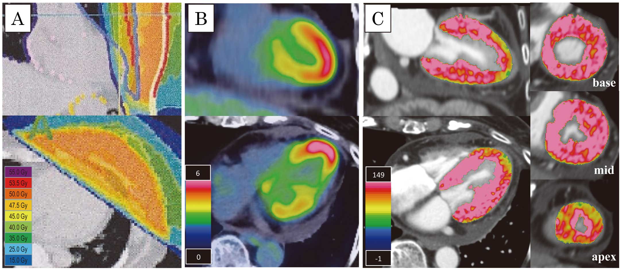
(A) Radiation fields and dose distribution for residual breast cancer in (Upper) coronal view and (Bottom) axial view. Both (B) 18F-fluorodeoxyglucose uptake and (C) reduced myocardial perfusion on computed tomography myocardial perfusion imaging (CTP) correspond to the irradiated field. (C, Right side) Short-axis view of the left ventricle at the (Upper) base, (Middle) midsection, and (Bottom) apex on CTP.
To the best of our knowledge, this is the first report of RIMI detected on CTP. CTP costs less, and has higher spatial resolution than single-photon emission CT, and is not contraindicated for patients with cardiovascular implanted electronic devices.1 Furthermore, CTP can simultaneously evaluate the recurrence of malignant tumor and RIMI in a single examination. This suggests that CTP may be a useful diagnostic tool for RIMI.
The authors declare no conflicts of interest.