Images in Cardiovascular Medicine
Pace-and-Ablate Technique for Atrial Tachycardia Originating From the Left Atrial Appendage
2020 年 84 巻 6 号 p. 1046-

詳細
2020 年 84 巻 6 号 p. 1046-
Simultaneous pacing and ablation through the tip of a single mapping/ablation catheter (pace-and-ablate technique) has been reported as a simple and novel technique to avoid right phrenic nerve injury for an electrical superior vena cava isolation and to shorten total fluoroscopy time.1
A 17-year-old man underwent endocardial ablation of a paroxysmal atrial tachycardia (AT) originating from the left atrial appendage (LAA); however, the AT could not be eliminated, so he was referred for a combined endo- and epicardial ablation of the AT.
Given the P wave morphology of the AT, its origin was assumed to be the epicardial LAA, as Yamada et al have reported.2 Preprocedural computed tomography revealed that the left phrenic nerve coursed along the apex of the LAA (Figure). The earliest site of the AT was at the endocardial apex of the LAA, which was near the left phrenic nerve. The pacing (output 10 mA; pulse width 2 ms; pacing interval 1,000 ms) from the ablation catheter captured the left phrenic nerve to contract the diaphragm. We applied radiofrequency (RF) ablation at that site (5–40 W, 20–30 s, contact force 10–20 g) during pacing from the ablation catheter to confirm phrenic nerve stimulation (pace-and-ablate technique). We monitored the left diaphragmatic movement with fluoroscopy and palpation. The AT was eliminated without any complications, including phrenic nerve palsy. The patient has been free from any AT recurrence during a follow-up period of 12 months. A limitation of this technique is that the lower site of the phrenic nerve could be injured by RF energy if the pacing output is too high.
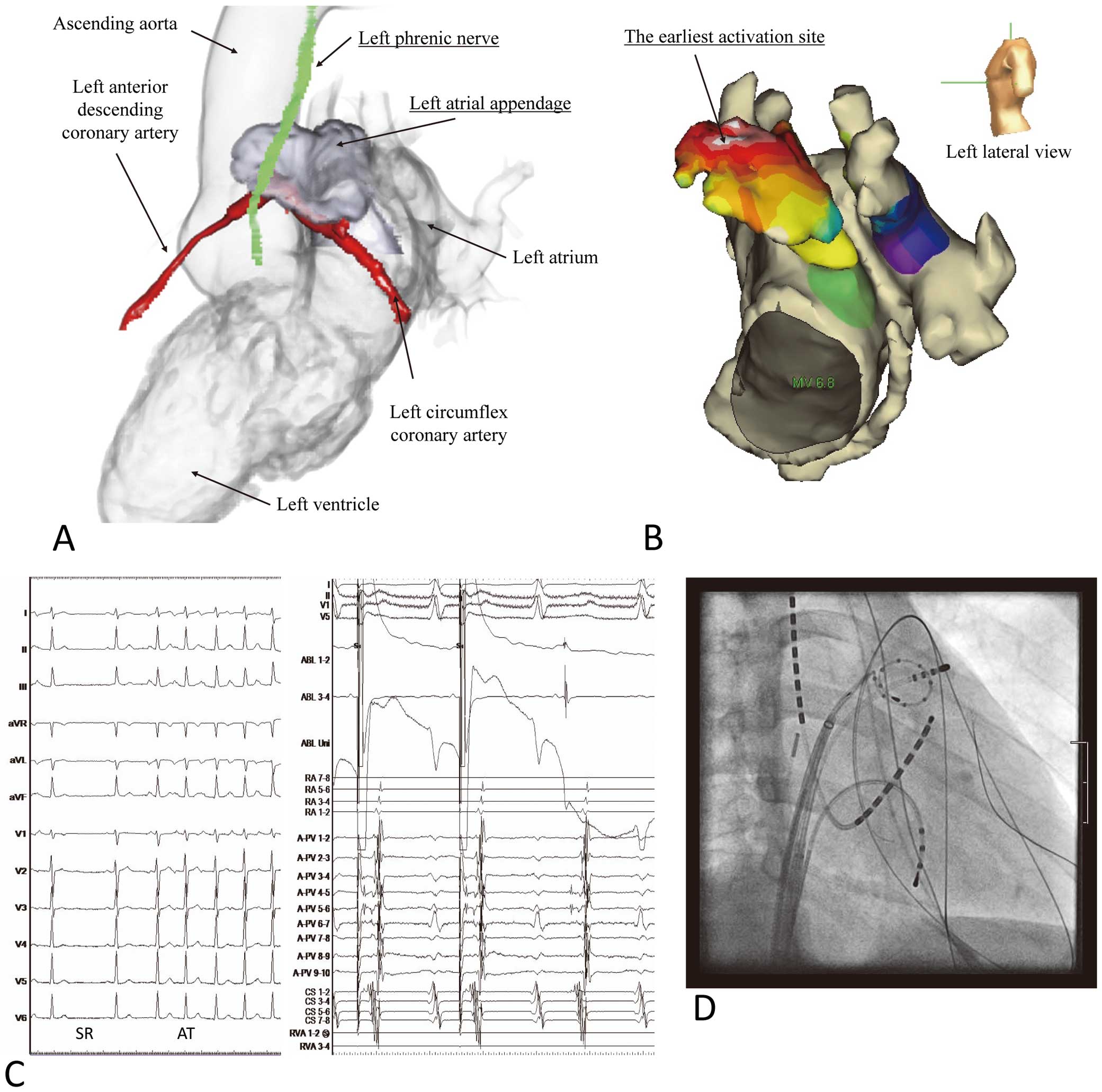
(A) Reconstructed image from computed tomography (left lateral view). (B) Activation map of atrial tachycardia (AT) originating from the left atrial appendage (LAA). (C) 12-lead electrogram during sinus rhythm (SR) and AT, and intracardiac electrogram during pacing from a successful site (good intra-cardiac pacing map). (D) Catheter positions during ablation of AT using pace-and-ablate technique. A ring catheter in the LAA is shown. Radiofrequency energy is applied at the site of the earliest activation, which is the apex of the LAA (right anterior oblique 30° view).
A novel pace-and-ablate technique was useful for avoiding left phrenic nerve injury during ablation of an AT originating from the LAA.
None.
Name: IRB of the National Cerebral and Cardiovascular Center. Number: M26-148-6.