Images in Cardiovascular Medicine
Optical Coherence Tomography-Guided Stent Implantation for Woven Coronary Artery Anomaly
2020 年 84 巻 6 号 p. 1044-

詳細
2020 年 84 巻 6 号 p. 1044-
Woven coronary artery anomaly (WCAA) is a rare congenital anomaly in which the epicardial coronary artery is divided into multiple channels that later merge in the distal segment. Pathologically, each channel of the WCAA has an individual vascular structure and preserved arterial integrity without a thrombus or dissection flaps.1
A 53-year-old male underwent cardiac catheterization because of significant ischemia in the inferior wall by stress myocardial perfusion imaging. Coronary angiography showed a double-lumen structure and tight stenosis in the mid-right coronary artery (Figure A). On optical coherence tomography (OCT), the wires were found separately in each lumen, and preserved vascular structure was observed in both lumens (Figure C-c–e,c′–e′; Supplementary Movies 1,2). A 3.0-mm×26-mm drug-eluting stent was implanted. OCT after stent implantation showed multiple lumens in the adventitia, which may be compressed by stent expansion (Figure C-i,j).
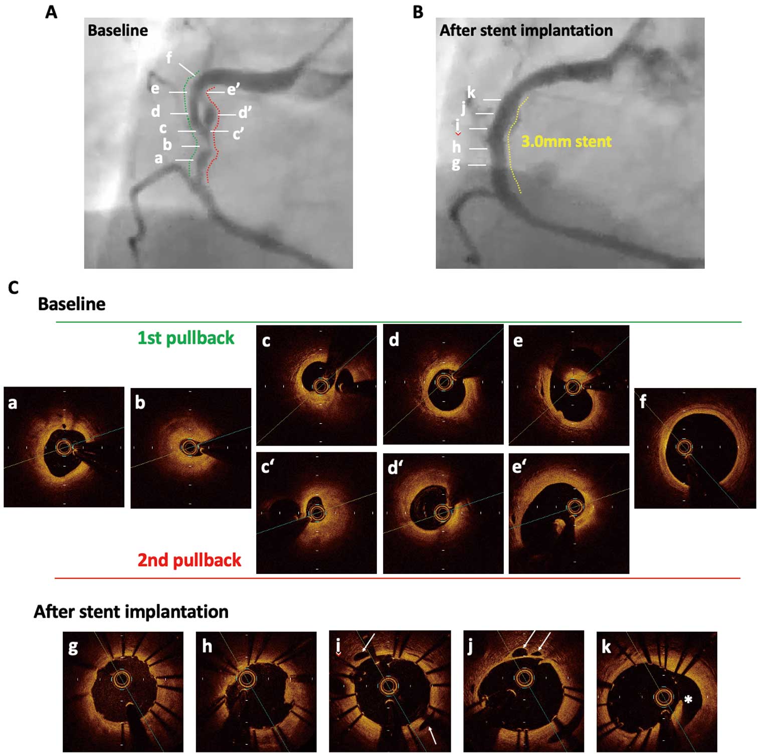
Angiography and optical coherence tomography (OCT) images. (A,B) Coronary angiograms at baseline (A) and after stent implantation (B). (C) Cross-sectional OCT images. (i,j) White arrows indicate compressed lumens in the adventitia. (k) The asterisk indicates the proximal orifice of the other lumen.
In this case, the septum between the 2 lumens was thick and had a preserved vascular structure, partly with fibroatheroma-like images. Furthermore, OCT after stent implantation showed the remaining oppressed lumens with a demilune appearance in the adventitia, not in the intima. These findings clearly demonstrated the anatomical and pathological features of WCAA, and enabled us to differentiate it from other etiologies, such as recanalized thrombus, which is reported to have a “lotus root” or “honeycomb-like” appearance, coronary dissection, and bridging collaterals.
H.I., T.M. are members of Circulation Journal ’ Editorial Team. The other authors have no conflicts of interest to declare.
Supplementary Movie 1. First pullback of OCT at baseline.
Supplementary Movie 2. Second pullback of OCT at baseline.
Please find supplementary file(s);
http://dx.doi.org/10.1253/circj.CJ-20-0011