Abstract
The aim of this study was to identify and determine the qualitative and quantitative composition of furanocoumarins in anatomical parts (blade and petiole) of two cultivars of ribbed celery, depending on the age of the plant. Two accurate, precise and inexpensive techniques for analytical detection of coumarin in a relatively short period of time were used in the present study. TLC - the method considered as preliminary, showed the presence of psolaren, bergapten, xanthotoxin and simple coumarin - umbelliferone. Additionally, isopimpinellin was detected by HPLC method. Chromatographic analysis of ribbed celery leaves showed the presence of four furanocoumarins. Psoralen and isopimpinellin are dominant in the leaf blades, wherein content is correlated with the term of harvesting. Bergapten was dominant in petioles regardless of the harvesting time and cultivar.
Introduction
Leafy vegetables are an important group of plants in the daily diet. Less known species like endive, chicory, arugula, rochet and chard has become popular among consumers. Also ribbed celery (Apium graveolens L. var. Dulce Mill. / Pers.), valued for its attractive appearance, taste and the nutritional composition resulting from the rich mineral and vitamin composition is classified to this group (Vogel, 1996). In part for consumption (petioles and partially leaf blades) contains many valuable biologically active substances such as phenolic acids, flavonoids, tannins, mono-and sesquiterpenes which make them useful in the treatment of certain dermatological diseases, arthritic-rheumatic, urinary and digestive system (Williamson, 1999; Wolski et al., 2002).
Plants from the family Apiaceae contain compounds with multidirectional biological activity: antimicrobial, anti-cancer, for the liver agents and cyclooxygenase inhibitors (Chen et al., 1995; Strohl, 2000). The presence of these substances is responsible for the sealing properties and capillary walls strengthens, improving blood circulation in the myocardium, spasmolytic, diuretic, platelet anticoagulation, anti-ulcer, anti-allergic, anti-inflammatory, anti-hepatotoxic, antifungal and antiviral effects (Trumble and Quiros, 1988, Zheng et al., 1993).
Coumarins are fairly common in many fruits and vegetables, especially in the families Apiaceae and Rutaceae (Zobel and Brown, 1990; Ojala, 2001). These group of compounds are important from the therapeutic point of view (Leal et al., 2000; Waksmundzka-Hajnos et al., 2004). Currenly, furanocoumarins of natural origin are used in PUVA therapy of vitiligo and psoriasis; furthermore, they are active Ca+2 channel blockers (Murray et al., 1982; Vuorela et al., 1988; Coven et al., 1999). Furanocoumarins are characterized by a broad spectrum of biological activity. They reduce the platelet agregation in in vitro test (Chen et al., 1996), they induce apoptosis in human promyelocytic leukemia, HL-60 cells (Bogucka-Kocka and Kocki, 2002; Pae et al., 2002) and they work as hepatoprotective agents (Jagiełło-Wójtowicz et al., 2004). Photosensitizer effect of these substances (particularly psoralen derivative) is used in medicine and the treatment of many dermatological disorders (Towers and Abramowski, 1983; Bhatnagar et al., 2007; Serrano-Perez et al., 2008). Furanocoumarins content in the plant varies depending on the degree of its development, and is different in different vegetative phases (Borkowski, 1973; Bogucka-Kocka and Kocki, 2002). Increase in the content of furanocoumarins in different organs was observed in many species of plants under stress conditions due to various factors: biotic (fungi, bacteria, viruses), abiotic (heavy metals, detergents, acid rain) or physical (UV radiation, low temperature). The diverse biological activity of furanocoumarins and the role they fulfill in the life of the plant tend to clarify the relationship between the plant defense system and external conditions. Stress metabolites named phytoalexins including furanocoumarins are formed as a result of defense property (Hain et al., 1993; Dixon and Lamb, 1999).
The aim of this study was to identify and determine the qualitative and quantitative composition of furanocoumarins in anatomical parts (blade and petiole) of two cultivars of ribbed celery, depending on the age of the plant. Two accurate, precise and inexpensive techniques for analytical detection of coumarin in a relatively short period of time were used in the present study.
Material and Methods
Plant material Two original Polish varieties of celery Zefir and Helios (company PlantiCo Zielonka) were tested in this study. Plants for analysis were obtained from outdoor experience of the Department of Vegetable and Medicinal Plants, University of Life Sciences in Lublin, Poland (N 51 ° 16′ E 22 ° 34′). Cultivation was carried out in the ecological system on luvisol soil formed from loess, containing 1.6% organic matter. Crops harvesting was carried out on two dates i. e. I — after 100 (August) and II — after 120 days (September) of two growing seasons: in 2011 and 2012. In a two-year cycle, the experimental average air temperature in the growing seasons of 2011 and 2012 showed a little varied. The average air temperature in the period May-September was 17.2°C (Table 1). The range of rainfall in years of research was at a similar level. Average monthly temperatures in May and June were higher than the perennial average, it has the second decade of July to the end of the growing season showed a gradual decrease in temperatures that were higher than the average perennial. Research material consisted blades and petioles of celery; 25 plants from each cultivar (5 plants × 5 replications) in each year. Samples were dried in a conventional dryer at 35°C to dry mass of raw material. 85 g of dry raw material were obtained from 1000 g of fresh petioles and 170 g from 1000 g of fresh blades. Dry plant material was pulverized and extracted.
Table 1.
Mean monthly air temperatures and total amount of precipitation at ES Felin in the years 2011–2012
| Month |
Temperature °C |
Amount of precipitation mm |
| 2011 |
2012 |
Mean for 1951–2000 |
2011 |
2012 |
Mean for 1951–2000 |
| V |
14.9 |
15.0 |
13.0 |
80.5 |
81.5 |
58.3 |
| VI |
18.1 |
18.1 |
16.5 |
87.8 |
87.8 |
65.8 |
| VII |
19.1 |
19.2 |
17.9 |
87.0 |
87.0 |
78.0 |
| VIII |
18.8 |
19.2 |
17.1 |
65.3 |
37.6 |
68.6 |
| IX |
15.2 |
15.0 |
12.6 |
5.4 |
35.5 |
51.6 |
| Mean |
17.2 |
17.3 |
15.4 |
65.2 |
65.88 |
64.46 |
Chemical reagents Solvents for extraction were purchased from POCh SA (Gliwice, Poland) or J.T. Baker (USA). Standards of high purity were purchased from Sigma-Aldrich Chemie GmbH (Munich, Germany), Merck (Darmstadt, Germany) and from other suppliers.
Solid phase extraction Analysis of free furanocoumarin arrangements was performed in dry and powdered whole leaves, leaf blades, and petioles of leaf celery plants. Extraction was conducted in liquid-solid system, while extract purification by means of SPE method (Solid Phase Extraction).
Samples of 50 g powdered and dried leaves, leaf blades, and petioles of two leaf celery cultivars were weighed and put into the tubes for extraction. After preliminary maceration for 24 hours, material was extracted using petroleum ether (1:10 v/v) and processed in Soxhlet's apparatus for 36 hours. The petroleum ether was evaporated till dry then. Dry residue of petroleum ether were purified on a SPE column (BackerBond RP 18). Microcolumns were mounted in a set of vacuum filtration (J Beaker) and further conditioned successively with 5 mL of methanol, 5 mL of double-distilled water, then dissolved extracts were applied (100 mg / 3 mL of double-distilled water). Elution was carried out by washing the bed of pollutant 30 mL of double-distilled water for removal of the ballast. Furanocoumarin compounds elution was performed 10 mL of methanol solution containing 0.3 mL of 25% ammonia. The eluents were evaporated to dryness and the residue was dissolved in 10 mL of methanol. The thus obtained samples were analyzed by TLC and HPLC for the presence of furanocoumarins.
Thin layer chromatography TLC was performed on 60G Si gel, cellulose and polyamide plates (Merck, Germany). Samples were applied using Autosampler3 (Camag, Switzerland). The plates were developed in the chromatographic chambers: flat type DS (Chromdes, Poland) and vertical (Camag, Switzerland). Developing phase was n-heptane:dichloromethane:ethyl acetate (40:40:20 v/v/v). Visualization of the chromatograms was carried out in visible light, under UV light Emita VP 60 (Famed, Poland) at the wavelength λ = 254 nm and 366 nm, and using VideoScanner Reprostar3 (Camag, Switzerland). The identification of compounds was performed by comparing their Rf values and color stain patterns, as well as by using a “co-chromatography”.
High-performance liquid chromatography The qualitative and quantitative HPLC determination was made in reversed-phase system applying liquid chromatograph of LaChrom-Merck type equipped with diode array detector DAD (L-7450), pump (L-7100), degasser (L-7612), injection loop 20 µL, thermostat (L-7360), injector Rheodyne, steel column LiChrospher 100 RP C 18 (250 mm × 4 mm dimensions) filled with stationery phase (dp = 5 µm). The analyses were conducted at 25°C. A stepwise mobile phase gradient was prepared from methanol (A) and water (B). The gradient was: 0 – 10 min 40 – 50% A; 10 – 25 min 50 – 75% A; 25 – 35 min isocratic 75% A; 35 – 40 min 75 – 100% A. The flow rate was 0.8 mL/min., while injected sample volume 20 µL. Coumarins identification was made by comparing their retention times (tR) with those for standards and spectroscopically by recording their spectra within UV range (220 – 400 nm) (Najda, 2009). Contents of particular furanocoumarins in studied material were then calculated on a base of calibration curve.
Statistical analysis The statistical analysis of data was performed using SAS “for Windows” version 9.1 and Statistica® version 7.0 software. Results were submitted to analysis of variance (ANOVA), Tukey test (p < 0.05) and Principal Component Analysis (PCA). All analyses were carried out in triplicate.
Results and Discussion
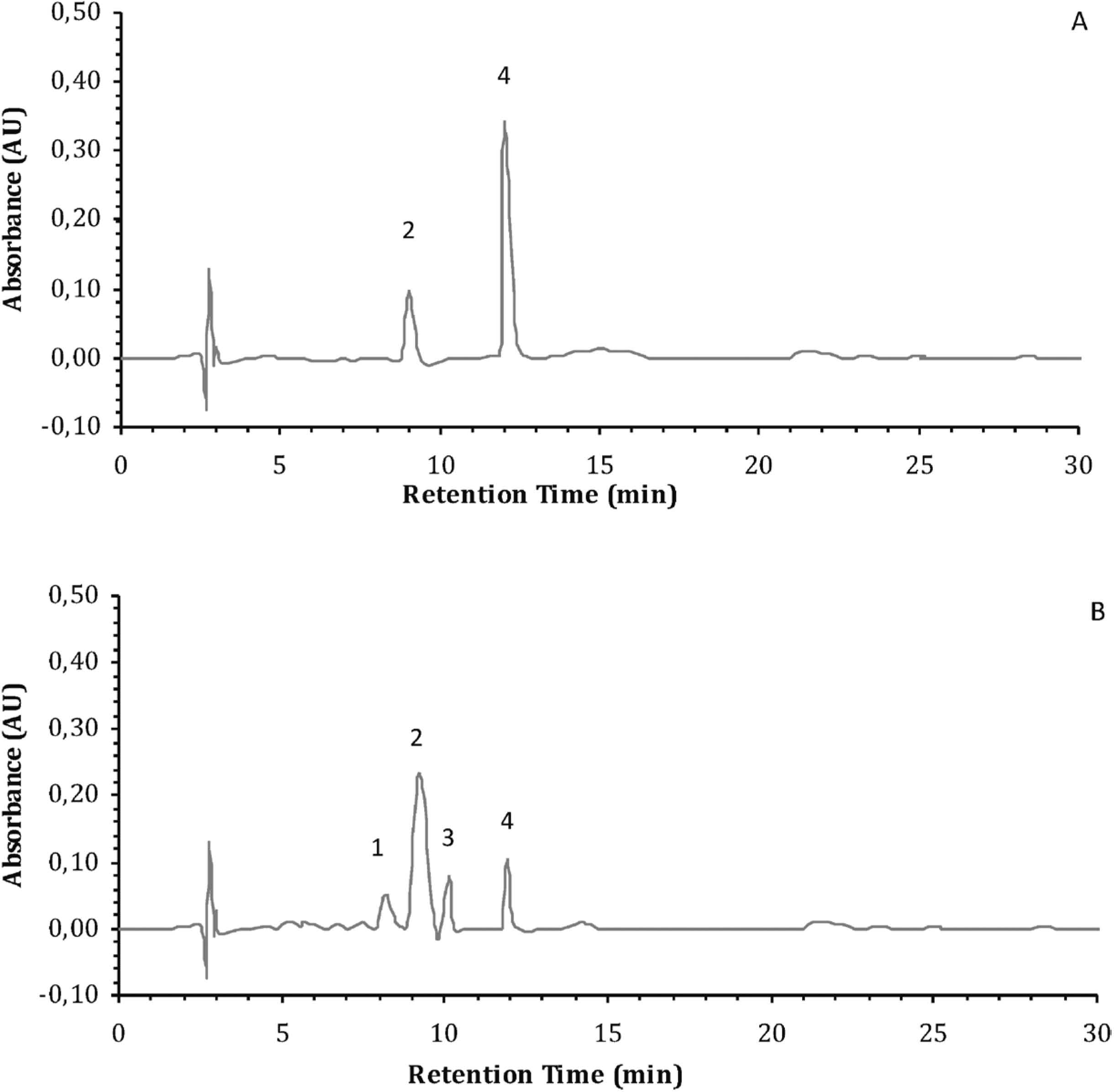
Our findings concerning the presence and the qualitative and quantitative furanocoumarin compounds were carried out by TLC and HPLC methods. TLC - the method considered as preliminary, showed the presence of psolaren, bergapten, xanthotoxin and simple coumarin — umbelliferone. The Fig. 1 shows the developed TLC plate of methanolic extracts from leaf blades and petioles of two ribbed celery cultivars. As shown in Fig. 1., sample purification method on octadecyl C18 columns eliminated ballast substances. This allowed the identification of coumarin compounds in the extracts. Additionally, isopimpinellin was detected by HPLC method. HPLC chromatogram of standards furanocoumarins distribution is presented in Fig. 2. Fig. 3 shows the UV spectroscopic spectrums. Tab. 2. contains the average retention times for the different standards of furanocoumarins and furanocoumarins isolated from extracts of the tested materials. The presence of three furanocoumarins in the petioles of both studied cultivars and four furanocoumarins such as psoralen, bergapten, isopimpinellin and xanthotoxin in the leaf blades were found. Fig. 4 and 5 show chromatograms representing furanocoumarins content in the tested raw materials. Analysed petioles of tested varieties had 2 to 3 furanocoumarin compounds. In contrast, the leaf blades of both studied cultivars contain 4 basic furanocoumarins: xanthotoxin, psoralen, bergapten and isopimpinellin. The quantitative content of furanocoumarins in whole leaves demonstrated slightly lower content of these compounds due to their lower concentration in petioles.

Table 2.
Comparison of average retention times (min) for the different furanocoumarin standards and furanocoumarins isolated from the petioles and leaf blades of both studied cultivars of ribbed celery (based on HPLC analysis)
| Furanocoumarin |
The average retention time tR (min) |
| standard |
‘ZEFIR’ |
‘HELIOS’ |
| petioles |
leaf blades |
petioles |
leaf blades |
| 1. Xanthotoxin |
8.86 |
8.97 |
8.87 |
8.97 |
9.05 |
| 2. Psoralen |
9.37 |
9.41 |
9.49 |
9.26 |
9.39 |
| 3. Isopimpinellin |
10.01 |
10.12 |
10.0 |
10.15 |
10.10 |
| 4. Bergapten |
13.71 |
14.01 |
13.78 |
13.83 |
14.00 |
Quantitative content of individual furanocoumarins occurring in the studied raw materials was calculated by assuming the surface area of each peak as a quantitative relation of the furanocoumarins concentrations and using D 7000 HPLC system Manager program.
Data on the content of furanocoumarins in anatomical parts (blades and petiole) of each variety in the two years of the study as the mean of all varieties in each year of the study is presented in Table 3. The results show that the differences between cultivars × anatomical parts, cultivars x year and anatomical parts × year were not significant (P < 0.05). Data variation source for all measured varieties highlights that the seasonal variability plays an important role in parts of the anatomy of these varieties. All factors (variety, anatomical part and year of harvest) show a significant effect, with the exception of the year effect for isopimpinellin (P = 0.610) and xanthotoxin (P = 0.470) content and cultivar effect for bergapten (P = 0.619) and isopimpinellin (P = 0.410). The content of psoralen, bergapten and isopimpinellin was higher in 2012, with the exception of xanthotoxin. Psoralen content was higher in the leaf blades, in which also xanthotoksin and isopinpinellin were identified, while in petioles bergapten content was higher. Zefir, irrespective of the anatomical part, contained more of all the tested furanocoumarins compounds. Higher presence of psoralen, bergapten and isopimpinellin was observed in 2012, while xsanthotoxin content was higher in 2011. There was significant differences between individual furanocoumarins in a cultivar Zefir, in the analyzed anatomical parts and in 2012. The results indicate a similar profile of furanocoumarins in the analyzed anatomical parts of two cultivars of ribbed celery.
Table 3.
Furanocoumarins composition (µg · 100 g
−1 DM).
|
|
Xanthotoxin |
Psoralen |
Isopimpinellin |
Bergapten |
P-value |
| Cultivar |
Zefir |
0.139bA |
0.172bB |
0.127aA |
0.113aA |
0.0346 |
|
Helios |
0.088aA |
0.126aC |
0.112aB |
0.104aB |
0.2101 |
|
P-value |
0.0354 |
0.048 |
0.4103 |
0.619 |
|
| Anatomical parts |
Petioles |
- |
0.077aA |
- |
0.124bB |
0.0001 |
|
Leaf blades |
0.158aB |
0.238bC |
0.177aB |
0.063aA |
0.0001 |
|
P-value |
0.0055 |
0.0002 |
0.0022 |
0.0269 |
|
| Year |
2011 |
0.120aC |
0.118aC |
0.098aB |
0.080aA |
0.1194 |
|
2012 |
0.107aA |
0.179bC |
0.107aA |
0.137bB |
0.0067 |
|
P-value |
0.4706 |
0.0202 |
0.6109 |
0.0251 |
|
| Cultivar x anatomical parts |
P-value |
0.8556 |
0.8970 |
0.9493 |
0.4307 |
|
| Cultivar x year |
P-value |
0.7624 |
0.8528 |
0.2321 |
0.9617 |
|
| Anatomical parts x year |
P-value |
0.9999 |
0.9880 |
0.5400 |
0.4457 |
|
| Cultivar x anatomical parts x year |
P-value |
0.9685 |
0.9885 |
0.7411 |
0.6330 |
|
The results are presented as mean ± SD (n = 20 for each year; n = 15 for each cultivar; n = 15 for each anatomical parts).
Explanatory notes: different letters a, b, c… and A, B, C… in the same column and line indicate statistically significant differences (P < 0.05). In each column and for each cultivar different letters mean significant differences (P < 0.05).
Literature data indicate the occurrence of psoralen, 5-methoxypsoralen, bergapten and xsanthotoxin in ribbed celery leaf (Surico et al., 1987). Beier et al (1983) found that the psoralen, bergapten and trimethylpsoralen content in ribbed celery plants infected by fungi: Sclerotiora sclerotiorum and Ervin carotovara was significantly higher than in healthy plants, which follows from the properties of plant defense (biotic stress). Research conducted by Zobel and Brown (1990) on the furanocoumarins concentration occurring outside plants of the Apiaceae and Rutaceae family have shown that these compounds can be secreted on the leaf surface by the action of UV rays. Secretion of furanocoumarins on the leaf surface is a defense mechanism of plants against adverse environmental conditions arising as a result of the ultraviolet light stress (Afek and Carmeli, 1995; Afek et al., 1996). On the surface of celery leaves, xsanthotoxin content was 0.006 µg · g−1 of fresh weight, psoralen in a greater concentration −0.010 µg · g−1, and bergapten in trace amounts. In addition, the literature (Trumble et al., 1990; Trumble et al., 1992) reported that the contents of furanocoumarins in the ribbed celery leaves are on the level from 0.34 to 1.80 µg · g−1 of fresh weight, and depending on the age of the leaves, cultivar and climatic conditions. Quantitative content of furanocoumarins occurring in the studied raw materials varied in the petioles from 0,121 µg · g−1 to 0.287 µg · g−1 of dry weight in the cultivars Zefir and Helios, and in leaf blades from 0.522 µg · g−1 to 0.966 µg · g−1 of dry weight. These amounts may be higher for fresh weight of tested plants leaves, depending on the intensity of UV light. The obtained data confirm the xanthotoxin and bergapten content on the leaf surface, this can be inferred that the agricultural treatments in the cultivation of ribbed celery should be carried out on cloudy days with low insolation level. Otherwise, photodermatoses might occur. Considering the percentage of the identified furanocoumarins it has been demonstrated that psoralen varied from 29% to 55%, and bergapten from 45% to 71% (Fig. 6). Bergapten was dominant in petioles regardless of the harvesting time and cultivar. Petioles whose harvest was carried out after 100 days of growing season accumulated more bergapten (I harvest date). With the increase of growing season length, its amount in the analyzed parts of the plants in both studied cultivars decreased. In contrast, percentage of psoralen reached a maximum in petioles whose harvest was carried out after 120 days of growing season (II date).

Chromatographic analysis of ribbed celery leaves showed the presence of four furanocoumarins. Assessing the percentage of identified furanocoumarins, it has been reported that psoralen is dominant in leaf blades, wherein its content is correlated with the harvest date. Isopimpinellin is the second dominant compound regardless of the leaves harvest time with 24% and 30% content for Zefir and Helios respectively. Contents of psoralen and bergapten in the leaf blades decreased with the increasing growing season length, while the content of isopimpinellin and xanthotoxin slightly increased (Fig. 7).

In Table 4, the correlation coefficients between the posted furanocoumarins identified. Very strong correlation (>0.96) were observed between the content and xanthotoxin and isopimpinellin. At the same time a strong correlation has been shown between the content of a psoralen and xanthotoxin and isopimpinellin. Other correlation coefficients between the content of furanocoumarins in ribbed celery are not significant.
Table 4.
The correlation coefficients between the content of identified furanocoumarins in ribbed celery
| Cultivar |
Xanthotoxin |
Psoralen |
Isopimpinellin |
Bergapten |
| Xanthotoxin |
1 |
0.8433 |
0.9587 |
−0.2449 |
| Psoralen |
0.8433 |
1 |
0.8909 |
−0.1140 |
| Isopimpinellin |
0.9587 |
0.8909 |
1 |
−0.2629 |
| Bergapten |
−0.2449 |
−0.1140 |
−0.2629 |
1 |
Conclusions
In conclusion, the obtained results demonstrated that the cultivar of ribbed celery affects both quantitative and qualitative profile of furanocoumarins. Evaluating the data referring to anatomical parts it can be concluded that seasonal variability plays an important role. Leaf blades are more abundant in furanocoumarins. The presence of bergapten, psolaren, isopimpinellin and xanthotoxin makes the furanocoumarins more interesting source of compounds from therapeutic point of view.
References
- Afek, U., Aharon, I.N., and Carmeli, S. (1996). The involvement of marmesin in celery resistance to pathogens during storage and the effect of temperature on its concentration. American Phytopath. Soc., 85, 711-714.
- Afek, U. and Carmeli, S. (1995). Increasing celery resistance to pathogens during storage and reducing high-risk psoralen concentration by treatment with GA3. J. Amer. Soc. Hort. Sci., 120, 562-565.
- Beier, R.C., Ivie, G.W., Oertli, E.H., and Holt, D.L. (1983). HPLC analysis of linear furocoumarins (psoralens) in healthy celery (Apium graveolens). Food Chem. Toxicol., 21, 163-165.
- Bhatnagar, A., Kanwar, A. J., Parsad, D., and De, D. (2007). Psoralen and ultraviolet A and narrowband ultraviolet B in inducing stability in vitiligo, assessed by vitiligo disease activity score: an open prospective comparative study. J. Eur. Acad. Dermatol. Venerol., 21, 1381-1385.
- Bogucka-Kocka, A. and Kocki, J. (2002). Influence of two furocoumarins: bergapten and xanthotoxin from meadow cow parsnip (Heracleum sibiricum L.) on apoptosis induction and apoptotic genes expression in HL-60 human leukaemic cell line. Bull. Vet. Inst. Puławy Suppl., 1, 111-116.
- Borkowski, B. (Rd.) (1973). Thin layer chromatography in the pharmaceutical analysis. PZWL, Warsaw, Poland (in Polish).
- Chen, I., Chang, C., and Sheen, W. (1996). Coumarins and antiplatelet aggregation constituents from Formosan Peucedanum japonicum. Phytochem., 41, 525-530.
- Chen, I.S., Lin, Y.C., Tsai, I.L., Teng, C.M., Ko, F.N., Ishikawa, T., and Ishii, H. (1995). Coumarins and anti-plateled aggregation constituents from Zanthoxylium schinifolium. Phytochem., 39, 1091-1097.
- Coven, T.R., Walters, I.B., Cardinale, I., and Krueger, J.G. (1999). PUVA-induced lymphocyte apoptosis: mechanism of action in psoriasis. Photoderm. Photoimmun. Photomed., 15, 22-27.
- Dixon, R.A. and Lamb, C. J. (1999). Molecular communication in interactions between plants and microbial pathogens. Ann. Rev. Plant Physiol. Plant Mol. Biol. 41, 229-367.
- Hain, R., Reif, H. J., Krause, E., Langebartels, R., and Kndle, H. (1993). Disease resistance results from foreign Phytoalexin expression in a novel plant. Nature, 361, 153-156.
- Jagiełło-Wojtowicz, E., Chodkowska, A., Madej, A., Głowniak, P., and Burczyk, J. (2004). Comparison of CNS activity of imperatorine with fraction of furanocoumarins from Angelica archangelica fruit in mice. Herba Pol., 50, 106-111.
- Leal, L.K.A.M., Ferreira, A.A.G., Bezerra, G.A., Matos, F.J.A., and Viana, G.S.B. (2000). Antinociceptive, anti-inflammatory and bronchodilator activities of Brazilian medicinal plants containing coumarin: a comparative study. J. Ethnopharm., 70, 151-159.
- Murray, R., Mendez, J., and Brown, S. (1982). The Natural Coumarins, Occurrence. Chem. and Biochem., John Wiley & Sons Ltd., Chichester, UK.
- Najda, A. (2009). Furanocoumarins in the anatomical parts of celery (Apium graveolens L. var. dulce Mill./Pers.). Allium and Umbelliferae Improv. Newsletter, Madison USA, 19, 30-34.
- Ojala, T. (2001). Biological screening of plant coumarins. Acad. Diss. University of Helsinki, Yliopistopaino, Helsinki, 2001.
- Pae, H.O., Yun, Y.G., Och, G.S., Jang, S.I., Hwang, K.M., Kwon, D.O., Lee, H.S., and Chung, H.T. (2002). Imperatorin, a furanocoumarin from Angelica dahurica (Umbelliferae), induces cytochrome c-dependent apoptosis in human promyelocytic leukaemia, HL-60 cells. Pharm. Toxic., 91, 40-48.
- Serrano-Pérez, J. J., González-Luque, R., Merchán, M., and Serrano-Andrés, L. (2008). The family of furocoumarins: Looking for the best photosensitizer for phototherapy. J. Photochem. Photobiol. A: Chem., 199, 34-41.
- Strohl, W.R. (2000). The role of natural products in a modern drug discovery program. Edit. Drug Discovery Today, 5, 39-41.
- Surico, G., Varvaro, L., and Solfrizzo, M. (1987). Linear furocoumarin accumulation in celery plants infected with Erwinia carotovora pv. carotovora. Am. Chem. Soc., 35, 406-409.
- Towers, G.H. and Abramowski, Z. (1983). UV — mediated genotoxicity of furanoquinoline and of certain tryptophan — derived alkaloids. J. Nat. Prod. and Plant Res., 46, 576-581.
- Trumble, J.T., Derks, W., Quiros, C.F., and Beier, R.C. (1990). The furanocumarns isolated from Apium species. J. Plant Nutr., 21, 1779-1789.
- Trumble, J.T., Milla, R, J.G., Ott, D.E., and Carson, W.C. (1992). Seasonal patterns and pesticidal effects on the phototoxic linear furanocoumarins in celery (Apium graveolens L.). J. Agric. Food Chem., 40, 1501-1506.
- Trumble, J.T. and Quiros, C.F. (1988). Antixenotic and antibiotic resistance in Apium species to Liryomiza trifolii (Burgess). J. Economic Entomol., 81, 602-607.
- Vogel, G. (1996). Handbuch des speziellen Gemüsebaues. Eugen Ulmer. Stuttgart, Germany, 975-990.
- Vuorela, H., Törnquist, K., Sticher, O., and Hiltunen, R. (1988): Effects of furocoumarins in Peucedanum palustre on prolactin releasse from GH3 rat pituitary cells. Acta Pharm. Fennica, 97, 167-174.
- Waksmundzka-Hajnos, M., Petruczynik, A., Dragan, A. Wianowska, D., and Dawidowicz, A.L. 2004. Effect of extraction method on the yield of furanocoumarins from fruits of Archangelica officinalis Hoffm. Phytochem. Analysis, 15, 313-319.
- Williamson, G. (1999). Functional Foods-A new challenge for the food chemists. Proceed. Euro Food Chem. X, Budapeszt, 1, 192.
- Wolski, T., Dyduch, J., and Najda, A. (2002). Evaluation of content and composition of phenolic acids and tannins in leaf dry matter of two celery cultivars (Apium graveolens L. var. dulce Mill. Pers.). www.ejpan.media.pl/series/volumine5/issue1/horticulture/art-01.html.
- Zheng, G.Q., Kenney, P.M., Zhang, J., and Lam, L.K. (1993). Chemoprevention of benzo-α-pyrene — induced forestomach cancer in mice by natural phthalides from celery seed oil. Nutr. and Cancer, 19, 77-86.
- Zobel, A.M. and Brown, S.A. (1990). Dermatitis-inducing furanocoumarins on leaf surfaces of eight species of rutaceous and umbelliferous plants. J. Chem. Ecology, 16, 693-700.