抄録
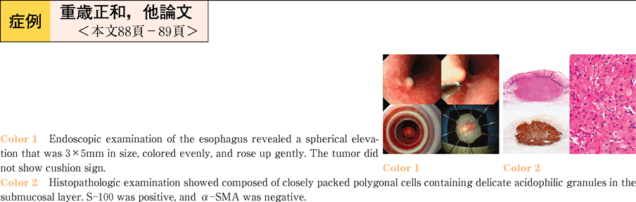
A 60-year-old man underwent endogastroduodenoscopy because of a close examination of the prostate cancer. Endoscopic examination of the esophagus revealed a spherical elevation that was 3×5 mm in size, colored evenly,and rose up gently. The tumor did not show cushion sign. He was admitted to our hospital for further evaluation and treatment. Physical examination on admission was unremarkable. Laboratory tests on admission revealed normocytic normochromic anemia. Endoscopic ultrasonography (EUS) was performed. Ultrasonic imaging showed a hyperechoic mass in the third layer. The differential diagnoses included leiomyoma, GIST, and lipoma in EUS. The patient desired endoscopic treatment, and endoscopic mucosal resection was performed. Histopathologic examination showed composed of closely packed polygonal cells containing delicate acidophilic granules in the submucosal layer. S-100 was positive,and α-SMA was negative. The tumor was diagnosed as granular cell tumor.
In our case,the esophageal lesion revealed hyperecoic tumor,although usual GCTs exhibit hypoechoic mass by EUS. We could not figure out the cause of the high echogenecity in spite of close analysis with histopathological evaluation. GCT of higecho is rare. We report a case with esophageal GCT as demonstrated as a hyperechoic mass by EUS, which is rare phenomenon, in addition to a discussion of the relevant literature.