抄録
Endoscopic ultrasonography (EUS) was widely performed according to the routine application of the ultrasonic probe. 3-dimentional endoscopic ultrasonography (3-D EUS) system was developed using ordinary ultrasonic probe (UM-3R, UM-3T) by Olympus Co. From 1995 July to March 1996, 4 cases of lipoma of the colon were examined using 3-D EUS to evaluate the usefulness and the visualization.
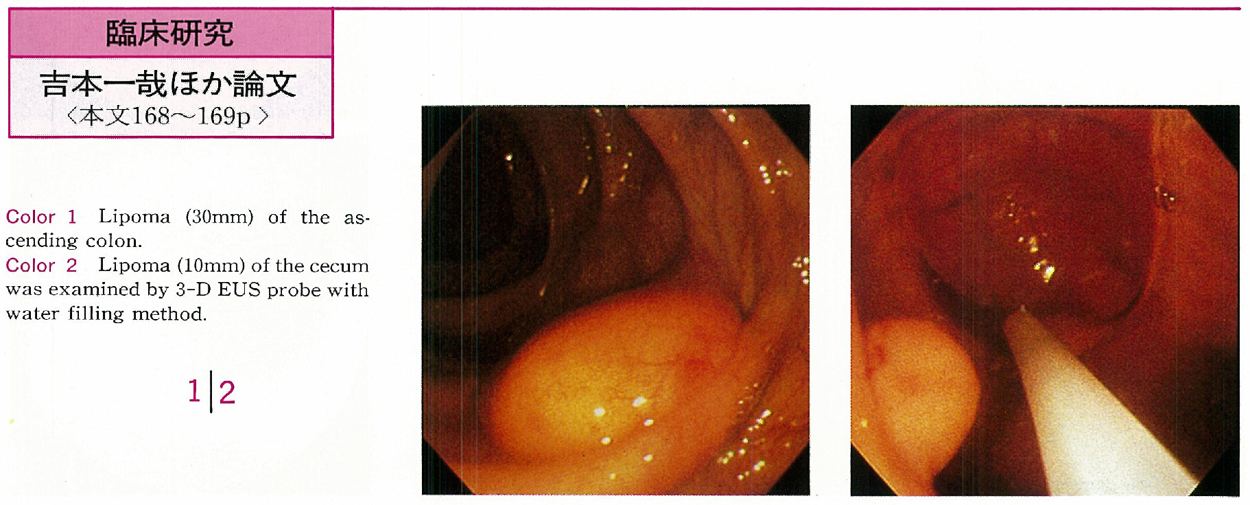
Four cases of lipoma were located in the right side colon. Tumor size were ranged from 10mm to 30mm and noted yellowish surface with cushion-sign endoscopically. EUS image were able to visualize in all cases. Ordinary radial image and linear image were also useful to diagose the lipoma in all cases. Reconstructed 3-D EUS image of the large lipoma provide much information for the diagnosis. But image of the samll lipoma was not so different from radial or linear image. Linear image sometimes seemed to be quite useful in the cases of the lesion on the fold or unvisible lesion.
3-D EUS image thought to be given diagnosable information by internal echo findings also three dimentional tumor recognition in many cases. 3-D EUS will be advanced much more in near future and will be given much information for the endoscopic diagnosis.