症例
胃腎シャント閉塞下内視鏡的硬化療法が有効であった胃静脈瘤の1例
1996 年 49 巻 p. 188-189
詳細
1996 年 49 巻 p. 188-189
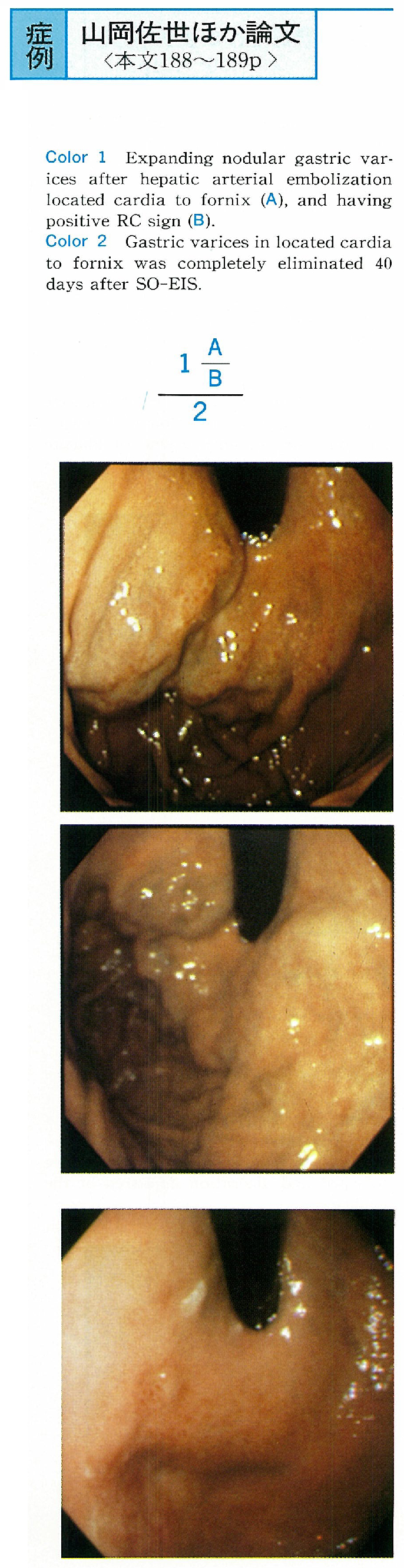
A 70-year-old male patient accompanying with liver cirrhosis complicated hepatoma was having solitary gastric varices enlarged after transcatheter hepatic arterial embolization. His endoscopic findings showed that varices were nodulary enlarged shape and having positive red color sign located from cardia to fornix. And also angiographic study showed gastrorenal shunt.
He was tried to BRTO. But we could not visualze varices on BRTV by using 40 ml contrast medium. We thought the method of BRTO was not indicated because of poor obliteration for gastric varices of this case. We performed SO-EIS. BRTV just after SO-EIS showed filling defect meant thrombosed gastric varices.
In this case endoscopic findings post 40 days since SO-EIS performed showed solitary gastric varices was completely eliminated. We thought SO-EIS had usefull for solitary gastric varices in cases where BRTO was not indicated.