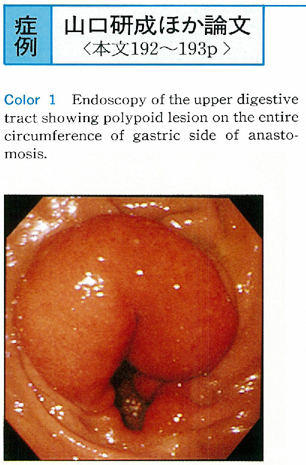
The patient was a 54-year-old male who had undergone distant partial gastrectomy with reconstruction by the Billroth II procedure 10 years earlier. Endoscopic examination of the upper digestive tract performed due to discomfort during swallowing showed a red tumorous lesion with a smooth surface on the entire circumference of the anastomosis site. Biopsy specimen revealed cystic dilatation and hyperplasia of the glands without atypia. Based on these findings, a diagnosis of gastritis cystica polyposa was made.
Endoscopic ultrasonography showed high echoic tumor image in the submucosal layer and an aechoic-low echo multiple small cystic image within, which was covered with nearly normal first and second layers. Compared with histological image, these findings of endoscopic ultrasonography appeared to be specific to gastritis cystica polyposa.