Biophysics and Physicobiology
Online ISSN : 2189-4779
ISSN-L : 2189-4779
Volume 20, Issue 2
Displaying 1-15 of 15 articles from this issue
Regular Article
  • Tomoyoshi Seto
    Article type: Regular Article
    2023 Volume 20 Issue 2 Article ID: e200005
    Published: 2023
    Released on J-STAGE: April 12, 2023
    Advance online publication: January 25, 2023
    JOURNAL OPEN ACCESS FULL-TEXT HTML

    The GABAA receptor (GABAAR) is a target channel for the loss of awareness of general anesthesia. General anesthetic (GA) spans a wide range of chemical structures, such as monatomic molecules, barbital acids, phenols, ethers, and alkanes. GA has a weak binding affinity, and the affinity has a characteristic that correlates with the solubility in olive oil rather than the molecular shape. The GA binding site of GABAAR is common to GAs and exists in the transmembrane domain of the GABAAR intersubunit. In this study, the mechanism of GA binding, which allows binding of various GAs with intersubunit selectivity, was elucidated from the hydration analysis of the binding site. Regardless of the diverse GA chemical structures, a strong correlation was observed between the binding free energy and total dehydration number of the binding process. The GA binding free energy was more involved in the binding dehydration and showed molecular recognition that allowed for the binding of various GA structures via binding site hydration. We regarded the GA substitution for the interfacial water molecule of the binding site as a dissolution into the interfacial hydration layer. The elucidation of the GA binding mechanism mediated by hydration at the GABAAR common binding site provides a rationale for the combined use of anesthetics in medical practice and its combination adjustments via drug interactions.

  • Tatsuki Negami, Tohru Terada
    Article type: Regular Article
    2023 Volume 20 Issue 2 Article ID: e200016
    Published: 2023
    Released on J-STAGE: April 12, 2023
    Advance online publication: March 25, 2023
    JOURNAL OPEN ACCESS FULL-TEXT HTML
    Supplementary material

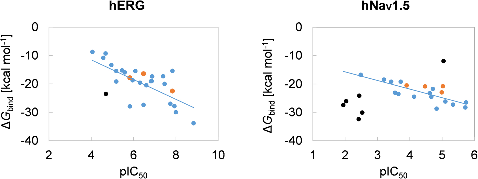
    The evaluation of the inhibitory activities of drugs on multiple cardiac ion channels is required for the accurate assessment of proarrhythmic risks. Moreover, the in silico prediction of such inhibitory activities of drugs on cardiac channels can improve the efficiency of the drug-development process. Here, we performed molecular docking simulations to predict the complex structures of 25 reference drugs that were proposed by the Comprehensive in vitro Proarrhythmia Assay consortium using two cardiac ion channels, the human ether-a-go-go-related gene (hERG) potassium channel and human NaV1.5 (hNaV1.5) sodium channel, with experimentally available structures. The absolute binding free energy (ΔGbind) values of the predicted structures were calculated by a molecular dynamics-based method and compared with the experimental half-maximal inhibitory concentration (IC50) data. Furthermore, the regression analysis between the calculated values and negative of the common logarithm of the experimental IC50 values (pIC50) revealed that the calculated values of four and ten drugs deviated significantly from the regression lines of the hERG and hNaV1.5 channels, respectively. We reconsidered the docking poses and protonation states of the drugs based on the experimental data and recalculated their ΔGbind values. Finally, the calculated ΔGbind values of 24 and 19 drugs correlated with their experimental pIC50 values (coefficients of determination=0.791 and 0.613 for the hERG and hNaV1.5 channels, respectively). Thus, the regression analysis between the calculated ΔGbind and experimental IC50 data ensured the realization of an increased number of reliable complex structures.

Review Article (Invited)
  • Toma Kashima, Akihiro Ishiwata, Kiyotaka Fujita, Shinya Fushinobu
    Article type: Review Article (Invited)
    2023 Volume 20 Issue 2 Article ID: e200017
    Published: 2023
    Released on J-STAGE: April 27, 2023
    Advance online publication: March 29, 2023
    JOURNAL OPEN ACCESS FULL-TEXT HTML

    Cooking with fire produces foods containing carbohydrates that are not naturally occurring, such as α-d-fructofuranoside found in caramel. Each of the hundreds of compounds produced by caramelization reactions is considered to possess its own characteristics. Various studies from the viewpoints of biology and biochemistry have been conducted to elucidate some of the scientific characteristics. Here, we review the composition of caramelized sugars and then describe the enzymatic studies that have been conducted and the physiological functions of the caramelized sugar components that have been elucidated. In particular, we recently identified a glycoside hydrolase (GH), GH172 difructose dianhydride I synthase/hydrolase (αFFase1), from oral and intestinal bacteria, which is implicated in the degradation of oligosaccharides in caramel. The structural basis of αFFase1 and its ligands provided many insights. This discovery opened the door to several research fields, including the structural and phylogenetic relationship between the GH172 family enzymes and viral capsid proteins and the degradation of cell membrane glycans of acid-fast bacteria by some αFFase1 homologs. This review article is an extended version of the Japanese article, Identification and Structural Basis of an Enzyme Degrading Oligosaccharides in Caramel, published in SEIBUTSU BUTSURI Vol. 62, p. 184–186 (2022).

  • Hiromi Miyoshi, Masashi Yamazaki, Hiromichi Fujie, Satoru Kidoaki
    Article type: Review Article (Invited)
    2023 Volume 20 Issue 2 Article ID: e200018
    Published: 2023
    Released on J-STAGE: May 19, 2023
    Advance online publication: April 19, 2023
    JOURNAL OPEN ACCESS FULL-TEXT HTML

    Mesenchymal stem cells (MSCs) have the potential for self-renewal and multipotency to differentiate into various lineages. Thus, they are of great interest in regenerative medicine as a cell source for tissue engineering. Substrate stiffness is one of the most extensively studied exogenous physical factors; however, consistent results have not always been reported for controlling MSCs. Conventionally used stiff culture substrates, such as tissue-culture polystyrene and glass, enhance nuclear localization of a mechanotransducer YAP and a pre-osteogenic transcription factor RUNX2, and bias MSCs towards the osteogenic lineage, even without osteogenic-inducing soluble factors. The mechanosensitive nature and intrinsic heterogeneity present challenges for obtaining reproducible results. This review summarizes the heterogeneity in human MSC response, specifically, nuclear/cytoplasmic localization changes in the mechanotransducer yes-associated protein (YAP) and the osteogenic transcription factor RUNX2, in response to substrate stiffness. In addition, a perspective on the intracellular factors attributed to response heterogeneity is discussed. The optimal range of stiffness parameters, Young’s modulus, for MSC expansion culture to suppress osteogenic differentiation bias through the suppression of YAP and RUNX2 nuclear localization, and cell cycle progression is likely to be surprisingly narrow for a cell population from an identical donor and vary among cell populations from different donors. We believe that characterization of the heterogeneity of MSCs and understanding their biological meaning is an exciting research direction to establish guidelines for the design of culture substrates for the sophisticated control of MSC properties.

  • Atsushi Mochizuki
    Article type: Review Article (Invited)
    2023 Volume 20 Issue 2 Article ID: e200019
    Published: 2023
    Released on J-STAGE: May 23, 2023
    Advance online publication: April 21, 2023
    JOURNAL OPEN ACCESS FULL-TEXT HTML

    Progress of molecular biology resulted in the accumulation of information on biomolecular interactions, which are complex enough to be termed as networks. Dynamical behavior generated by complex network systems is considered to be the origin of the biological functions. One of the largest missions in modern life science is to obtain logical understanding for the dynamics of complex systems based on experimentally identified networks. However, a network does not provide sufficient information to specify dynamics explicitly, i.e. it lacks information of mathematical formulae of functions or parameter values. One has to develop mathematical models under assumptions of functions and parameter values to know the detail of dynamics of network systems. In this review, on the other hand, we introduce our own mathematical theory to understand the behavior of biological systems from the information of regulatory networks alone. Using the theory, important aspects of dynamical properties can be extracted from networks. Namely, key factors for observing/controlling the whole dynamical system are determined from network structure alone. We also show an application of the theory to a real biological system, a gene regulatory network for cell-fate specification in ascidian. We demonstrate that the system was completely controllable by experimental manipulations of the key factors identified by the theory from the information of network alone. This review article is an extended version of the Japanese article, Controlling Cell-Fate Specification System Based on a Mathematical Theory of Network Dynamics, published in SEIBUTSU BUTSURI Vol. 60, p. 349–351 (2020).

Regular Article
  • Shingo Wakao, Noriko Saitoh, Akinori Awazu
    Article type: Regular Article
    2023 Volume 20 Issue 2 Article ID: e200020
    Published: 2023
    Released on J-STAGE: May 23, 2023
    Advance online publication: April 27, 2023
    JOURNAL OPEN ACCESS FULL-TEXT HTML
    Supplementary material

    Nuclear speckles are nuclear bodies consisting of populations of small and irregularly shaped droplet-like molecular condensates that contain various splicing factors. Recent experiments have revealed the following structural features of nuclear speckles: (I) Each molecular condensate contains SON and SRRM2 proteins, and MALAT1 non-coding RNA surrounds these condensates; (II) During normal interphase of the cell cycle in multicellular organisms, these condensates are broadly distributed throughout the nucleus. In contrast, when cell transcription is suppressed, the condensates fuse and form strongly condensed spherical droplets; (III) SON is dispersed spatially in MALAT1 knocked-down cells and MALAT1 is dispersed in SON knocked-down cells because of the collapse of the nuclear speckles. However, the detailed interactions among the molecules that are mechanistically responsible for the structural variation remain unknown. In this study, a coarse-grained molecular dynamics model of the nuclear speckle was developed by considering the dynamics of SON, SRRM2, MALAT1, and pre-mRNA as representative components of the condensates. The simulations reproduced the structural changes, which were used to predict the interaction network among the representative components of the condensates.

Database and Computer Program
  • Hiroshi Imamura
    Article type: Database and Computer Program
    2023 Volume 20 Issue 2 Article ID: e200021
    Published: 2023
    Released on J-STAGE: May 23, 2023
    Advance online publication: May 10, 2023
    JOURNAL OPEN ACCESS FULL-TEXT HTML
    J-STAGE Data

    Small-angle scattering (SAS) is a powerful tool for the detailed structural analysis of objects at the nanometer scale. In contrast to techniques such as electron microscopy, SAS data are presented as reciprocal space information, which hinders the intuitive interpretation of SAS data. This study presents a workflow: (1) creating objects, (2) 3D scanning, (3) the representation of the object as point clouds on a laptop, (4) computation of a distance distribution function, and (5) computation of SAS, executed via the computer program Phone2SAS. This enables us to realize SAS and perform the interactive modeling of SAS of the object of interest. Because 3D scanning is easily accessible through smartphones, this workflow driven by Phone2SAS contributes to the widespread use of SAS. The application of Phone2SAS for the structural assignment of SAS to Y-shaped antibodies is reported in this study.

Review Article (Invited)
  • Shigeyuki Matsumoto, Shoichi Ishida, Kei Terayama, Yasuhshi Okuno
    Article type: Review Article (Invited)
    2023 Volume 20 Issue 2 Article ID: e200022
    Published: 2023
    Released on J-STAGE: June 10, 2023
    Advance online publication: May 16, 2023
    JOURNAL OPEN ACCESS FULL-TEXT HTML

    Protein functions associated with biological activity are precisely regulated by both tertiary structure and dynamic behavior. Thus, elucidating the high-resolution structures and quantitative information on in-solution dynamics is essential for understanding the molecular mechanisms. The main experimental approaches for determining tertiary structures include nuclear magnetic resonance (NMR), X-ray crystallography, and cryogenic electron microscopy (cryo-EM). Among these procedures, recent remarkable advances in the hardware and analytical techniques of cryo-EM have increasingly determined novel atomic structures of macromolecules, especially those with large molecular weights and complex assemblies. In addition to these experimental approaches, deep learning techniques, such as AlphaFold 2, accurately predict structures from amino acid sequences, accelerating structural biology research. Meanwhile, the quantitative analyses of the protein dynamics are conducted using experimental approaches, such as NMR and hydrogen-deuterium mass spectrometry, and computational approaches, such as molecular dynamics (MD) simulations. Although these procedures can quantitatively explore dynamic behavior at high resolution, the fundamental difficulties, such as signal crowding and high computational cost, greatly hinder their application to large and complex biological macromolecules. In recent years, machine learning techniques, especially deep learning techniques, have been actively applied to structural data to identify features that are difficult for humans to recognize from big data. Here, we review our approach to accurately estimate dynamic properties associated with local fluctuations from three-dimensional cryo-EM density data using a deep learning technique combined with MD simulations.

Regular Article
  • Kenta Odagiri, Hiroshi Fujisaki, Hiroya Takada, Rei Ogawa
    Article type: Regular Article
    2023 Volume 20 Issue 2 Article ID: e200023
    Published: 2023
    Released on J-STAGE: June 10, 2023
    Advance online publication: May 24, 2023
    JOURNAL OPEN ACCESS FULL-TEXT HTML

    To computationally investigate the recent experimental finding such that extracellular ATP release caused by exogeneous mechanical forces promote wound closure, we introduce a mathematical model, the Cellular Potts Model (CPM), which is a popular discretized model on a lattice, where the movement of a “cell” is determined by a Monte Carlo procedure. In the experiment, it was observed that there is mechanosensitive ATP release from the leading cells facing the wound gap and the subsequent extracellular Ca2+ influx. To model these phenomena, the Reaction-Diffusion equations for extracellular ATP and intracellular Ca2+ concentrations are adopted and combined with CPM, where we also add a polarity term because the cell migration is enhanced in the case of ATP release. From the numerical simulations using this hybrid model, we discuss effects of the collective cell migration due to the ATP release and the Ca2+ influx caused by the mechanical forces and the consequent promotion of wound closure.

Review Article (Invited)
  • Yoshiaki Kinosita, Yoshiyuki Sowa
    Article type: Review Article (Invited)
    2023 Volume 20 Issue 2 Article ID: e200024
    Published: 2023
    Released on J-STAGE: June 14, 2023
    Advance online publication: May 30, 2023
    JOURNAL OPEN ACCESS FULL-TEXT HTML

    Most motile bacteria use supramolecular motility machinery called bacterial flagellum, which converts the chemical energy gained from ion flux into mechanical rotation. Bacterial cells sense their external environment through a two-component regulatory system consisting of a histidine kinase and response regulator. Combining these systems allows the cells to move toward favorable environments and away from their repellents. A representative example of flagellar motility is run-and-tumble swimming in Escherichia coli, where the counter-clockwise (CCW) rotation of a flagellar bundle propels the cell forward, and the clockwise (CW) rotation undergoes cell re-orientation (tumbling) upon switching the direction of flagellar motor rotation from CCW to CW. In this mini review, we focus on several types of chemotactic behaviors that respond to changes in flagellar shape and direction of rotation. Moreover, our single-cell analysis demonstrated back-and-forth swimming motility of an original E. coli strain. We propose that polymorphic flagellar changes are required to enhance bacterial movement in a structured environment as a colony spread on an agar plate.

  • Manabu Hori, Takashi Tominaga, Masaki Ishida, Mutsumi Kawano
    Article type: Review Article (Invited)
    2023 Volume 20 Issue 2 Article ID: e200025
    Published: 2023
    Released on J-STAGE: June 14, 2023
    Advance online publication: May 31, 2023
    JOURNAL OPEN ACCESS FULL-TEXT HTML

    In Paramecium, a mechanical stimulus applied to the posterior portion of the cell causes a transient increase in membrane permeability to potassium ions, transiently rendering the membrane in a hyperpolarized state. Hyperpolarization causes a transient increase in Cyclic adenosine monophosphate (cAMP) concentration in the cilia, resulting in a transient fast-forward swimming of the cell. Schultz and coworkers (1992) reported that a unique adenylate cyclase (AC)-coupled potassium channel is involved in the reaction underlying this response, which is known as the “escape response.” However, the AC responsible for this reaction remains to be identified. Moreover, the molecular linkage between mechanoreception and AC activation has not been elucidated adequately. Currently, we can perform an efficient and simple gene-knockdown technique in Paramecium using RNA interference (RNAi). Paramecium is one of the several model organisms for which whole-genome sequences have been elucidated. The RNAi technique can be applied to whole genome sequences derived from the Paramecium database (ParameciumDB) to investigate the types of proteins that elicit specific biological responses and compare them with those of other model organisms. In this review, we describe the applications of the RNAi technique in elucidating the molecular mechanism underlying the escape response and identifying the AC involved in this reaction. The findings of this study highlight the advantages of the RNAi technique and ParameciumDB.

  • Shunsuke Tagami
    Article type: Review Article (Invited)
    2023 Volume 20 Issue 2 Article ID: e200026
    Published: 2023
    Released on J-STAGE: June 20, 2023
    Advance online publication: June 02, 2023
    JOURNAL OPEN ACCESS FULL-TEXT HTML

    Is it a miracle that life exists on the Earth, or is it a common phenomenon in the universe? If extraterrestrial organisms exist, what are they like? To answer these questions, we must understand what kinds of molecules could evolve into life, or in other words, what properties are generally required to perform biological functions and store genetic information. This review summarizes recent findings on simple ancestral proteins, outlines the basic knowledge in textbooks, and discusses the generally required properties for biological molecules from structural biology viewpoints (e.g., restriction of shapes, and types of intra- and intermolecular interactions), leading to the conclusion that proteins and nucleic acids are at least one of the simplest (and perhaps very common) forms of catalytic and genetic biopolymers in the universe. This review article is an extended version of the Japanese article, On the Origin of Life: Coevolution between RNA and Peptide, published in SEIBUTSU BUTSURI Vol. 61, p. 232–235 (2021).

  • Yutaro Nagasawa, Hiromi H. Ueda, Haruka Kawabata, Hideji Murakoshi
    Article type: Review Article (Invited)
    2023 Volume 20 Issue 2 Article ID: e200027
    Published: 2023
    Released on J-STAGE: June 20, 2023
    Advance online publication: June 06, 2023
    JOURNAL OPEN ACCESS FULL-TEXT HTML

    Optogenetic techniques offer a high spatiotemporal resolution to manipulate cellular activity. For instance, Channelrhodopsin-2 with global light illumination is the most widely used to control neuronal activity at the cellular level. However, the cellular scale is much larger than the diffraction limit of light (<1 μm) and does not fully exploit the features of the “high spatial resolution” of optogenetics. For instance, until recently, there were no optogenetic methods to induce synaptic plasticity at the level of single synapses. To address this, we developed an optogenetic tool named photoactivatable CaMKII (paCaMKII) by fusing a light-sensitive domain (LOV2) to CaMKIIα, which is a protein abundantly expressed in neurons of the cerebrum and hippocampus and essential for synaptic plasticity. Combining photoactivatable CaMKII with two-photon excitation, we successfully activated it in single spines, inducing synaptic plasticity (long-term potentiation) in hippocampal neurons. We refer to this method as “Local Optogenetics”, which involves the local activation of molecules and measurement of cellular responses. In this review, we will discuss the characteristics of LOV2, the recent development of its derivatives, and the development and application of paCaMKII.

Regular Article
  • Kanji Takahashi, Tatsuro Nishikino, Hiroki Kajino, Seiji Kojima, Takay ...
    Article type: Regular Article
    2023 Volume 20 Issue 2 Article ID: e200028
    Published: 2023
    Released on J-STAGE: June 23, 2023
    Advance online publication: June 09, 2023
    JOURNAL OPEN ACCESS FULL-TEXT HTML
    Supplementary material

    The marine bacterium Vibrio alginolyticus has a single flagellum as a locomotory organ at the cell pole, which is rotated by the Na+-motive force to swim in a liquid. The base of the flagella has a motor composed of a stator and rotor, which serves as a power engine to generate torque through the rotor–stator interaction coupled to Na+ influx through the stator channel. The MS-ring, which is embedded in the membrane at the base of the flagella as part of the rotor, is the initial structure required for flagellum assembly. It comprises 34 molecules of the two-transmembrane protein FliF. FliG, FliM, and FliN form a C-ring just below the MS-ring. FliG is an important rotor protein that interacts with the stator PomA and directly contributes to force generation. We previously found that FliG promotes MS-ring formation in E. coli. In the present study, we constructed a fliF–fliG fusion gene, which encodes an approximately 100 kDa protein, and the successful production of this protein effectively formed the MS-ring in E. coli cells. We observed fuzzy structures around the ring using either electron microscopy or high-speed atomic force microscopy (HS-AFM), suggesting that FliM and FliN are necessary for the formation of a stable ring structure. The HS-AFM movies revealed flexible movements at the FliG region.

Review Article (Invited)
  • Hiroki Yasuga
    Article type: Review Article (Invited)
    2023 Volume 20 Issue 2 Article ID: e200029
    Published: 2023
    Released on J-STAGE: June 23, 2023
    Advance online publication: June 09, 2023
    JOURNAL OPEN ACCESS FULL-TEXT HTML

    Herein, I review our recent work toward developing methods for generating three-dimensional (3D) droplet arrays driven by capillarity. Microdroplet array-based systems are useful for bioassays and bioengineering because they require only small amounts of samples and reagents and provide the high throughput. Various methods have been developed for preparing droplet arrays, among which methods based on capillarity have attracted considerable attention owing to their simplicity. I and collaborators have developed such methods based on capillary flow, including a method for preparing droplet arrays via oil–water replacement. We recently proposed our own concept of “fluid–fluid interfacial energy driven 3D structure emergence in a micropillar scaffold (FLUID3EAMS)” and its application. FLUID3EAMS allows a 3D droplet (or hydrogel bead) array to be generated in a micropillar scaffold by passing a fluid–fluid interface through the scaffold. This approach is useful for applications requiring ordered or arrayed microdroplets in biosensors, biophysics, biology, and tissue engineering. This review is an extended version of the article “FLUID3EAMS: Fluid–Fluid Interfacial Energy Driven 3D Structure Emergence in a Micropillar Scaffold and Development in Bioengineering” published in Seibutsu Butsuri (vol. 62, p. 110–113, 2022).

feedback
Top