Biophysics and Physicobiology
Online ISSN : 2189-4779
ISSN-L : 2189-4779
Volume 14
Displaying 1-23 of 23 articles from this issue
Regular Article
  • Kazuya Suzuki, Takeshi Itabashi, Shin’ichi Ishiwata
    2017 Volume 14 Pages 1-11
    Published: 2017
    Released on J-STAGE: January 24, 2017
    JOURNAL FREE ACCESS
    Supplementary material

    The metaphase spindle is organized for accurate chromosome segregation. One of the fundamental features of the spindle across the species is its symmetrical shape; the spindle consists of two polar arrays of microtubules at both ends. Although it has been suggested that the formation of the bipolar shape requires force balance coordination by molecular motors, i.e., kinesins and dyneins, quantitative analysis for the pole mechanics has not been conducted. Here, we demonstrate that it is not only the shape but also the stiffness and microtubule density of the pairs of pole regions are symmetrically balanced in single spindles self-assembled in Xenopus egg extracts. We found that the inhibition of dynein functions dramatically reduced the stiffness and microtubule density in the pole region. By contrast, the inhibition of one of the kinesins, Eg5, which is the antagonistic motor protein of dynein, increased the value of these parameters. Moreover, the inhibition of both dynein and Eg5 recovered these parameter values to those of non-treated spindle poles. We also found that, when one pole structure was held widened with the use of two glass microneedles, the opposite pole structure spontaneously widened, resulting in the formation of the barrel-like shaped spindle. The values of stiffness and microtubule density in the manipulated pole region decreased, following the spontaneous decrement of those in the paired unmanipulated pole region. These results suggest that the spindle possesses a mechanism to dynamically maintain its symmetry in mechanical properties.

    Download PDF (4653K)
  • Alemeh Zamani, Shigeo Sakuragi, Toru Ishizuka, Hiromu Yawo
    2017 Volume 14 Pages 13-22
    Published: 2017
    Released on J-STAGE: January 24, 2017
    JOURNAL FREE ACCESS
    Supplementary material

    Channelrhodopsin (ChR)-1 and ChR2 were the first-identified members of ChRs which are a growing subfamily of microbial-type rhodopsins. Light absorption drives the generation of a photocurrent in cell membranes expressing ChR2. However, the photocurrent amplitude attenuates and becomes steady-state during prolonged irradiation. This process, called desensitization or inactivation, has been attributed to the accumulation of intermediates less conductive to cations. Here we provided evidence that the dark-adapted (DA) photocurrent before desensitization is kinetically different from the light-adapted (LA) one after desensitization, that is, the deceleration of both basal-to-conductive and conductive-to-basal transitions. When the kinetics were compared between the DA and LA photocurrents for the ChR1/2 chimeras, the transmembrane helices, TM1 and TM2, were the determinants of both basal-to-conductive and conductive-to-basal transitions, whereas TM4 may contribute to the basal-to-conductive transitions and TM5 may contribute to the conductive-to-basal transitions, respectively. The fact that the desensitization-dependent decrease of the basal-to-conductive and conductive-to-basal transitions was facilitated by the TM1 exchange from ChR2 to ChR1 and reversed by the further TM2 exchange suggests that the conformation change for the channel gating is predominantly regulated by the interaction between TM1 and TM2. Although the exchange of TM1 from ChR2 to ChR1 showed no obvious influence on the spectral sensitivity, this exchange significantly induced the desensitization-dependent blue shift. Therefore, the TM1 and 2 are the main structures involved in two features of the desensitization, the stabilization of protein conformation and the charge distribution around the retinal-Schiff base (RSB+).

    Download PDF (2271K)
  • Kazuki Takahashi, Masato Sunohara, Takuya Terai, Shigefumi Kumachi, Na ...
    2017 Volume 14 Pages 23-28
    Published: 2017
    Released on J-STAGE: February 11, 2017
    JOURNAL FREE ACCESS
    Supplementary material

    In vitro display technologies such as mRNA and cDNA display are powerful tools to create and select functional peptides. However, in some cases, efficiency of mRNA-protein fusion is very low, which results in decreased library size and lower chance of successful selection. In this study, to improve mRNA-protein fusion efficiency, we prepared an mRNA display library of a protein with random N- and C-terminal coding regions consisting of 12 nucleotides (i.e. four amino acids), and performed an electrophoresis mobility shift assay (EMSA)-based selection of successfully formed mRNA display molecules. A single-domain antibody (Nanobody, or VHH) was used as a model protein, and as a result, a pair of sequences was identified that increased mRNA-protein fusion efficiency of this protein by approximately 20%. Interestingly, enhancement of the fusion efficiency induced by the identified sequences was protein-specific, and different results were obtained for other proteins including VHHs with different CDRs. The results suggested that conformation of mRNA as a whole, rather than the amino acid sequence of the translated peptide, is an important factor to determine mRNA-protein fusion efficiency.

    Download PDF (1789K)
  • Yohei Murakami, Masanori Koyama, Shigeyuki Oba, Shinya Kuroda, Shin Is ...
    2017 Volume 14 Pages 29-40
    Published: 2017
    Released on J-STAGE: February 22, 2017
    JOURNAL FREE ACCESS
    Supplementary material

    The functions of intracellular signal transduction systems are determined by the temporal behavior of intracellular molecules and their interactions. Of the many dynamical properties of the system, the relationship between the dynamics of upstream molecules and downstream molecules is particularly important. A useful tool in understanding this relationship is a methodology to control the dynamics of intracellular molecules with an extracellular stimulus. However, this is a difficult task because the relationship between the levels of upstream molecules and those of downstream molecules is often not only stochastic, but also time-inhomogeneous, nonlinear, and not one-to-one. In this paper, we present an easy-to-implement model-based control method that makes the target downstream molecule to trace a desired time course by changing the concentration of a controllable upstream molecule. Our method uses predictions from Monte Carlo simulations of the model to decide the strength of the stimulus, while using a particle-based approach to make inferences regarding unobservable states. We applied our method to in silico control problems of insulin-dependent AKT pathway model and EGF-dependent Akt pathway model with system noise. We show that our method can robustly control the dynamics of the intracellular molecules against unknown system noise of various strengths, even in the absence of complete knowledge of the true model of the target system.

    Download PDF (1416K)
  • Noriyo Mitome, Hiroki Sato, Taishi Tomiyama, Katsuya Shimabukuro, Taku ...
    2017 Volume 14 Pages 41-47
    Published: 2017
    Released on J-STAGE: March 16, 2017
    JOURNAL FREE ACCESS
    Supplementary material

    The Fo-a subunit of the Na+-transporting FoF1 ATP synthase from Propionigenium modestum plays a key role in Na+ transport. It forms half channels that allow Na+ to enter and leave the buried carboxyl group on Fo-c subunits. The essential Arg residue R226, which faces the carboxyl group of Fo-c subunits in the middle of transmembrane helix 5 of the Fo-a subunit, separates the cytoplasmic side and periplasmic half-channels. To elucidate contributions of other amino acid residues of transmembrane helix 5 using hybrid FoF1 (Fo from P. modestum and F1 from thermophilic Bacillus PS3), 25 residues were individually mutated to Cys, and effects of modification with the SH-modifying agent N-ethylmaleimide (NEM) on ATP synthesis and hydrolysis activity were analyzed. NEM significantly inhibited ATP synthesis and hydrolysis as well as proton pumping activities of A214C, G215C, A218C, I223C (cytoplasmic side from R226), and N230C (periplasmic side from R226) mutants and inhibited ATP synthesis activity of the K219C mutant (cytoplasmic side from R226). Thus, these residues contribute to the integrity of the Na+ half channel, and both half channels are present in the Fo-a subunit.

    Download PDF (2084K)
  • Jun Tamogami, Takashi Kikukawa, Toshifumi Nara, Makoto Demura, Tomomi ...
    2017 Volume 14 Pages 49-55
    Published: 2017
    Released on J-STAGE: March 18, 2017
    JOURNAL FREE ACCESS

    A spectrally silent change is often observed in the photocycle of microbial rhodopsins. Here, we suggest the presence of two O intermediates in the photocycle of Acetabularia rhodopsin II (ARII or also called Ace2), a light-driven algal proton pump from Acetabularia acetabulum. ARII exhibits a photocycle including a quasi-equilibrium state of M, N, and O (M<=>N<=>O→) at near neutral and above pH values. However, acidification of the medium below pH ~5.5 causes no accumulation of N, resulting in that the photocycle of ARII can be described as an irreversible scheme (M→O→). This may facilitate the investigation of the latter part of the photocycle, especially the rise and decay of O, during which molecular events have not been sufficiently understood. Thus we analyzed the photocycle under acidic conditions (pH ≤ 5.5). Analysis of the absorbance change at 610 nm, which mainly monitors the fractional concentration changes of K and O, was performed and revealed a photocycle scheme containing two sequential O-states with the different molar extinction coefficients. These photoproducts, termed O1 and O2, may be even produced at physiological pH, although they are not clearly observed under this condition due to the existence of a long M-N-O equilibrium.

    Download PDF (2203K)
  • Yumeka Yamauchi, Masae Konno, Shota Ito, Satoshi P. Tsunoda, Keiichi I ...
    2017 Volume 14 Pages 57-66
    Published: 2017
    Released on J-STAGE: May 20, 2017
    JOURNAL FREE ACCESS
    Supplementary material

    Microbial rhodopsins are membrane proteins found widely in archaea, eubacteria and eukaryotes (fungal and algal species). They have various functions, such as light-driven ion pumps, light-gated ion channels, light sensors and light-activated enzymes. A light-driven proton pump bacteriorhodopsin (BR) contains a DTD motif at positions 85, 89, and 96, which is unique to archaeal proton pumps. Recently, channelrhodopsins (ChRs) containing the DTD motif, whose sequential identity is ~20% similar to BR and to cation ChRs in Chlamydomonas reinhardtii (CrCCRs), were found. While extensive studies on ChRs have been performed with CrCCR2, the molecular properties of DTD ChRs remain an intrigue. In this paper, we studied a DTD rhodopsin from G. theta (GtCCR4) using electrophysiological measurements, flash photolysis, and low-temperature difference FTIR spectroscopy. Electrophysiological measurements clearly showed that GtCCR4 functions as a light-gated cation channel, similar to other G. theta DTD ChRs (GtCCR1-3). Light-driven proton pump activity was also suggested for GtCCR4. Both electrophysiological and flash photolysis experiments showed that channel closing occurs upon reprotonation of the Schiff base, suggesting that the dynamics of retinal and channels are tightly coupled in GtCCR4. From Fourier transform infrared (FTIR) spectroscopy at 77 K, we found that the primary reaction is an all-trans to a 13-cis photoisomerization, like other microbial rhodopsins, although perturbations in the secondary structure were much smaller in GtCCR4 than in CrCCR2.

    Download PDF (1437K)
  • Kotomi Shibata, Tsubasa Koyama, Shohei Inde, Sosuke Iwai, Shigeru Chae ...
    2017 Volume 14 Pages 67-73
    Published: 2017
    Released on J-STAGE: May 31, 2017
    JOURNAL FREE ACCESS

    The myosin II SH1 helix is a joint that links the converter subdomain to the rest of the myosin motor domain and possibly plays a key role in the arrangement of the converter/lever arm. Several point mutations within the SH1 helix in human myosin IIs have been shown to cause diseases. To reveal whether these SH1 helix mutations affect not only motile activities but also thermal properties of myosin II, here we introduced the E683K or R686C point mutation into the SH1 helix in Dictyostelium myosin II. Thermal inactivation as well as thermal aggregation rates of these mutant proteins demonstrated that these mutations decreased the thermal stability of myosin II. Temperature dependence of sliding velocities of actin filaments showed that these mutations also reduced the activation energy of a rate-limiting process involved in actin movement. Given that these mutations are likely to alter coupling between the subdomains, and thus their thermal fluctuations, we propose that the SH1 helix is a key structural element that determines the flexibility and thermal properties of the myosin motor. These characteristics of the SH1 helix may contribute to the pathogenesis of the human diseases caused by mutations within this structural element.

    Download PDF (1402K)
  • Ryo Yoshizawa, Nobuhisa Umeki, Masataka Yanagawa, Masayuki Murata, Yas ...
    2017 Volume 14 Pages 75-84
    Published: 2017
    Released on J-STAGE: June 03, 2017
    JOURNAL FREE ACCESS
    Supplementary material

    RalGDS is one of the Ras effectors and functions as a guanine nucleotide exchange factor for the small G-protein, Ral, which regulates membrane trafficking and cytoskeletal remodeling. The translocation of RalGDS from the cytoplasm to the plasma membrane is required for Ral activation. In this study, to understand the mechanism of Ras–Ral signaling we performed a single-molecule fluorescence analysis of RalGDS and its functional domains (RBD and REMCDC) on the plasma membranes of living HeLa cells. Increased molecular density of RalGDS and RBD, but not REMCDC, was observed on the plasma membrane after EGF stimulation of the cells to induce Ras activation, suggesting that the translocation of RalGDS involves an interaction between the GTP-bound active form of Ras and the RBD of RalGDS. Whereas the RBD played an important role in increasing the association rate constant between RalGDS and the plasma membrane, the REMCDC domain affected the dissociation rate constant from the membrane, which decreased after Ras activation or the hyperexpression of Ral. The Y64 residue of Ras and clusters of RalGDS molecules were involved in this reduction. From these findings, we infer that Ras activation not merely increases the cell-surface density of RalGDS, but actively stimulates the RalGDS–Ral interaction through a structural change in RalGDS and/or the accumulation of Ral, as well as the GTP–Ras/RalGDS clusters, to induce the full activation of Ral.

    Download PDF (2089K)
Review Article
  • Souhei Sakata, Makoto Matsuda, Akira Kawanabe, Yasushi Okamura
    2017 Volume 14 Pages 85-97
    Published: 2017
    Released on J-STAGE: June 23, 2017
    JOURNAL FREE ACCESS

    Voltage-sensing phosphatase (VSP) consists of a transmembrane voltage sensor and a cytoplasmic enzyme region. The enzyme region contains the phosphatase and C2 domains, is structurally similar to the tumor suppressor phosphatase PTEN, and catalyzes the dephosphorylation of phosphoinositides. The transmembrane voltage sensor is connected to the phosphatase through a short linker region, and phosphatase activity is induced upon membrane depolarization. Although the detailed molecular characteristics of the voltage sensor domain and the enzyme region have been revealed, little is known how these two regions are coupled. In addition, it is important to know whether mechanism for coupling between the voltage sensor domain and downstream effector function is shared among other voltage sensor domain-containing proteins. Recent studies in which specific amino acid sites were genetically labeled using a fluorescent unnatural amino acid have enabled detection of the local structural changes in the cytoplasmic region of Ciona intestinalis VSP that occur with a change in membrane potential. The results of those studies provide novel insight into how the enzyme activity of the cytoplasmic region of VSP is regulated by the voltage sensor domain.

    Download PDF (1786K)
Note
  • Akira R. Kinjo
    2017 Volume 14 Pages 99-110
    Published: 2017
    Released on J-STAGE: July 12, 2017
    JOURNAL FREE ACCESS

    A grand canonical Monte Carlo (MC) algorithm is presented for studying the lattice gas model (LGM) of multiple protein sequence alignment, which coherently combines long-range interactions and variable-length insertions. MC simulations are used for both parameter optimization of the model and production runs to explore the sequence subspace around a given protein family. In this Note, I describe the details of the MC algorithm as well as some preliminary results of MC simulations with various temperatures and chemical potentials, and compare them with the mean-field approximation. The existence of a two-state transition in the sequence space is suggested for the SH3 domain family, and inappropriateness of the mean-field approximation for the LGM is demonstrated.

    Download PDF (3986K)
Regular Article
  • Yutaro Yamada, Hiroki Konno, Katsuya Shimabukuro
    2017 Volume 14 Pages 111-117
    Published: 2017
    Released on J-STAGE: July 28, 2017
    JOURNAL FREE ACCESS
    Supplementary material

    In this study, we present a new technique called correlative atomic force and transmission electron microscopy (correlative AFM/TEM) in which a targeted region of a sample can be observed under AFM and TEM. The ultimate goal of developing this new technique is to provide a technical platform to expand the fields of AFM application to complex biological systems such as cell extracts. Recent advances in the time resolution of AFM have enabled detailed observation of the dynamic nature of biomolecules. However, specifying molecular species, by AFM alone, remains a challenge. Here, we demonstrate correlative AFM/TEM, using actin filaments as a test sample, and further show that immuno-electron microscopy (immuno-EM), to specify molecules, can be integrated into this technique. Therefore, it is now possible to specify molecules, captured under AFM, by subsequent observation using immuno-EM. In conclusion, correlative AFM/TEM can be a versatile method to investigate complex biological systems at the molecular level.

    Download PDF (3017K)
  • Hiroaki Machiyama, Takamitsu J. Morikawa, Kazuko Okamoto, Tomonobu M. ...
    2017 Volume 14 Pages 119-125
    Published: 2017
    Released on J-STAGE: August 19, 2017
    JOURNAL FREE ACCESS

    We evaluated usability of a previously developed genetically encoded molecular crowding sensor in various biological phenomena. Molecular crowding refers to intracellular regions that are occupied more by proteins and nucleotides than by water molecules and is thought to have a strong effect on protein function. To evaluate intracellular molecular crowding, usually the diffusion coefficient of a probe is used because it is related to mobility of the surrounding molecular crowding agents. Recently, genetically encoded molecular crowding sensors based on Förster resonance energy transfer were reported. In the present study, to evaluate the usability of a genetically encoded molecular crowding sensor, molecular crowding was monitored during several biological events. Changes in molecular crowding during stem cell differentiation, cell division, and focal adhesion development and difference in molecular crowding in filopodia locations were examined. The results show usefulness of the genetically encoded molecular crowding sensor for understanding the biological phenomena relating to molecular crowding.

    Download PDF (5209K)
Review Article
  • Mikihiro Shibata, Hiroki Watanabe, Takayuki Uchihashi, Toshio Ando, Ry ...
    2017 Volume 14 Pages 127-135
    Published: 2017
    Released on J-STAGE: August 23, 2017
    JOURNAL FREE ACCESS

    Direct imaging of morphological dynamics of live mammalian cells with nanometer resolution under physiological conditions is highly expected, but yet challenging. High-speed atomic force microscopy (HS-AFM) is a unique technique for capturing biomolecules at work under near physiological conditions. However, application of HS-AFM for imaging of live mammalian cells was hard to be accomplished because of collision between a huge mammalian cell and a cantilever during AFM scanning. Here, we review our recent improvements of HS-AFM for imaging of activities of live mammalian cells without significant damage to the cell. The improvement of an extremely long (~3 μm) AFM tip attached to a cantilever enables us to reduce severe damage to soft mammalian cells. In addition, a combination of HS-AFM with simple fluorescence microscopy allows us to quickly locate the cell in the AFM scanning area. After these improvements, we demonstrate that developed HS-AFM for live mammalian cells is possible to image morphogenesis of filopodia, membrane ruffles, pits open-close formations, and endocytosis in COS-7, HeLa cells as well as hippocampal neurons.

    Download PDF (9580K)
Regular Article
  • Ayumi Hashimoto, Toyohiro Sawada, Kiyohisa Natsume
    2017 Volume 14 Pages 137-146
    Published: 2017
    Released on J-STAGE: September 05, 2017
    JOURNAL FREE ACCESS

    The study aimed to determine whether and how the activation of the acetylcholine receptor affects epileptiform discharges in the CA3 region in a rat hippocampus. Picrotoxin (100 μM), a GABAA receptor antagonist, was applied to a hippocampal slice to induce epileptiform discharges. The effects of the cholinergic agonist, carbachol, on the discharges were examined at the several concentrations (1–30 μM). Carbachol had different impacts on epileptiform discharges at the different concentrations. Relatively low concentrations of carbachol (<10 μM) increased the frequency but decreased the amplitude of the discharges. At 10 μM, carbachol induced the discharges, including bursts of theta frequency oscillations. At 30 μM, carbachol could induce bursts of beta frequency oscillations instead of epileptiform discharges. The amplitudes of the oscillations were smaller than those of the discharges. Carbachol suppressed the evoked population EPSPs (pEPSPs) in a dose-dependent manner. These effects were blocked by the muscarinic cholinergic receptor antagonist atropine sulfate. The high level of muscarinic receptor activation can replace epileptiform discharges with theta or beta oscillation. These results suggest that the dose-dependent alternation of the acetylcholine receptor activation may provide the three different stages the epileptiform discharges, the bursts of theta oscillation, and the bursts of the beta oscillation.

    Download PDF (834K)
  • Toshifumi Kumai
    2017 Volume 14 Pages 147-152
    Published: 2017
    Released on J-STAGE: September 14, 2017
    JOURNAL FREE ACCESS
    Supplementary material

    It is established knowledge that the action potential event of nerves is formed by the combination of a phasic inward Na+ current and a following outward K+ current which increases gradually. These changes in current are commonly referred to as conductance changes of channels for Na+ and K+ with time. On the other hand, electric requirements for action potential generation in phenomena such as anode break excitation, hyperpolarizing break stimulation and accommodation, strongly suggest an existence of an inductance factor in the plasma membrane of nerves. In this study, the possibility that the Na+ channel could be simulated by a circuit composed serially of resistance (R), inductance (L), and capacitance (C) was examined using a computer simulation. Electric responses of the RLC circuit (R2/4L21/LC) to step voltages are as followings: (1) A transient potential is produced on the inductor, (2) the circuit current simulates well the Na+ current manner, and (3) time course of the capacitor potential resembles the K+ current change.

    Download PDF (2732K)
Review Article
  • Eiji Yamamoto
    2017 Volume 14 Pages 153-160
    Published: 2017
    Released on J-STAGE: October 26, 2017
    JOURNAL FREE ACCESS

    Many cellular functions, including cell signaling and related events, are regulated by the association of peripheral membrane proteins (PMPs) with biological membranes containing anionic lipids, e.g., phosphatidylinositol phosphate (PIP). This association is often mediated by lipid recognition modules present in many PMPs. Here, I summarize computational and theoretical approaches to investigate the molecular details of the interactions and dynamics of a lipid recognition module, the pleckstrin homology (PH) domain, on biological membranes. Multiscale molecular dynamics simulations using combinations of atomistic and coarse-grained models yielded results comparable to those of actual experiments and could be used to elucidate the molecular mechanisms of the formation of protein/lipid complexes on membrane surfaces, which are often difficult to obtain using experimental techniques. Simulations revealed some modes of membrane localization and interactions of PH domains with membranes in addition to the canonical binding mode. In the last part of this review, I address the dynamics of PH domains on the membrane surface. Local PIP clusters formed around the proteins exhibit anomalous fluctuations. This dynamic change in protein-lipid interactions cause temporally fluctuating diffusivity of proteins, i.e., the short-term diffusivity of the bound protein changes substantially with time, and may in turn contribute to the formation/dissolution of protein complexes in membranes.

    Download PDF (6793K)
Regular Article
  • Naoki Arai, Tadaomi Furuta, Minoru Sakurai
    2017 Volume 14 Pages 161-171
    Published: 2017
    Released on J-STAGE: December 05, 2017
    JOURNAL FREE ACCESS
    Supplementary material

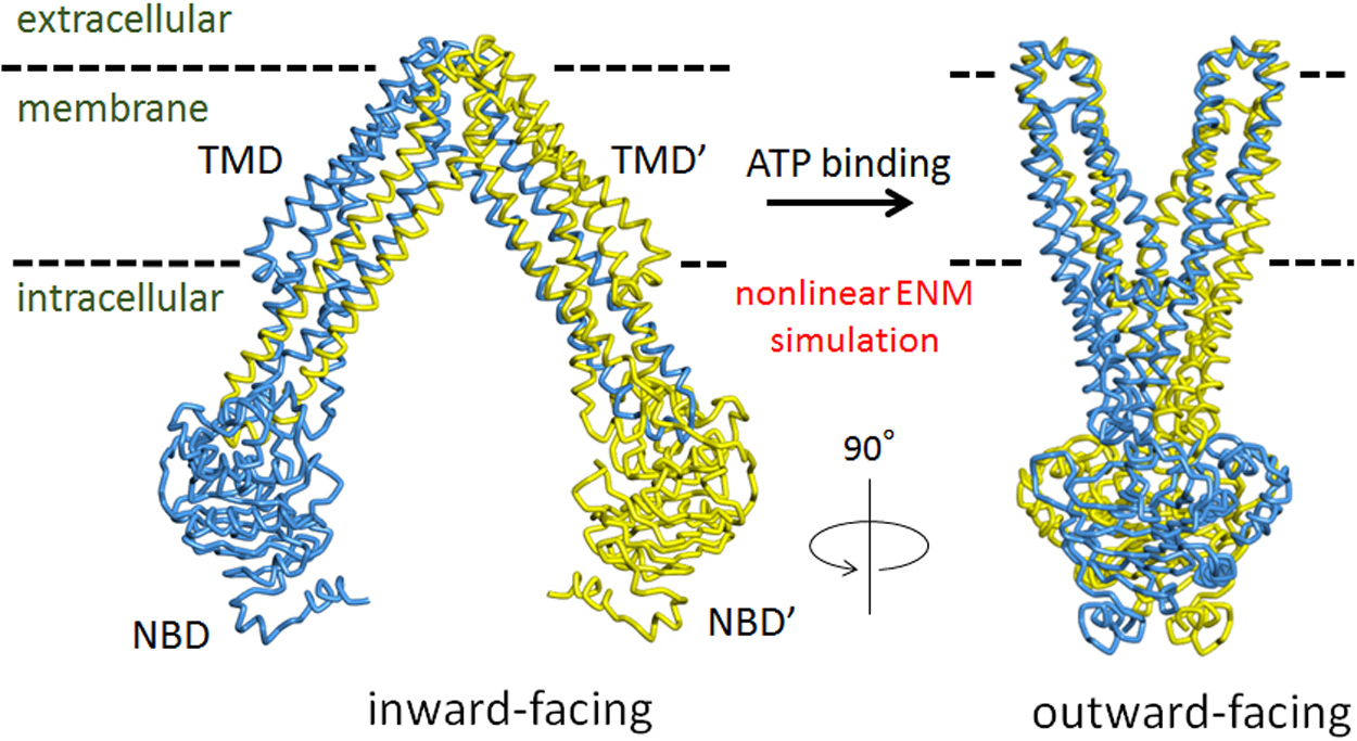
    Upon the binding of ATP molecules to nucleotide binding domains (NBDs), ATP-binding cassette (ABC) exporters undergo a conformational transition from an inward-facing (IF) to an outward-facing (OF) state. This molecular event is a typical example of chemo-mechanical coupling. However, the underlying mechanism remains unclear. In this study, we analyzed the IF→OF transition of a representative ABC exporter, MsbA, by solving the equation of motion under an elastic network model (ENM). ATP was represented as a single node in ENM or replaced by external forces. When two ATP nodes were added to the ENM of the IF state protein, the two NBDs dimerized; subsequently, the two transmembrane domains opened toward the extracellular side, resulting in the formation of the OF structure. Such a conformational transition was also reproduced by applying external forces, which caused the rotational motion of the NBDs instead of the addition of ATP nodes. The process of the conformational transition was analyzed in detail using cross-correlation maps for node-node interactions. More importantly, it was revealed that the ATP binding energy is converted into distortion energy of several transmembrane helices. These results are useful for understanding the chemo-mechanical coupling in ABC transporters.

    Download PDF (6388K)
  • Shota Kondo, Michio Homma, Seiji Kojima
    2017 Volume 14 Pages 173-181
    Published: 2017
    Released on J-STAGE: December 05, 2017
    JOURNAL FREE ACCESS
    Supplementary material

    Vibrio alginolyticus normally has a single polar flagellum whose number and placement are regulated positively by FlhF. FlhF is a GTPase and homolog of a signal recognition particle (SRP) protein called Ffh and SRP receptor FtsY. FlhF is located at the cell pole and directs formation of the flagellum. To study the mechanism of FlhF localization, we introduced random mutations into flhF by means of hydroxylamine and isolated mutants that could not generate the flagellum at the cell pole. The novel mutations were only mapped to the GTPase motif of FlhF. The mutant FlhF proteins showed reduced polar localization as compared to the wild type and still could associate with the membrane. These results support the assumption that the GTPase motif of FlhF plays a critical role in the polar localization of this protein during formation of the flagellum.

    Download PDF (3382K)
  • Kazuho Yoshida, Takahiro Yamashita, Kengo Sasaki, Keiichi Inoue, Yoshi ...
    2017 Volume 14 Pages 183-190
    Published: 2017
    Released on J-STAGE: December 19, 2017
    JOURNAL FREE ACCESS

    We previously showed that the chimeric proteins of microbial rhodopsins, such as light-driven proton pump bacteriorhodopsin (BR) and Gloeobacter rhodopsin (GR) that contain cytoplasmic loops of bovine rhodopsin, are able to activate Gt protein upon light absorption. These facts suggest similar protein structural changes in both the light-driven proton pump and animal rhodopsin. Here we report two trials to engineer chimeric rhodopsins, one for the inserted loop, and another for the microbial rhodopsin template. For the former, we successfully activated Gs protein by light through the incorporation of the cytoplasmic loop of β2-adrenergic receptor (β2AR). For the latter, we did not observe any G-protein activation for the light-driven sodium pump from Indibacter alkaliphilus (IndiR2) or a light-driven chloride pump halorhodopsin from Natronomonas pharaonis (NpHR), whereas the light-driven proton pump GR showed light-dependent G-protein activation. This fact suggests that a helix opening motion is common to G protein coupled receptor (GPCR) and GR, but not to IndiR2 and NpHR. Light-induced difference FTIR spectroscopy revealed similar structural changes between WT and the third loop chimera for each light-driven pump. A helical structural perturbation, which was largest for GR, was further enhanced in the chimera. We conclude that similar structural dynamics that occur on the cytoplasmic side of GPCR are needed to design chimeric microbial rhodopsins.

    Download PDF (1706K)
Review Article
  • Hiroyuki Terashima, Akihiro Kawamoto, Yusuke V. Morimoto, Katsumi Imad ...
    2017 Volume 14 Pages 191-198
    Published: 2017
    Released on J-STAGE: December 19, 2017
    JOURNAL FREE ACCESS

    The bacterial flagellum is a supramolecular motility machine consisting of the basal body as a rotary motor, the hook as a universal joint, and the filament as a helical propeller. Intact structures of the bacterial flagella have been observed for different bacterial species by electron cryotomography and subtomogram averaging. The core structures of the basal body consisting of the C ring, the MS ring, the rod and the protein export apparatus, and their organization are well conserved, but novel and divergent structures have also been visualized to surround the conserved structure of the basal body. This suggests that the flagellar motors have adapted to function in various environments where bacteria live and survive. In this review, we will summarize our current findings on the divergent structures of the bacterial flagellar motor.

    Download PDF (3760K)
  • Hiroshi Hashimoto, Asami Hishiki, Kodai Hara, Sotaro Kikuchi
    2017 Volume 14 Pages 199-205
    Published: 2017
    Released on J-STAGE: December 22, 2017
    JOURNAL FREE ACCESS

    DNA damage tolerance (DDT) is a cell function to avoid replication arrest by DNA damage during DNA replication. DDT includes two pathways, translesion DNA synthesis (TLS) and template-switched DNA synthesis (TS). DDT is regulated by ubiquitination of proliferating cell nuclear antigen that binds to double-stranded DNA and functions as scaffold protein for DNA metabolism. TLS is transient DNA synthesis using damaged DNA as a template by error-prone DNA polymerases termed TLS polymerases specialized for DNA damage. TS, in which one newly synthesized strand is utilized as an undamaged template for replication by replicative polymerases, is error-free process. Thus, DDT is not inherently a repair pathway. DDT is a mechanism to tolerate DNA damage, giving priority to DNA synthesis and enabling finish of DNA replication for cell survival and genome stability. DDT is associated with cancer development and thus is of great interest in drug discovery for cancer therapy. This review article describes recent progress in structural studies on protein-protein and protein-DNA complexes involved in TLS and TS, providing the molecular mechanisms of interactions in DDT.

    Download PDF (1761K)
Regular Article
  • Daisuke Miyashiro, Misato Ohtsuki, Yuta Shimamoto, Jun’ichi Wakayama, ...
    2017 Volume 14 Pages 207-220
    Published: 2017
    Released on J-STAGE: December 28, 2017
    JOURNAL FREE ACCESS

    We have studied the stiffness of myofilament lattice in sarcomeres in the pre-force generating state, which was realized by a relaxing reagent, BDM (butane dione monoxime). First, the radial stiffness for the overlap regions of sarcomeres of isolated single myofibrils was estimated from the resulting decreases in diameter by osmotic pressure applied with the addition of Dextran. Then, the radial stiffness was also estimated from force-distance curve measurements with AFM technology. The radial stiffness for the overlap regions thus obtained was composed of a soft and a rigid component. The soft component visco-elastically changed in a characteristic fashion depending on the physiological conditions of myofibrils, suggesting that it comes from cross-bridge structures. BDM treatments significantly affected the soft radial component of contracting myofibrils depending on the approach velocity of cantilever: It was nearly equal to that in the contracting state at high approach velocity, whereas as low as that in the relaxing state at low approach velocity. However, comparable BDM treatments greatly suppressed the force production and the axial stiffness in contracting glycerinated muscle fibers and also the sliding velocity of actin filaments in the in vitro motility assay. Considering that BDM shifts the cross-bridge population from force generating to pre-force generating states in contracting muscle, the obtained results strongly suggest that cross-bridges in the pre-force generating state are visco-elastically attached to the thin filaments in such a binding manner that the axial stiffness is low but the radial stiffness significantly high similar to that in force generating state.

    Download PDF (1980K)
feedback
Top