Biophysics and Physicobiology
Online ISSN : 2189-4779
ISSN-L : 2189-4779
Volume 19
Displaying 1-48 of 48 articles from this issue
Review Article (Invited)
  • Takashi Fujishiro, Ryosuke Nakamura, Kouhei Kunichika, Yasuhiro Takaha ...
    Article type: Review Article (Invited)
    2022 Volume 19 Article ID: e190001
    Published: 2022
    Released on J-STAGE: February 22, 2022
    Advance online publication: February 08, 2022
    JOURNAL OPEN ACCESS FULL-TEXT HTML

    Cysteine desulfurases are pyridoxal-5'-phosphate (PLP)-dependent enzymes that mobilize sulfur derived from the l-cysteine substrate to the partner sulfur acceptor proteins. Three cysteine desulfurases, IscS, NifS, and SufS, have been identified in ISC, NIF, and SUF/SUF-like systems for iron-sulfur (Fe-S) cluster biosynthesis, respectively. These cysteine desulfurases have been investigated over decades, providing insights into shared/distinct catalytic processes based on two types of enzymes (type I: IscS and NifS, type II: SufS). This review summarizes the insights into the structural/functional varieties of bacterial and eukaryotic cysteine desulfurases involved in Fe-S cluster biosynthetic systems. In addition, an inactive cysteine desulfurase IscS paralog, which contains pyridoxamine-5'-phosphate (PMP), instead of PLP, is also described to account for its hypothetical function in Fe-S cluster biosynthesis involving this paralog. The structural basis for cysteine desulfurase functions will be a stepping stone towards understanding the diversity and evolution of Fe-S cluster biosynthesis.

Regular Article
  • Ryohei Kondo, Kota Kasahara, Takuya Takahashi
    Article type: Regular Article
    2022 Volume 19 Article ID: e190002
    Published: 2022
    Released on J-STAGE: February 22, 2022
    Advance online publication: February 08, 2022
    JOURNAL OPEN ACCESS FULL-TEXT HTML
    Supplementary material

    Elucidating the principles of sequence–structure relationships of proteins is a long-standing issue in biology. The nature of a short segment of a protein is determined by both the subsequence of the segment itself and its environment. For example, a type of subsequence, the so-called chameleon sequences, can form different secondary structures depending on its environments. Chameleon sequences are considered to have a weak tendency to form a specific structure. Although many chameleon sequences have been identified, they are only a small part of all possible subsequences in the proteome. The strength of the tendency to take a specific structure for each subsequence has not been fully quantified. In this study, we comprehensively analyzed subsequences consisting of four to nine amino acid residues, or N-gram (4≤N≤9), observed in non-redundant sequences in the Protein Data Bank (PDB). Tendencies to form a specific structure in terms of the secondary structure and accessible surface area are quantified as information quantities for each N-gram. Although the majority of observed subsequences have low information quantity due to lack of samples in the current PDB, thousands of N-grams with strong tendencies, including known structural motifs, were found. In addition, machine learning partially predicted the tendency of unknown N-grams, and thus, this technique helps to extract knowledge from the limited number of samples in the PDB.

  • Yuka Oka, Shota Ushiba, Naruto Miyakawa, Madoka Nishio, Takao Ono, Yas ...
    Article type: Regular Article
    2022 Volume 19 Article ID: e190003
    Published: 2022
    Released on J-STAGE: February 26, 2022
    Advance online publication: February 09, 2022
    JOURNAL OPEN ACCESS FULL-TEXT HTML

    C-reactive protein (CRP) is an important biomarker of infection and inflammation, as CRP is one of the most prominent acute-phase proteins. CRP is usually detected using anti-CRP antibodies (Abs), where the intermolecular interactions between CRP and the anti-CRP Ab are largely affected by the pH and ionic strength of environmental solutions. Therefore, it is important to understand the environmental effects of CRP–anti-CRP Ab interactions when designing highly sensitive biosensors. Here, we investigated the efficiency of fluorescently labeled CRP–anti-CRP monoclonal antibody (mAb) interactions at different pHs and ionic strengths. Our results indicate that the affinity was insensitive to pH changes in the range of 5.9 to 8.1, while it was significantly sensitive to ionic strength changes. The binding affinity decreased by 55% at an ionic strength of 1.6 mM, when compared to that under a physiological condition (~150 mM). Based on the isoelectric focusing results, both the labeled CRP and anti-CRP mAb were negatively charged in the studied pH range, which rendered the system insensitive to pH changes, but sensitive to ionic strength changes. The decreased ionic strength led to a significant enhancement of the repulsive force between CRP and the anti-CRP mAb. Although the versality of the findings is not fully studied yet, the results provide insights into designing highly sensitive CRP sensors, especially field-effect transistor-based sensors.

Commentary and Perspective
Review Article (Invited)
  • Taro Furubayashi, Norikazu Ichihashi
    Article type: Review Article (Invited)
    2022 Volume 19 Article ID: e190005
    Published: 2022
    Released on J-STAGE: February 26, 2022
    Advance online publication: February 15, 2022
    JOURNAL OPEN ACCESS FULL-TEXT HTML

    How can evolution assemble lifeless molecules into a complex living organism? The emergent process of biological complexity in the origin of life is a big mystery in biology. In vitro evolution of artificial molecular replication systems offers unique experimental opportunities to probe possible pathways of a simple molecular system approaching a complex life-like system. This review focuses on experimental efforts to examine evolvability of molecules in vitro from the pioneering Spiegelman’s experiment to our latest research on an artificial RNA self-replication system. Genetic translation and compartmentalization are shown to enable sustainable replication and evolution. Latest studies are revealing that coevolution of self-replicating “host replicators” and freeloading “parasitic replicators” is crucial to extend evolvability of a molecular replication system for continuous evolution and emergence of diversity. Intense competition between hosts and parasites would have existed even before the origin of life and contributed to generating complex molecular ecosystems. This review article is an extended version of the Japanese article “An in vitro evolutionary journey of an artificial RNA replication system towards biological complexity” published in SEIBUTSU-BUTSURI Vol.61, p.240–244 (2021).”

  • Hiroaki Yokota
    Article type: Review Article (Invited)
    2022 Volume 19 Article ID: e190006
    Published: 2022
    Released on J-STAGE: March 25, 2022
    Advance online publication: March 10, 2022
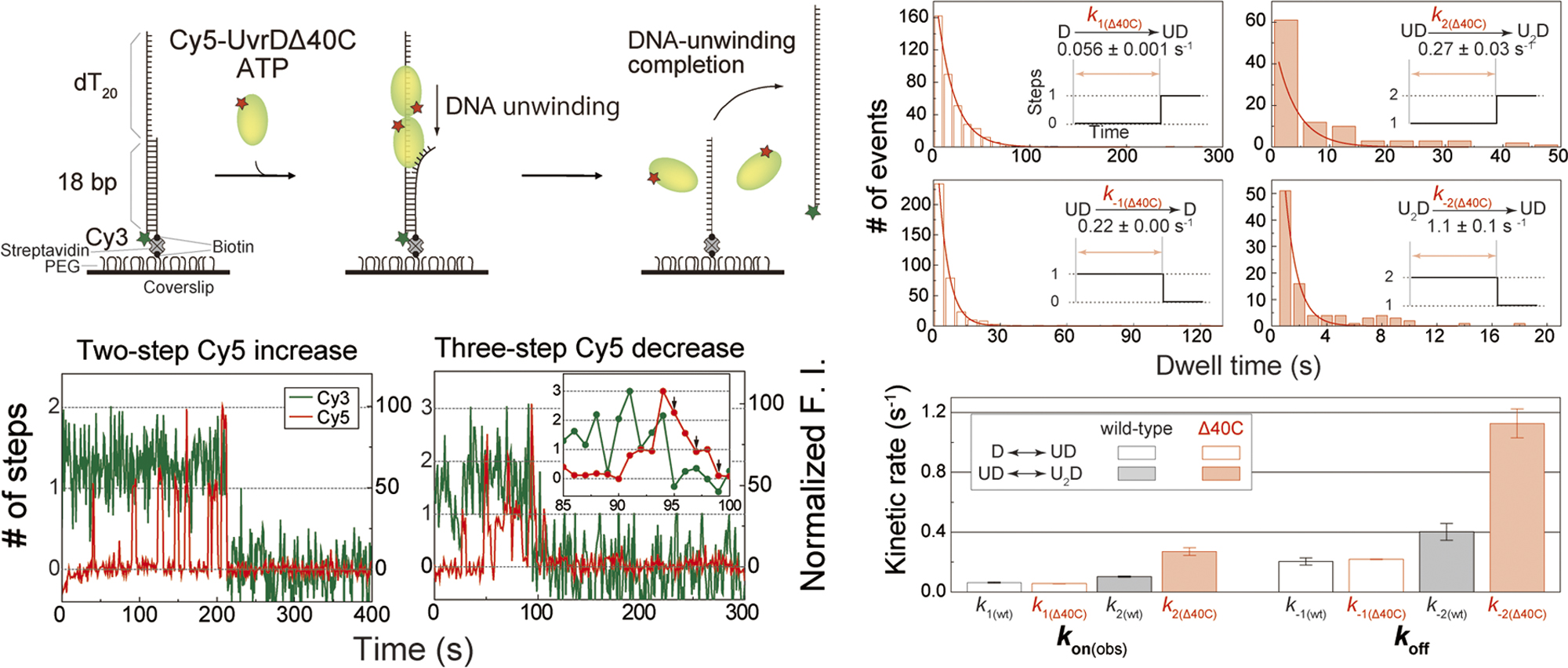
    JOURNAL OPEN ACCESS FULL-TEXT HTML

    Helicases are nucleic acid-unwinding enzymes involved in the maintenance of genome integrity. Helicases share several “helicase motifs” that are highly conserved amino acid sequences and are classified into six superfamilies (SFs). The helicase SFs are further grouped into two classes based on their functional units. One class that includes SFs 3–6 functions as a hexamer that can form a ring around DNA. Another class that includes SFs 1 and 2 functions in a non-hexameric form. The high homology in the primary and tertiary structures among SF1 helicases suggests that SF1 helicases have a common underlying mechanism. However, two opposing models for the functional unit, monomer and dimer models, have been proposed to explain DNA unwinding by SF1 helicases. This paper briefly describes the classification of helicase SFs and discusses the structural homology and the two opposing non-hexameric helicase models of SF1 helicases by focusing on Escherichia coli SF1 helicase UvrD, which plays a significant role in both nucleotide-excision repair and methyl-directed mismatch repair. This paper reviews past and recent studies on UvrD, including the author's single-molecule direct visualization of wild-type UvrD and a UvrD mutant lacking the C-terminal 40 amino acids (UvrDΔ40C), the latter of which was used in genetic and biochemical assays that supported the monomer model. The visualization revealed that multiple UvrDΔ40C molecules jointly unwind DNA, presumably in an oligomeric form, similar to wild-type UvrD. Therefore, single-molecule direct visualization of nucleic acid-binding proteins can provide quantitative and kinetic information to reveal their fundamental mechanisms.

  • Hideaki Yoshimura
    Article type: Review Article (Invited)
    2022 Volume 19 Article ID: e190007
    Published: 2022
    Released on J-STAGE: March 26, 2022
    Advance online publication: March 11, 2022
    JOURNAL OPEN ACCESS FULL-TEXT HTML

    Membrane receptors provide interfaces of various extracellular stimuli to transduce the signal into the cell. Receptors are required to possess such conflicting properties as high sensitivity and noise reduction for the cell to keep its homeostasis and appropriate responses. To understand the mechanisms by which these functions are achieved, single-molecule monitoring of the motilities of receptors and signaling molecules on the plasma membrane is one of the most direct approaches. This review article introduces several recent single-molecule imaging studies of receptors, including the author’s recent work on triple-color single-molecule imaging of G protein-coupled receptors. Based on these researches, advantages and perspectives of the single-molecule imaging approach to solving the mechanisms of receptor functions are illustrated.

Regular Article
  • Damien Simon, Atsushi Mukaiyama, Yoshihiko Furuike, Shuji Akiyama
    Article type: Regular Article
    2022 Volume 19 Article ID: e190008
    Published: 2022
    Released on J-STAGE: April 14, 2022
    Advance online publication: March 30, 2022
    JOURNAL OPEN ACCESS FULL-TEXT HTML

    KaiC is the central pacemaker of the circadian clock system in cyanobacteria and forms the core in the hetero-multimeric complexes, such as KaiB–KaiC and KaiA–KaiB–KaiC. Although the formation process and structure of the binary and ternary complexes have been studied extensively, their disassembly dynamics have remained elusive. In this study, we constructed an experimental system to directly measure the autonomous disassembly of the KaiB–KaiC complex under the condition where the dissociated KaiB cannot reassociate with KaiC. At 30°C, the dephosphorylated KaiB–KaiC complex disassembled with an apparent rate of 2.1±0.3 d–1, which was approximately twice the circadian frequency. Our present analysis using a series of KaiC mutants revealed that the apparent disassembly rate correlates with the frequency of the KaiC phosphorylation cycle in the presence of KaiA and KaiB and is robustly temperature-compensated with a Q10 value of 1.05±0.20. The autonomous cancellation of the interactions stabilizing the KaiB–KaiC interface is one of the important phenomena that provide a link between the molecular-scale and system-scale properties.

Review Article (Invited)
  • Fumiaki Kono, Kazuo Kurihara, Taro Tamada
    Article type: Review Article (Invited)
    2022 Volume 19 Article ID: e190009
    Published: 2022
    Released on J-STAGE: April 16, 2022
    Advance online publication: April 01, 2022
    JOURNAL OPEN ACCESS FULL-TEXT HTML

    Hydrogen atoms and hydration water molecules in proteins are essential for many biochemical processes, especially enzyme catalysis. Neutron crystallography enables direct observation of hydrogen atoms, and reveals molecular recognition through hydrogen bonding and catalytic reactions involving proton-coupled electron transfer. The use of neutron crystallography is still limited for proteins, but its popularity is increasing owing to an increase in the number of diffractometers for structural biology at neutron facilities and advances in sample preparation. According to the characteristics of the neutrons, monochromatic or quasi-Laue methods and the time-of-flight method are used in nuclear reactors and pulsed spallation sources, respectively, to collect diffraction data. Growing large crystals is an inevitable problem in neutron crystallography for structural biology, but sample deuteration, especially protein perdeuteration, is effective in reducing background levels, which shortens data collection time and decreases the crystal size required. This review also introduces our recent neutron structure analyses of copper amine oxidase and copper-containing nitrite reductase. The neutron structure of copper amine oxidase gives detailed information on the protonation state of dissociable groups, such as the quinone cofactor, which are critical for catalytic reactions. Electron transfer via a hydrogen-bond jump and a hydroxide ion ligation in copper-containing nitrite reductase are clarified, and these observations are consistent with the results from the quantum chemical calculations. This review article is an extended version of the Japanese article, Elucidation of Enzymatic Reaction Mechanism by Neutron Crystallography, published in SEIBUTSU-BUTSURI Vol. 61, p.216–222 (2021).

  • Yuhei Tachi, Satoru G. Itoh, Hisashi Okumura
    Article type: Review Article (Invited)
    2022 Volume 19 Article ID: e190010
    Published: 2022
    Released on J-STAGE: April 20, 2022
    Advance online publication: April 02, 2022
    JOURNAL OPEN ACCESS FULL-TEXT HTML

    Alzheimer’s disease is thought to be caused by the aggregation of amyloid-β (Aβ) peptides. Their aggregation is accelerated at hydrophilic/hydrophobic interfaces such as the air–water interface and the surface of monosialotetrahexosylganglioside (GM1) clusters on neuronal cell membranes. In this review, we present recent studies of full-length Aβ (Aβ40) peptides and Aβ(16–22) fragments in such heterogeneous environments by molecular dynamics (MD) simulations. These peptides have both hydrophilic and hydrophobic amino-acid residues and tend to exist at the hydrophilic/hydrophobic interface. Therefore, the peptide concentration increases at the interface, which is one of the factors that promote aggregation. Furthermore, it was found that Aβ40 forms an α-helix structure and then a β-hairpin structure at the interface. The β-hairpin promotes the formation of oligomers with intermolecular β-sheets. It means that not only the high concentration of Aβ40 at the interface but also the structure of Aβ40 itself promotes aggregation. In addition, MD simulations of Aβ40 on recently-developed GM1-glycan clusters showed that the HHQ (13–15) segment of Aβ40 is important for the recognition of GM1-glycan clusters. It was also elucidated that Aβ40 forms a helix structure in the C-terminal region on the GM1-glycan cluster. This result suggests that the helix formation, which is the first step in the conformational changes toward pathological aggregation, is initiated at the GM1-glycan moieties rather than at the lipid-ceramide moieties. These studies will enhance the physicochemical understanding of the structural changes of Aβ at the heterogeneous interfaces and the mechanism of Alzheimer’s disease pathogenesis.

  • Takao K. Suzuki
    Article type: Review Article (Invited)
    2022 Volume 19 Article ID: e190011
    Published: 2022
    Released on J-STAGE: April 20, 2022
    Advance online publication: April 05, 2022
    JOURNAL OPEN ACCESS FULL-TEXT HTML

    Design principles of phenotypes in organisms are fundamental issues in physical biology. So far, understanding “systems” of living organisms have been chiefly promoted by understanding the underlying biomolecules such as genes and proteins, and their intra- and inter-relationships and regulations. After a long period of sophistication, biophysics and molecular biology have established a general framework for understanding ‘molecular systems’ in organisms without regard to species, so that the findings of fly studies can be applied to mouse studies. However, little attention has been paid to exploring “phenotypic systems” in organisms, and thus its general framework remains poorly understood. Here I review concepts, methods, and case studies using butterfly and moth wing patterns to explore phenotypes as systems. First, I present a unifying framework for phenotypic traits as systems, termed multi-component systems. Second, I describe how to define components of phenotypic systems, and also show how to quantify interactions among phenotypic parts. Subsequently, I introduce the concept of the macro-evolutionary process, which illustrates how to generate complex traits. In this point, I also introduce mathematical methods, “phylogenetic comparative methods”, which provide stochastic processes along molecular phylogeny as bifurcated paths to quantify trait evolution. Finally, I would like to propose two key concepts, macro-evolutionary pathways and genotype-phenotype loop (GP loop), which must be needed for the next directions. I hope these efforts on phenotypic biology will become one major target in biophysics and create the next generations of textbooks. This review article is an extended version of the Japanese article, Biological Physics in Phenotypic Systems of Living Organisms, published in SEIBUTSU-BUTSURI Vol. 61, p. 31–35 (2021).

Regular Article
  • Shinjiro Nakahata, Tetsushi Komoto, Masashi Fujii, Akinori Awazu
    Article type: Regular Article
    2022 Volume 19 Article ID: e190012
    Published: 2022
    Released on J-STAGE: April 20, 2022
    Advance online publication: April 05, 2022
    JOURNAL OPEN ACCESS FULL-TEXT HTML
    Supplementary material

    During the repair of double-strand breaks (DSBs) in DNA, active mobilizations for conformational changes in chromosomes have been widely observed in eukaryotes, from yeast to animal and plant cells. DSB-damaged loci in the yeast genome showed increased mobility and relocation to the nuclear periphery. However, the driving forces behind DSB-induced chromatin dynamics remain unclear. In this study, mathematical models of normal and DSB-damaged yeast chromosomes were developed to simulate their structural dynamics. The effects of histone degradation in the whole nucleus and the change in the physical properties of damaged loci due to the binding of SUMOylated repair proteins were considered in the model of DSB-induced chromosomes based on recent experimental results. The simulation results reproduced DSB-induced changes to structural and dynamical features by which the combination of whole nuclear histone degradation and the rigid structure formation of repair protein accumulations on damaged loci were suggested to be primary contributors to the process by which damaged loci are relocated to the nuclear periphery.

Review Article (Invited)
  • Toru Kondo, Yutaka Shibata
    Article type: Review Article (Invited)
    2022 Volume 19 Article ID: e190013
    Published: 2022
    Released on J-STAGE: April 28, 2022
    Advance online publication: April 08, 2022
    JOURNAL OPEN ACCESS FULL-TEXT HTML

    Photosynthetic light-harvesting complexes (LHCs) play a crucial role in concentrating the photon energy from the sun that otherwise excites a typical pigment molecule, such as chlorophyll-a, only several times a second. Densely packed pigments in the complexes ensure efficient energy transfer to the reaction center. At the same time, LHCs have the ability to switch to an energy-quenching state and thus play a photoprotective role under excessive light conditions. Photoprotection is especially important for oxygenic photosynthetic organisms because toxic reactive oxygen species can be generated through photochemistry under aerobic conditions. Because of the extreme complexity of the systems in which various types of pigment molecules strongly interact with each other and with the surrounding protein matrixes, there has been long-standing difficulty in understanding the molecular mechanisms underlying the flexible switching between the light-harvesting and quenching states. Single-molecule spectroscopy studies are suitable to reveal the conformational dynamics of LHCs reflected in the fluorescence properties that are obscured in ordinary ensemble measurements. Recent advanced single-molecule spectroscopy studies have revealed the dynamical fluctuations of LHCs in their fluorescence peak position, intensity, and lifetime. The observed dynamics seem relevant to the conformational plasticity required for the flexible activations of photoprotective energy quenching. In this review, we survey recent advances in the single-molecule spectroscopy study of the light-harvesting systems of oxygenic photosynthesis.

Review Article
  • Etsuro Ito, Kotaro Oka, Fusako Koshikawa
    Article type: Review Article
    2022 Volume 19 Article ID: e190014
    Published: 2022
    Released on J-STAGE: April 28, 2022
    Advance online publication: April 08, 2022
    JOURNAL OPEN ACCESS FULL-TEXT HTML

    Chronic pain often has an unknown cause, and many patients with chronic pain learn to accept that their pain is incurable and pharmacologic treatments are only temporarily effective. Complementary and integrative health approaches for pain are thus in high demand. One such approach is soft touch, e.g., adhesion of pyramidal thorn patches in a pain region. The effects of patch adhesion on pain relief have been confirmed in patients with various types of pain. A recent study using near-infrared spectroscopy revealed that the dorsolateral prefrontal cortex (DLPFC), especially the left side, is likely to be inactivated in patients experiencing pain relief during patch treatment. Mindfulness meditation is another well-known complementary and integrative approach for achieving pain relief. The relation between pain relief due to mindfulness meditation and changes in brain regions, including the DLPFC, has long been examined. In the present review article, we survey the literature describing the effects of the above-mentioned complementary and integrative treatments on pain relief, and outline the important brain regions, including the DLPFC, that are involved in analgesia. We hope that the present article will provide clues to researchers who hope to advance neurosensory treatments for pain relief without medication.

Regular Article
  • Keisuke Inoue, Shoji Takada, Tsuyoshi Terakawa
    Article type: Regular Article
    2022 Volume 19 Article ID: e190015
    Published: 2022
    Released on J-STAGE: May 11, 2022
    Advance online publication: April 14, 2022
    JOURNAL OPEN ACCESS FULL-TEXT HTML
    Supplementary material

    DNA mismatches are frequently generated by various intrinsic and extrinsic factors including DNA replication errors, oxygen species, ultraviolet, and ionizing radiation. These mismatches should be corrected by the mismatches repair (MMR) pathway to maintain genome integrity. In the Escherichia coli (E. coli) MMR pathway, MutS searches and recognizes a base-pair mismatch from millions of base-pairs. Once recognized, ADP bound to MutS is exchanged with ATP, which induces a conformational change in MutS. Previous single-molecule fluorescence microscopy studies have suggested that ADP-bound MutS temporarily slides along double-stranded DNA in a rotation-coupled manner to search a base-pair mismatch and so does ATP-bound MutS in a rotation-uncoupled manner. However, the detailed structural dynamics of the sliding remains unclear. In this study, we performed coarse-grained molecular dynamics simulations of the E. coli MutS bound on DNA in three different conformations: ADP-bound (), ATP-bound open clamp (), and ATP-bound closed clamp () conformations. In the simulations, we observed conformation-dependent diffusion of MutS along DNA. and diffused along DNA in a rotation-coupled manner with rare and frequent groove-crossing events, respectively. In the groove-crossing events, MutS overcame an edge of a groove and temporarily diffused in a rotation-uncoupled manner. It was also indicated that mismatch searches by is inefficient in terms of mismatch checking even though it diffuses along DNA and reaches unchecked regions more rapidly than .

  • Damien Hall, Adam S. Foster
    Article type: Regular Article
    2022 Volume 19 Article ID: e190016
    Published: 2022
    Released on J-STAGE: May 18, 2022
    Advance online publication: April 15, 2022
    JOURNAL OPEN ACCESS FULL-TEXT HTML
    J-STAGE Data Supplementary material

    High speed atomic force microscopy (HS-AFM) is, in principle, capable of yielding nanometer level detail about the surface of static structures. However, for highly dynamic samples HS-AFM may struggle with the correct feature assignment both within and between frames. Feature assignment in HS-AFM is dependent on (i) the intrinsic sampling rate, and (ii) the rate of internal redistribution of the sample. Whilst the first quantity (the sampling rate) is defined by the device parameters, the second quantity is frequently unknown, and is often the desired target of the measurement. This work examines how, even in the absence of gross cell morphological change, the rapid dynamics of living cell membranes, may impose an upper spatial limit to the frame-to-frame assignment of cell micro-topography and other related properties (such as local elasticity) whose motion may be described stochastically. Such a practical maximum may prove useful in the setup of HS-AFM experiments involving dynamic surfaces thereby facilitating selection of the most parsimonious relationship between observation size, image pixilation and sampling rates. To assist with performing the described calculations a graphical user interface-based software package called HS-AFM UGOKU is made freely available.

Review Article (Invited)
  • Naoki Yamamoto, Eri Chatani
    Article type: Review Article (Invited)
    2022 Volume 19 Article ID: e190017
    Published: 2022
    Released on J-STAGE: June 01, 2022
    Advance online publication: May 10, 2022
    JOURNAL OPEN ACCESS FULL-TEXT HTML

    It is crucial to understand the mechanism of amyloid fibril formation for the development of the therapeutic ways against amyloidoses and neurodegenerative diseases. Prefibrillar intermediates, which emerge prior to the fibril formation, seem to play a key role to the occurrence of nuclei of amyloid fibrils. We have focused on an insulin-derived peptide, B chain, to precisely clarify the mechanism of the fibril formation via prefibrillar intermediates. Various kinds of methods such as circular dichroism spectroscopy, dynamic light scattering, small-angle X-ray scattering, and atomic force microscopy were employed to track the structural changes in prefibrillar intermediates. The prefibrillar intermediates possessing rod-shaped structures elongated as a function of time, which led to fibril formation. We have also found that a blood clotting protein, fibrinogen, inhibits the amyloid fibril formation of B chain. This was caused by the stabilization of prefibrillar intermediates and thus the suppression of their elongation by fibrinogen. These findings have not only shed light on detailed mechanisms about how prefibrillar intermediates convert to the amyloid fibril, but also demonstrated that inhibiting the structural development of prefibrillar intermediates is an effective strategy to develop therapeutic ways against amyloid-related diseases. This review article is an extended version of the Japanese article, Observing Development of Amyloid Prefibrillar Intermediates and their Interaction with Chaperones for Inhibiting the Fibril Formation, published in SEIBUTSU BUTSURI Vol. 61, p. 236–239 (2021).

Regular Article
  • Tetsushi Komoto, Masashi Fujii, Akinori Awazu
    Article type: Regular Article
    2022 Volume 19 Article ID: e190018
    Published: 2022
    Released on J-STAGE: June 01, 2022
    Advance online publication: May 10, 2022
    JOURNAL OPEN ACCESS FULL-TEXT HTML
    Supplementary material

    X chromosome inactivation center (Xic) pairing occurs during the differentiation of embryonic stem (ES) cells from female mouse embryos, and is related to X chromosome inactivation, the circadian clock, intra-nucleus architecture, and metabolism. However, the mechanisms underlying the identification and approach of X chromosome pairs in the crowded nucleus are unclear. To elucidate the driving force of Xic pairing, we developed a coarse-grained molecular dynamics model of intranuclear chromosomes in ES cells and in cells 2 days after the onset of differentiation (2-day cells) by considering intrachromosomal epigenetic-structural feature-dependent mechanics. The analysis of the experimental data showed that X-chromosomes exhibit the rearrangement of their distributions of open/closed chromatin regions on their surfaces during cell differentiation. By simulating models where the excluded volume effects of closed chromatin regions are stronger than those of open chromatin regions, such rearrangement of open/closed chromatin regions on X-chromosome surfaces promoted the mutual approach of the Xic pair. These findings suggested that local intrachromosomal epigenetic features may contribute to the regulation of cell species-dependent differences in intranuclear architecture.

  • Nobutaka Numoto, Seiko Onoda, Yoshiaki Kawano, Hideo Okumura, Seiki Ba ...
    Article type: Regular Article
    2022 Volume 19 Article ID: e190019
    Published: 2022
    Released on J-STAGE: June 01, 2022
    Advance online publication: May 12, 2022
    JOURNAL OPEN ACCESS FULL-TEXT HTML
    Supplementary material

    Ever since the historic discovery of the cooperative oxygenation of its multiple subunits, hemoglobin (Hb) has been among the most exhaustively studied allosteric proteins. However, the lack of structural information on the intermediates between oxygenated and deoxygenated forms prevents our detailed understanding of the molecular mechanism of its allostery. It has been difficult to prepare crystals of intact oxy-deoxy intermediates and to individually identify the oxygen saturation for each subunit. However, our recent crystallographic studies have demonstrated that giant Hbs from annelids are suitable for overcoming these problems and can provide abundant information on oxy-deoxy intermediate structures. Here, we report the crystal structures of oxy-deoxy intermediates of a 400 kDa Hb (V2Hb) from the annelid Lamellibrachia satsuma, following up on a series of previous studies of similar giant Hbs. Four intermediate structures had average oxygen saturations of 78%, 69%, 55%, and 26%, as determined by the occupancy refinement of the bound oxygen based on ambient temperature factors. The structures demonstrate that the cooperative oxygen dissociation is weaker, large ternary and quaternary changes are induced at a later stage of the oxygen dissociation process, and the ternary and quaternary changes are smaller with local perturbations. Nonetheless, the overall structural transition seemed to proceed in the manner of the MWC two-state model. Our crystallographic snapshots of the allosteric transition of V2Hb provide important experimental evidence for a more detailed understanding of the allostery of Hbs by extension of the Monod–Wyman–Changeux (MWC) model.

Review Article (Invited)
  • Kazusa Beppu, Yusuke T. Maeda
    Article type: Review Article (Invited)
    2022 Volume 19 Article ID: e190020
    Published: 2022
    Released on J-STAGE: June 03, 2022
    Advance online publication: May 12, 2022
    JOURNAL OPEN ACCESS FULL-TEXT HTML

    Ordered collective motion emerges in a group of autonomously motile elements (known as active matter) as their density increases. Microswimmers, such as swimming bacteria, have been extensively studied in physics and biology. A dense suspension of bacteria forms seemingly chaotic turbulence in viscous fluids. Interestingly, this active turbulence driven by bacteria can form a hidden ensemble of many vortices. Understanding the active turbulence in a bacterial suspension can provide physical principles for pattern formation and insight into the instability underlying biological phenomena. This review presents recent findings regarding ordered structures causing active turbulence and discusses a physical approach for controlling active turbulence via geometric confinement. When the active matter is confined in a compartment with a size comparable to the correlation length of the collective motion, vortex-like rotation appears, and the vortex pairing order is indicated by the patterns of interacting vortices. Additionally, we outline the design principle for controlling collective motions via the geometric rule of the vortex pairing, which may advance engineering microdevices driven by a group of active matter. This article is an extended version of the Japanese article, Ordered Structure and Geometric Control of Active Matter in Dense Bacterial Suspensions, published in SEIBUTSU BUTSURI Vol. 60, p. 13–18 (2020).

  • Keisuke Fujiyama, Tomoya Hino, Shingo Nagano
    Article type: Review Article (Invited)
    2022 Volume 19 Article ID: e190021
    Published: 2022
    Released on J-STAGE: June 21, 2022
    Advance online publication: June 01, 2022
    JOURNAL OPEN ACCESS FULL-TEXT HTML

    Steroid hormones modulate numerous physiological processes in various higher organisms. Research on the physiology, biosynthesis, and metabolic degradation of steroid hormones is crucial for developing drugs, agrochemicals, and anthelmintics. Most steroid hormone biosynthetic pathways, excluding those in insects, have been elucidated, and the roles of several cytochrome P450s (CYPs, P450s), heme (iron protoporphyrin IX)-containing monooxygenases, have been identified. Specifically, P450s of the animal steroid hormone biosynthetic pathways and their three dimensional structures and reaction mechanisms have been extensively studied; however, the mechanisms of several uncommon P450 reactions involved in animal steroid hormone biosynthesis and structures and reaction mechanisms of various P450s involved in plant and insect steroid hormone biosynthesis remain unclear. Recently, we determined the crystal structure of P450 responsible for the first and rate-determining step in brassinosteroids biosynthesis and clarified the regio- and stereo-selectivity in the hydroxylation reaction mechanism. In this review, we have outlined the general catalytic cycle, reaction mechanism, and structure of P450s. Additionally, we have described the recent advances in research on the reaction mechanisms of steroid hormone biosynthesis-related P450s, some of which catalyze unusual P450 reactions including C–C bond cleavage reactions by utilizing either a heme–peroxo anion species or compound I as an active oxidizing species. This review article is an extended version of the Japanese article, Structure and mechanism of cytochrome P450s involved in steroid hormone biosynthesis, published in SEIBUTSU BUTSURI Vol. 61, p. 189–191 (2021).

  • Yuhei Araiso, Toshiya Endo
    Article type: Review Article (Invited)
    2022 Volume 19 Article ID: e190022
    Published: 2022
    Released on J-STAGE: June 30, 2022
    Advance online publication: June 07, 2022
    JOURNAL OPEN ACCESS FULL-TEXT HTML

    Most mitochondrial proteins are synthesized as precursor proteins (preproteins) in the cytosol and imported into mitochondria. The translocator of the outer membrane (TOM) complex functions as a main entry gate for the import of mitochondrial proteins. The TOM complex is a multi-subunit membrane protein complex composed of a β-barrel channel Tom40 and six single-pass membrane proteins. Recent cryo-EM studies have revealed high-resolution structures of the yeast and human TOM complexes, which enabled us to discuss the mechanism of protein import at an amino-acid residue level. The cryo-EM structures show that two Tom40 β-barrels are surrounded by two sets of small Tom subunits to form a dimeric structure. The intermembrane space (IMS) domains of Tom40, Tom22, and Tom7 form a binding site for presequence-containing preproteins in the middle of the dimer to achieve their efficient transfer of to the downstream translocase, the TIM23 complex. The N-terminal segment of Tom40 spans the channel from the cytosol to the IMS to interact with Tom5 at the periphery of the dimer, where downstream components of presequence-lacking preproteins are recruited. Structure-based biochemical analyses together with crosslinking experiments revealed that each Tom40 channel possesses two distinct paths and exit sites for protein translocation of different sets of mitochondrial preproteins. Here we summarize the current knowledge on the structural features, protein translocation mechanisms, and remaining questions for the TOM complexes, with particular emphasis on their determined cryo-EM structures. This article is an extended version of the Japanese article, Structural basis for protein translocation by the translocase of the outer mitochondrial membrane, published in SEIBUTSU BUTSURI Vol. 60, p. 280-283 (2020).

Commentary and Perspective
Note
  • Ha T. T. Duong, Hirofumi Suzuki, Saki Katagiri, Mayu Shibata, Misae Ar ...
    Article type: Note
    2022 Volume 19 Article ID: e190025
    Published: 2022
    Released on J-STAGE: August 20, 2022
    Advance online publication: July 28, 2022
    JOURNAL OPEN ACCESS FULL-TEXT HTML
    Supplementary material

    Sequencing of individual human genomes enables studying relationship among nucleotide variations, amino acid substitutions, effect on protein structures and diseases. Many studies have found general tendencies, for instance, that pathogenic variations tend to be found in the buried regions of the protein structures, that benign variations tend to be found on the surface of the proteins, and that variations on evolutionary conserved residues tend to be pathogenic. These tendencies were deduced from globular proteins with standard evolutionary changes in amino acid sequences. In this study, we investigated the variation distribution on actin, one of the highly conserved proteins. Many nucleotide variations and three-dimensional structures of actin have been registered in databases. By combining those data, we found that variations buried inside the protein were rather benign and variations on the surface of the protein were pathogenic. This idiosyncratic distribution of the variation impact is likely ascribed to the extensive use of the surface of the protein for protein-protein interactions in actin.

Review Article (Invited)
  • Takuya Ohmura, Yukinori Nishigami, Masatoshi Ichikawa
    Article type: Review Article (Invited)
    2022 Volume 19 Article ID: e190026
    Published: 2022
    Released on J-STAGE: August 26, 2022
    Advance online publication: August 09, 2022
    JOURNAL OPEN ACCESS FULL-TEXT HTML

    Ciliates are swimming microorganisms in aquatic environments. Habitats where ciliates accumulate include nutrient-rich solid–liquid interfaces such as pond bottom walls and waterweed surfaces. The ciliates stay near the walls to survive. We investigated the dynamics of the near-wall behavior of ciliates. In experiments, the ciliates were made to slide on a flat wall of glass substrate. When encountering the wall, the wall-side cilia of the cells stop their motion and lose their propelling activity, which indicates that the ciliates have a mechano-sensing system for cilia beating. Based on the experimental results, we hypothesized that the ciliary thrust force that propels the cell body becomes asymmetric, and the asymmetry of the thrust force generates a head-down torque to keep the cell sliding on the wall. To prove this hypothesis, we performed numerical simulations by using a developed hydrodynamic model for swimming ciliates. The model revealed that the loss of cilia activity on the wall side physically induces a sliding motion, and the aspect ratio of the cell body and effective cilium area are critical functions for the sliding behavior on a wall. In addition, we investigated the stability of the sliding motion against an external flow. We found that ciliates slide upstream on a wall. Interestingly, the dynamics of this upstream sliding, called rheotaxis, were also explained by the identical physical conditions for no-flow sliding. Only two simple physical conditions are required to explain the dynamics of ciliate survival behavior. This review article is an extended version of the Japanese article, Fluid Dynamic Model Reveals a Mechano-sensing System Underlying the Behavior of Ciliates, published in SEIBUTSU BUTSURI Vol. 61, p. 16–19 (2021).

  • Takeru Kameda, Akinori Awazu, Yuichi Togashi
    Article type: Review Article (Invited)
    2022 Volume 19 Article ID: e190027
    Published: 2022
    Released on J-STAGE: September 08, 2022
    Advance online publication: August 23, 2022
    JOURNAL OPEN ACCESS FULL-TEXT HTML
    Supplementary material

    With the recent progress in structural biology and genome biology, structural dynamics of molecular systems that include nucleic acids has attracted attention in the context of gene regulation. The structure–function relationship is an important topic that highlights the importance of the physicochemical properties of nucleotides, as well as that of amino acids in proteins. Simulations are a useful tool for the detailed analysis of molecular dynamics that complement experiments in molecular biology; however, molecular simulation of nucleic acids is less well developed than that of proteins partly due to the physical nature of nucleic acids. In this review, we briefly describe the current status and future directions of the field as a guide to promote collaboration between experimentalists and computational biologists.

Commentary and Perspective
Review Article
  • Ryo Iizuka, Hirohito Yamazaki, Sotaro Uemura
    Article type: Review Article
    2022 Volume 19 Article ID: e190032
    Published: 2022
    Released on J-STAGE: September 14, 2022
    Advance online publication: August 30, 2022
    JOURNAL OPEN ACCESS FULL-TEXT HTML

    Single-molecule technologies can provide detailed information regarding molecular mechanisms and interactions that cannot easily be studied on the bulk scale; generally, individual molecular behaviors cannot be distinguished, and only average characteristics can be measured. Nevertheless, the development of the single-molecule sequencer had a significant impact on conventional in vitro single-molecule research, featuring automated equipment, high-throughput chips, and automated analysis systems. However, the utilization of sequencing technology in in vitro single-molecule research is not yet globally prevalent, owing to the large gap between highly organized single-molecule sequencing and manual-based in vitro single-molecule research. Here, we describe the principles of zero-mode waveguides (ZMWs) and nanopore methods used as single-molecule DNA sequencing techniques, and provide examples of functional biological measurements beyond DNA sequencing that contribute to a global understanding of the current applications of these sequencing technologies. Furthermore, through a comparison of these two technologies, we discuss future applications of DNA sequencing technologies in in vitro single-molecule research.

Commentary and Perspective
Review Article (Invited)
  • Shingo Sotoma, Hirotaka Okita, Shunsuke Chuma, Yoshie Harada
    Article type: Review Article (Invited)
    2022 Volume 19 Article ID: e190034
    Published: 2022
    Released on J-STAGE: September 29, 2022
    Advance online publication: September 08, 2022
    JOURNAL OPEN ACCESS FULL-TEXT HTML

    Measuring physical quantities in the nanometric region inside single cells is of great importance for understanding cellular activity. Thus, the development of biocompatible, sensitive, and reliable nanobiosensors is essential for progress in biological research. Diamond nanoparticles containing nitrogen-vacancy centers (NVCs), referred to as fluorescent nanodiamonds (FNDs), have recently emerged as the sensors that show great promise for ultrasensitive nanosensing of physical quantities. FNDs emit stable fluorescence without photobleaching. Additionally, their distinctive magneto-optical properties enable an optical readout of the quantum states of the electron spin in NVC under ambient conditions. These properties enable the quantitative sensing of physical parameters (temperature, magnetic field, electric field, pH, etc.) in the vicinity of an FND; hence, FNDs are often described as “quantum sensors”. In this review, recent advancements in biosensing applications of FNDs are summarized. First, the principles of orientation and temperature sensing using FND quantum sensors are explained. Next, we introduce surface coating techniques indispensable for controlling the physicochemical properties of FNDs. The achievements of practical biological sensing using surface-coated FNDs, including orientation, temperature, and thermal conductivity, are then highlighted. Finally, the advantages, challenges, and perspectives of the quantum sensing of FND are discussed. This review article is an extended version of the Japanese article, In Situ Measurement of Intracellular Thermal Conductivity Using Diamond Nanoparticle, published in SEIBUTSU BUTSURI Vol. 62, p. 122–124 (2022).

Commentary and Perspective
Review Article (Invited)
  • Hiroyuki Ebata, Satoru Kidoaki
    Article type: Review Article (Invited)
    2022 Volume 19 Article ID: e190036
    Published: 2022
    Released on J-STAGE: October 05, 2022
    Advance online publication: September 13, 2022
    JOURNAL OPEN ACCESS FULL-TEXT HTML

    In living tissues where cells migrate, the spatial distribution of mechanical properties, especially matrix stiffness, is generally heterogeneous, with cell scales ranging from 10 to 1000 μm. Since cell migration in the body plays a critical role in morphogenesis, wound healing, and cancer metastasis, it is essential to understand the migratory dynamics on the matrix with cell-scale stiffness heterogeneity. In general, cell migration is driven by the extension and contraction of the cell body owing to the force from actin polymerization and myosin motors in the actomyosin cytoskeleton. When a cell is placed on a matrix with a simple stiffness gradient, directional migration called durotaxis emerges because of the asymmetric extension and contraction of the pseudopodia, which is accompanied by the asymmetric distribution of focal adhesions. Similarly, to determine cell migration on a matrix with cell-scale stiffness heterogeneity, the interaction between cell-scale stiffness heterogeneity and cellular responses, such as the dynamics of the cell-matrix adhesion site, intracellular prestress, and cell shape, should play a key role. In this review, we summarize systematic studies on the dynamics of cell migration, shaping, and traction force on a matrix with cell-scale stiffness heterogeneity using micro-elastically patterned hydrogels. We also outline the cell migration model based on cell-shaping dynamics that explains the general durotaxis induced by cell-scale stiffness heterogeneity. This review article is an extended version of the Japanese article, Dynamics of Cell Shaping and Migration on the Matrix with Cell-scale Stiffness-heterogeneity, published in SEIBUTSU BUTSURI Vol. 61, p. 152–156 (2021).

Commentary and Perspective
Editorial
Commentary and Perspective
Regular Article
  • Masami Lintuluoto, Mitsumasa Abe, Yota Horioka, Yoshifumi Fukunishi, H ...
    Article type: Regular Article
    2022 Volume 19 Article ID: e190040
    Published: 2022
    Released on J-STAGE: October 07, 2022
    Advance online publication: September 22, 2022
    JOURNAL OPEN ACCESS FULL-TEXT HTML
    Supplementary material

    Neuropsin is one of serine proteases mainly found at the hippocampus and the amygdala, where it contributes to the long-term potentiation and memory acquisition by rebuilding of synaptic connections. Despite of the importance of neuropsin, the substrate specificity and regulation mechanisms of neuropsin have been unclear. Thus, we investigated the substrate specificity and the catalytic activity of neuropsin by the protein-ligand docking and molecular dynamics (MD) simulations and succeeded to reproduce the trend of the experimental results. Our study revealed that the substrate specificity and the activity of neuropsin depended on multiple factors: the substrate charge, the substrate orientation, the hydrogen bond network within the catalytic triad and the substrate, and the formation of the oxyanion hole. The apo neuropsin was not reactive without proper alignment of catalytic triad. The substrate binding induced the reactive alignment of catalytic triad. Then the substrate-neuropsin interaction forms the oxyanion hole that stabilizes the transition state and reduces the free-energy barrier of the following scission reaction.

  • Chiaki Seto, Kenta Toyoda, Kousuke Inada, Kotaro Oka, Etsuro Ito
    Article type: Regular Article
    2022 Volume 19 Article ID: e190041
    Published: 2022
    Released on J-STAGE: October 07, 2022
    Advance online publication: September 22, 2022
    JOURNAL OPEN ACCESS FULL-TEXT HTML

    Epidermal cells, such as keratinocytes, are regarded as the first sensory cells to transmit nociception and mechanoreception to free nerve endings extended from the dorsal root ganglion (DRG). Previous studies suggested that this transmission occurs as Ca2+ propagation via ATP receptors. Conversely, the influence of gap junctions on this Ca2+ propagation is largely unknown. Thus, we examined the localization and the role of connexin 43 among keratinocytes and DRG neurons. We co-cultured keratinocytes and DRG neurons and investigated the effect of pharmacological blockade of gap junctions on Ca2+ propagation upon stimulation of a single keratinocyte. Immunocytochemical experiments showed that connexin 43 is localized between keratinocytes and between keratinocytes and DRG neurons. Octanol, a gap junction inhibitor, significantly suppressed the concentrical Ca2+ propagation. Therefore, we conclude that the Ca2+ propagation mechanism via gap junctions from stimulated keratinocytes to free nerve endings should be taken into account.

Editorial
Commentary and Perspective
Regular Article
  • Tsutomu Yamane, Toru Ekimoto, Mitsunori Ikeguchi
    Article type: Regular Article
    2022 Volume 19 Article ID: e190045
    Published: 2022
    Released on J-STAGE: December 07, 2022
    Advance online publication: November 19, 2022
    JOURNAL OPEN ACCESS FULL-TEXT HTML
    Supplementary material

    Membrane permeability of cyclic peptides is an important factor in drug design. To investigate the membrane permeability of cyclic peptides using molecular dynamics (MD) simulations, the accurate force fields for unnatural amino acids present in the cyclic peptides are required. Therefore, we developed the CHARMM force fields of the unnatural amino acids present in cyclosporin A (CsA), a cyclic peptide used as an immune suppressor. Especially for N-methyl amino acids, which contribute to the membrane permeability of cyclic peptides, we developed a grid correction map (CMAP) of the energy surface using the φ and ψ dihedral angles in the main chain of CsA. To validate the developed force field, we performed MD simulations, including the generalized replica exchange with solute tempering method, of CsA in water and chloroform solvents. The conformations of CsA in water and chloroform sampled using the developed force field were consistent with those of the experimental results of the solution nuclear magnetic resonance spectroscopy.

Review Article (Invited)
  • Tohru Minamino, Miki Kinoshita, Yusuke V. Morimoto, Keiichi Namba
    Article type: Review Article (Invited)
    2022 Volume 19 Article ID: e190046
    Published: 2022
    Released on J-STAGE: December 07, 2022
    Advance online publication: November 19, 2022
    JOURNAL OPEN ACCESS FULL-TEXT HTML

    Bacteria employ the flagellar type III secretion system (fT3SS) to construct flagellum, which acts as a supramolecular motility machine. The fT3SS of Salmonella enterica serovar Typhimurium is composed of a transmembrane export gate complex and a cytoplasmic ATPase ring complex. The transmembrane export gate complex is fueled by proton motive force across the cytoplasmic membrane and is divided into four distinct functional parts: a dual-fuel export engine; a polypeptide channel; a membrane voltage sensor; and a docking platform. ATP hydrolysis by the cytoplasmic ATPase complex converts the export gate complex into a highly efficient proton (H+)/protein antiporter that couples inward-directed H+ flow with outward-directed protein export. When the ATPase ring complex does not work well in a given environment, the export gate complex will remain inactive. However, when the electric potential difference, which is defined as membrane voltage, rises above a certain threshold value, the export gate complex becomes an active H+/protein antiporter to a considerable degree, suggesting that the export gate complex has a voltage-gated activation mechanism. Furthermore, the export gate complex also has a sodium ion (Na+) channel to couple Na+ influx with flagellar protein export. In this article, we review our current understanding of the activation mechanism of the dual-fuel protein export engine of the fT3SS. This review article is an extended version of a Japanese article, Membrane voltage-dependent activation of the transmembrane export gate complex in the bacterial flagellar type III secretion system, published in SEIBUTSU BUTSURI Vol. 62, p165–169 (2022).

Commentary and Perspective
Review Article (Invited)
  • Sristilekha Nath, Satoshi Toda, Satoru Okuda
    Article type: Review Article (Invited)
    2022 Volume 19 Article ID: e190048
    Published: 2022
    Released on J-STAGE: January 12, 2023
    Advance online publication: December 21, 2022
    JOURNAL OPEN ACCESS FULL-TEXT HTML

    Organoid, an organ-like tissue reproduced in a dish, has specialized, functional structures in three-dimensional (3D) space. Organoid development replicates the self-organizing process of each tissue development during embryogenesis but does not necessarily require external tissues, illustrating the autonomy of multicellular systems. Herein, we review the developmental processes of epithelial organoids, namely, the intestine, and optic-cup, with a focus on their mechanical aspects. Recent organoid studies have advanced our understanding of the mechanisms of 3D tissue deformation, including appropriate modes of deformation and factors controlling them. In addition, the autonomous nature of organoid development has also allowed us to access the stepwise mechanisms of deformation as organoids proceed through distinct stages of development. Altogether, we discuss the potential of organoids in unveiling the autonomy of multicellular self-organization from a mechanical point of view. This review article is an extended version of the Japanese article, Mechanics in Self-organizing Organoid Morphogenesis, published in SEIBUTSU BUTSURI Vol. 60, p.31–36 (2020).

feedback
Top