Biophysics and Physicobiology
Online ISSN : 2189-4779
ISSN-L : 2189-4779
Volume 18
Displaying 1-38 of 38 articles from this issue
Regular Article
  • Ryo Yoshizawa, Nobuhisa Umeki, Akihiro Yamamoto, Masayuki Murata, Yasu ...
    Article type: Regular Article
    2021 Volume 18 Pages 1-12
    Published: 2021
    Released on J-STAGE: February 05, 2021
    Advance online publication: January 08, 2021
    JOURNAL OPEN ACCESS FULL-TEXT HTML

    RTK-RAS-MAPK systems are major signaling pathways for cell fate decisions. Among the several RTK species, it is known that the transient activation of ERK (MAPK) stimulates cell proliferation, whereas its sustained activation induces cell differentiation. In both instances however, RAS activation is transient, suggesting that the strict temporal regulation of its activity is critical in normal cells. RAS on the cytoplasmic side of the plasma membrane is activated by SOS through the recruitment of GRB2/SOS complex to the RTKs that are phosphorylated after stimulation with growth factors. The adaptor protein GRB2 recognizes phospho-RTKs both directly and indirectly via another adaptor protein, SHC. We here studied the regulation of GRB2 recruitment under the SHC pathway using single-molecule imaging and fluorescence correlation spectroscopy in living cells. We stimulated MCF7 cells with a differentiation factor, heregulin, and observed the translocation, complex formation, and phosphorylation of cell signaling molecules including GRB2 and SHC. Our results suggest a biphasic regulation of the GRB2/SOS-RAS pathway by SHC: At the early stage (<10 min) of stimulation, SHC increased the amplitude of RAS activity by increasing the association sites for the GRB2/SOS complex on the plasma membrane. At the later stage however, SHC suppressed RAS activation and sequestered GRB2 molecules from the membrane through the complex formation in the cytoplasm. The latter mechanism functions additively to other mechanisms of negative feedback regulation of RAS from MEK and/or ERK to complete the transient activation dynamics of RAS.

Commentary and Perspective
Method and Protocol
  • Aya Okuda, Rintaro Inoue, Ken Morishima, Tomohide Saio, Yasuhiro Yunok ...
    Article type: Method and Protocol
    2021 Volume 18 Pages 16-27
    Published: 2021
    Released on J-STAGE: March 18, 2021
    Advance online publication: February 06, 2021
    JOURNAL OPEN ACCESS FULL-TEXT HTML

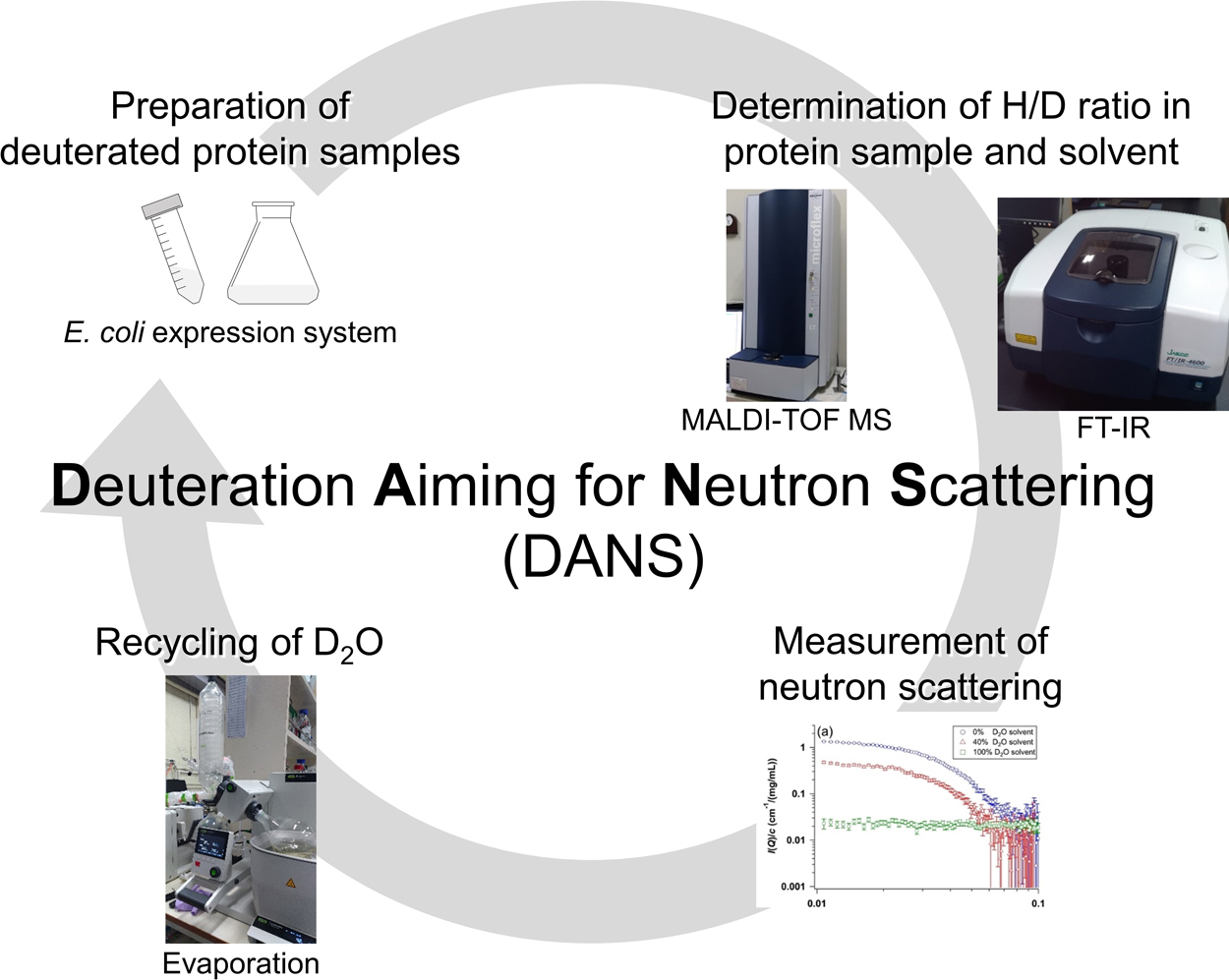
    The distinguished feature of neutron as a scattering probe is an isotope effect, especially the large difference in neutron scattering length between hydrogen and deuterium. The difference renders the different visibility between hydrogenated and deuterated proteins. Therefore, the combination of deuterated protein and neutron scattering enables the selective visualization of a target domain in the complex or a target protein in the multi-component system. Despite of this fascinating character, there exist several problems for the general use of this method: difficulty and high cost for protein deuteration, and control and determination of deuteration ratio of the sample. To resolve them, the protocol of protein deuteration techniques is presented in this report. It is strongly expected that this protocol will offer more opportunity for conducting the neutron scattering studies with deuterated proteins.

Review Article (Invited)
  • Yuta Kyosei, Sou Yamura, Mayuri Namba, Teruki Yoshimura, Satoshi Watab ...
    Article type: Review Article (Invited)
    2021 Volume 18 Pages 28-39
    Published: 2021
    Released on J-STAGE: March 18, 2021
    Advance online publication: February 10, 2021
    JOURNAL OPEN ACCESS FULL-TEXT HTML
    J-STAGE Data

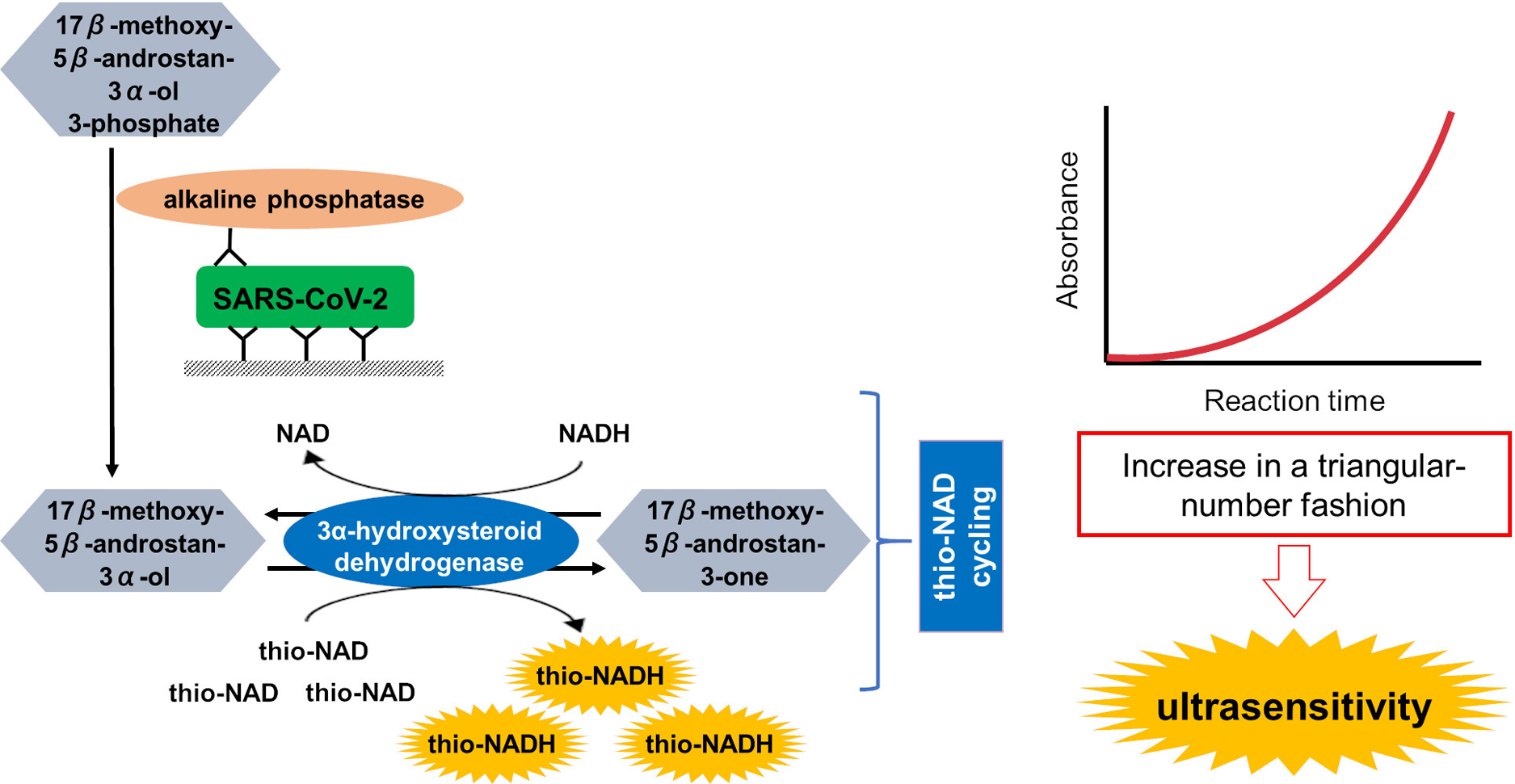
    PCR diagnosis has been considered as the gold standard for coronavirus disease 2019 (COVID-19) and other many diseases. However, there are many problems in using PCR, such as non-specific (i.e., false-positive) and false-negative amplifications, the limits of a target sample volume, deactivation of the enzymes used, complicated techniques, difficulty in designing probe sequences, and the expense. We, thus, need an alternative to PCR, for example an ultrasensitive antigen test. In the present review, we summarize the following three topics. (1) The problems of PCR are outlined. (2) The antigen tests are surveyed in the literature that was published in 2020, and their pros and cons are discussed for commercially available antigen tests. (3) Our own antigen test on the basis of an ultrasensitive enzyme-linked immunosorbent assay (ELISA) is introduced. Finally, we discuss the possibility that our antigen test by an ultrasensitive ELISA technique will become the gold standard for diagnosis of COVID-19 and other diseases.

Regular Article
  • Shunpei Hanai, Kota Katayama, Hiroo Imai, Hideki Kandori
    Article type: Regular Article
    2021 Volume 18 Pages 40-49
    Published: 2021
    Released on J-STAGE: March 24, 2021
    Advance online publication: February 13, 2021
    JOURNAL OPEN ACCESS FULL-TEXT HTML
    Supplementary material

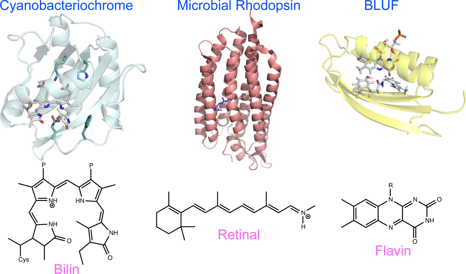
    Structural studies of color visual pigments lag far behind those of rhodopsin for scotopic vision. Using difference FTIR spectroscopy at 77 K, we report the first structural data of three primate color visual pig­ments, monkey red (MR), green (MG), and blue (MB), where the batho-intermediate (Batho) exhibits photo­equilibrium with the unphotolyzed state. This photo­chromic property is highly advantageous for limited samples since the signal-to-noise ratio is improved, but may not be applicable to late intermediates, because of large structural changes to proteins. Here we report the photochromic property of MB at 163 K, where the BL intermediate, formed by the relaxation of Batho, is in photoequilibrium with the initial MB state. A comparison of the difference FTIR spectra at 77 and 163 K provided information on what happens in the process of transition from Batho to BL in MB. The coupled C11=C12 HOOP vibration in the planer structure in MB is decoupled by distortion in Batho after retinal photoisomerization, but returns to the coupled C11=C12 HOOP vibration in the all-trans chromophore in BL. The Batho formation accompanies helical structural perturbation, which is relaxed in BL. Protein-bound water molecules that form an extended water cluster near the retinal chromophore change hydrogen bonds differently for Batho and BL, being stronger in the latter than in the initial state. In addition to structural dynamics, the present FTIR spectra show no signals of protonated carboxylic acids at 77 and 163 K, suggesting that E181 is deprotonated in MB, Batho and BL.

  • Yasumichi Takase, Yoichi Yamazaki, Yugo Hayashi, Sachiko Toma-Fukai, H ...
    Article type: Regular Article
    2021 Volume 18 Pages 50-59
    Published: 2021
    Released on J-STAGE: March 24, 2021
    Advance online publication: February 18, 2021
    JOURNAL OPEN ACCESS FULL-TEXT HTML
    Supplementary material

    Previously, the structure elements of dihydrofolate reductase (DHFR) were determined using comprehen­sive Ala-insertion mutation analysis, which is assumed to be a kind of protein “building blocks.” It is hypo­thesized that our comprehension of the structure elements could lead to understanding how an amino acid sequence dictates its tertiary structure. However, the comprehensive Ala-insertion mutation analysis is a time- and cost-consuming process and only a set of the DHFR structure elements have been reported so far. Therefore, developing a computational method to predict structure elements is an urgent necessity. We focused on intramolecular residue–residue contacts to predict the structure elements. We introduced a simple and effective parameter: the overlapped contact volume (CV) among the residues and calculated the CV along the DHFR sequence using the crystal structure. Our results indicate that the CV profile can recapitulate its precipitate ratio profile, which was used to define the structure elements in the Ala-insertion mutation analysis. The CV profile allowed us to predict structure elements like the experimentally determined structure elements. The strong correlation between the CV and precipitate ratio profiles indicates the importance of the intramolecular residue–residue contact in maintaining the tertiary structure. Additionally, the CVs between the structure elements are considerably more than those between a structure element and a linker or two linkers, indicating that the structure elements play a funda­mental role in increasing the intramolecular adhesion. Thus, we propose that the structure elements can be considered a type of “building blocks” that maintain and dictate the tertiary structures of proteins.

Commentary and Perspective (Invited)
Regular Article
  • Katsumi Omagari, Kaori Asamitsu, Yasuhito Tanaka
    Article type: Regular Article
    2021 Volume 18 Pages 67-77
    Published: 2021
    Released on J-STAGE: April 13, 2021
    Advance online publication: March 17, 2021
    JOURNAL OPEN ACCESS FULL-TEXT HTML
    Supplementary material

    The hepatitis B virus X protein (HBx) and the V protein of paramyxovirus simian virus 5 (SV5-V) interact with DNA damage-binding protein 1 (DDB1), a cellular enzyme involved in DNA repair and cell cycle regulation, to stimulate viral activity. DDB1 has several cellular substrates, and the amino acid sequences of the binding sites in the viral proteins and their substrates are notably dissimilar. To determine whether HBx binds preferentially to DDB1, despite differences in the amino acid sequences, we developed a system to monitor DDB1 binding in living cells through a protein-protein visuali­zation system, designated fluorescent-based technology detecting protein-protein interactions (Fluoppi). HBx in association with DDB1 formed clear fluorescent puncta. The number of these fluorescent puncta increased with an increase in the amount of HBx. The binding of HBx to DDB1 inhibited the cellular substrate DDB1-CUL4A-associated factor 9 (DCAF9) from binding to DDB1. The inhibitor nitazoxanide prevented the viral proteins HBx and SV5-V from binding to DDB1 but did not inhibit the binding of DCAF9 or HBx(ΔNC), which constitutes the binding site of HBx. Our results demonstrate that the Fluoppi system is useful for monitoring the binding of HBx to DDB1 as well as for examining the effect of drugs on DDB1-Hbx binding.

  • Maki Kawasaki, Masayuki Oda
    Article type: Regular Article
    2021 Volume 18 Pages 78-84
    Published: 2021
    Released on J-STAGE: April 13, 2021
    Advance online publication: March 25, 2021
    JOURNAL OPEN ACCESS FULL-TEXT HTML
    J-STAGE Data Supplementary material

    The minimum DNA-binding domain of the transcrip­tional factor c-Myb R2R3 remarkably fluctuates in the solution. In the present study, we evaluated the protein fluctuation of R2R3 C130I mutant, R2R3*, on its DNA-binding and folding thermodynamics. DNA-binding analysis using isothermal titration calorimetry revealed that the heat capacity change determined from the correlation between temperature and binding enthalpy change is highly negative above 35°C, indicating that the fluctuation increases with increasing temperature and elevates the conformational change on DNA binding. The results were in accordance with those of differential scanning calorimetry, which revealed that the heat capacity corresponding to thermal denatu­ration gradually increased above 35°C, followed by the broad transition peak. In contrast, the transition peak of R2R3* in the DNA-bound state was sharper and larger than that in the DNA-unbound state. The fluctuating form could transform into lesser fluctuating form upon DNA binding, resulting in a larger enthalpy change for denaturation of R2R3* in the DNA-bound state. It should also be noted that R2R3* could specifi­cally bind to DNA around thermal denaturation temperature. This would be due to proteins with numerous fluctuations. Moreover, we discuss specific and non-specific DNA binding accompanied by the conformational change between well-ordered and disordered forms of R2R3* observed around the denaturation temperature.

  • Seine A. Shintani
    Article type: Regular Article
    2021 Volume 18 Pages 85-95
    Published: 2021
    Released on J-STAGE: April 16, 2021
    Advance online publication: April 01, 2021
    JOURNAL OPEN ACCESS FULL-TEXT HTML
    J-STAGE Data Supplementary material

    The effects of high pressure (40–70 MPa) on the structure and function of myofibrils were investigated by high pressure microscopy. When this pressure was applied to myofibrils immersed in relaxing solution, the sarcomere length remained almost unchanged, and the A band became shorter and wider. The higher the applied pressure, the faster the change. However, shortening and widening of the A band were not observed when pressure was applied to myofibrils immersed in a solution obtained by omitting ATP from the relaxing solution. However, even under these conditions, structural loss, such as loss of the Z-line structure, occurred. In order to evaluate the consequences of this pressure-treated myofibril, the oscillatory movement of sarcomere (sarcomeric oscillation) was evoked and observed. It was possible to induce sarcomeric oscillation even in pressure-treated myofibrils whose structure was destroyed. The pressurization reduced the total power of the sarcomeric oscillation, but did not change the average frequency. The average frequency did not change even when a pressure of about 40 MPa was applied during sarcomeric oscillation. The average frequency returned to the original when the pressure was returned to the original value after applying stronger pressure to prevent the sarcomere oscillation from being observed. This result suggests that the decrease in the number of myosin molecules forming the crossbridge does not affect the average frequency of sarcomeric oscillation. This fact will help build a mechanical hypothesis for sarcomeric oscillation. The pressurization treatment is a unique method for controlling the structure of myofibrils as described above.

  • Ryota Kojima, Takashi Yoshidome
    Article type: Regular Article
    2021 Volume 18 Pages 96-107
    Published: 2021
    Released on J-STAGE: May 13, 2021
    Advance online publication: April 16, 2021
    JOURNAL OPEN ACCESS FULL-TEXT HTML
    Supplementary material

    Cryo-electron microscopy (cryo-EM) is an important experimental technique for the structural analysis of biomolecules that are difficult or impossible to crystallize. The three-dimensional structure of a biomolecule can be reconstructed using two-dimensional electron-density maps, which are experimentally sampled via the electron beam irradiation of vitreous ice in which the target biomolecules are embedded. One assumption required for this reconstruction is that the orientation of the biomolecules in the vitreous ice is isotropic. However, this is not always the case and two-dimensional electron-density maps are often sampled using preferred biomolecular orientations, which can make reconstruction difficult or impossible. Compensation for under-represented views is computationally feasible for the reconstruction of three-dimensional electron density maps, but one must know whether or not there is any missing information in the sampled two-dimensional electron density maps. Thus, a measure to identify whether a cryo-EM data is obtained from the bio­molecules adopting preferred orientations is required. In the present study, we propose a measure for which the geometry of manifold projected onto a low-dimensional space is used. To show the usefulness of the measure, we perform simulations for cryo-EM experiment of a protein. It is found that the geometry of manifold projected onto a two-dimensional space for a protein adopting a preferred biomolecular orientation is significantly different from that for a protein adopting a uniform orientation. This result suggests that the geometry of manifold projected onto a low-dimensional space can be used for the measure for the identification that the biomolecules adopt preferred orientations.

  • Teppei Sugimoto, Kota Katayama, Hideki Kandori
    Article type: Regular Article
    2021 Volume 18 Pages 108-115
    Published: 2021
    Released on J-STAGE: May 13, 2021
    Advance online publication: April 16, 2021
    JOURNAL OPEN ACCESS FULL-TEXT HTML
    Supplementary material

    Marine bacterial TAT rhodopsin possesses the pKa of the retinal Schiff base, the chromophore, at neutral pH, and photoexcitation of the visible protonated state forms the isomerized 13-cis state, but reverts to the original state within 10–5 sec. To understand the origin of these unique molecular properties of TAT rhodopsin, we mutated Thr82 into Asp, because many microbial rhodopsins contain Asp at the corresponding position as the Schiff base counterion. A pH titration study revealed that the pKa of the Schiff base increased considerably in T82D (>10.5), and that the pKa of the counterion, which is likely to be D82, is 8.1. It was thus concluded that T82 is the origin of the neutral pKa of the Schiff base in TAT rhodopsin. The photocycle of T82D TAT rhodopsin exhibited strong pH dependence. When pH is lower than the pKa of the counterion (pH <8.1), formation of the primary K intermediate was observed by low-temperature UV-visible spectroscopy, but flash photolysis failed to monitor photointermdiates at >10–5 sec. The results were identical for the wild-type TAT rhodopsin. In contrast, when pH was higher than the pKa of the counterion, we observed the formation of the M intermediate, which decayed with the time constants of 3.75 ms and 12.2 sec. It is likely that the protonation state of D82 dramatically switches the photoreaction dynamics of T82D, whose duration lies between <10–5 sec and >10 sec. It was thus concluded that T82 is one of the determinants of the unique photochemistry of TAT rhodopsin.

Review Article (Invited)
  • Yusuke Sato, Yuki Suzuki
    Article type: Review Article (Invited)
    2021 Volume 18 Pages 116-126
    Published: 2021
    Released on J-STAGE: May 26, 2021
    Advance online publication: April 27, 2021
    JOURNAL OPEN ACCESS FULL-TEXT HTML

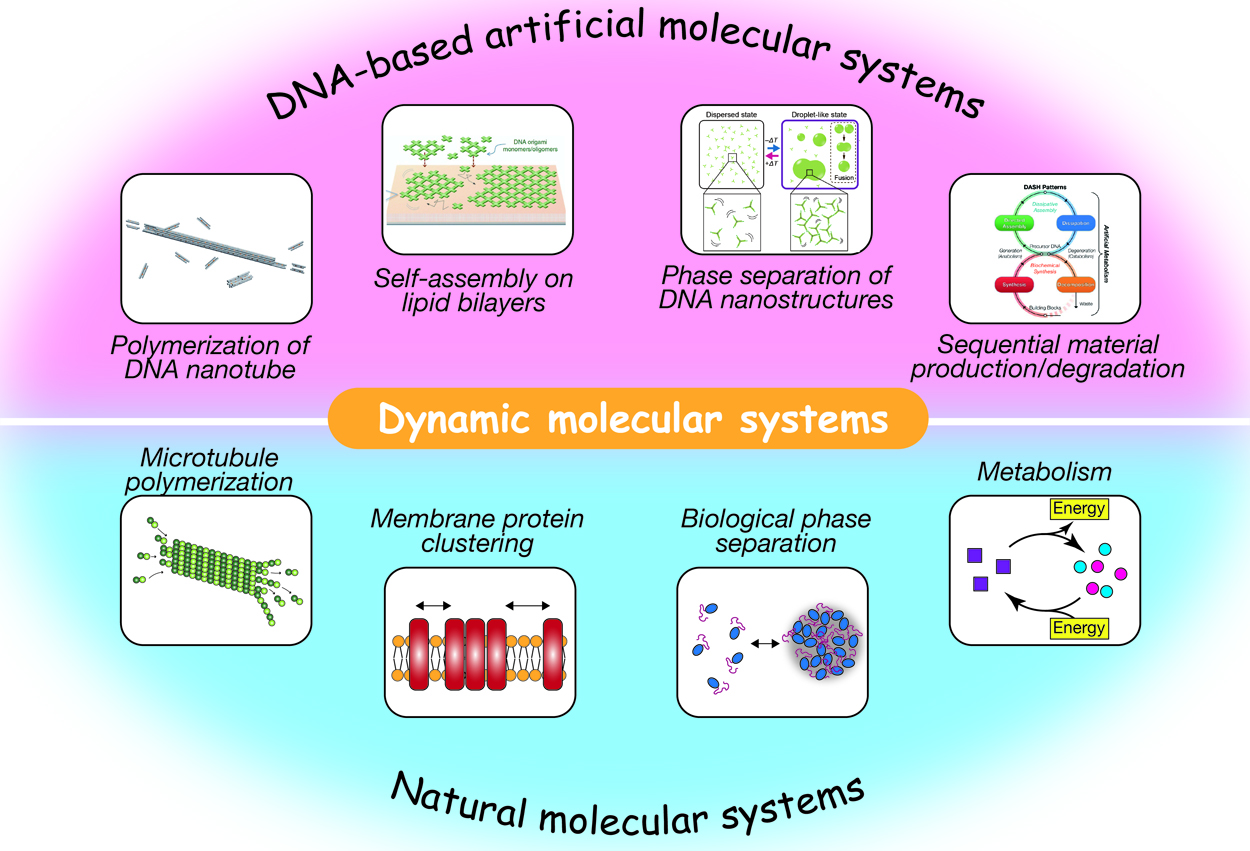
    Self-assembled supramolecular structures in living cells and their dynamics underlie various cellular events, such as endocytosis, cell migration, intracellular transport, cell metabolism, and gene expression. Spatiotemporally regulated association/dissociation and generation/degradation of assembly components is one of the remarkable features of biological systems. The significant advancement in DNA nanotechnology over the last few decades has enabled the construction of various-shaped nanostructures via programmed self-assembly of sequence-designed oligonucleotides. These nanostructures can further be assembled into micrometer-sized structures, including ordered lattices, tubular structures, macromolecular droplets, and hydrogels. In addition to being a structural material, DNA is adopted to construct artificial molecular circuits capable of activating/inactivating or producing/decomposing target DNA molecules based on strand displacement or enzymatic reactions. In this review, we provide an overview of recent studies on artificially designed DNA-based self-assembled systems that exhibit dynamic features, such as association/dis­sociation of components, phase separation, stimulus responsivity, and DNA circuit-regulated structural formation. These biomacromolecule-based, bottom-up approaches for the construction of artificial molecular systems will not only throw light on bio-inspired nano/micro engineering, but also enable us to gain insights into how autonomy and adaptability of living systems can be realized.

Commentary and Perspective (Invited)
Review Article (Invited)
  • Udoy S. Basak, Sulimon Sattari, Motaleb Hossain, Kazuki Horikawa, Tami ...
    Article type: Review Article (Invited)
    2021 Volume 18 Pages 131-144
    Published: 2021
    Released on J-STAGE: June 18, 2021
    Advance online publication: May 15, 2021
    JOURNAL OPEN ACCESS FULL-TEXT HTML

    Synchronized movement of (both unicellular and multicellular) systems can be observed almost everywhere. Understanding of how organisms are regulated to synchronized behavior is one of the challenging issues in the field of collective motion. It is hypothesized that one or a few agents in a group regulate(s) the dynamics of the whole collective, known as leader(s). The identification of the leader (influential) agent(s) is very crucial. This article reviews different mathematical models that represent different types of leadership. We focus on the improvement of the leader-follower classification problem. It was found using a simulation model that the use of interaction domain information significantly improves the leader-follower classification ability using both linear schemes and information-theoretic schemes for quantifying influence. This article also reviews different schemes that can be used to identify the interaction domain using the motion data of agents.

Regular Article
  • Takamitsu J Morikawa, Masayoshi Nishiyama, Keiko Yoshizawa, Hideaki Fu ...
    Article type: Regular Article
    2021 Volume 18 Pages 145-158
    Published: 2021
    Released on J-STAGE: June 18, 2021
    Advance online publication: May 21, 2021
    JOURNAL OPEN ACCESS FULL-TEXT HTML
    Supplementary material

    The green fluorescent protein (GFP) derived from Pacific Ocean jellyfish is an essential tool in biology. GFP-solvent interactions can modulate the fluorescent property of GFP. We previously reported that glycine insertion is an effective mutation in the yellow variant of GFP, yellow fluorescent protein (YFP). Glycine insertion into one of the β-strands comprising the barrel structure distorts its structure, allowing water molecules to invade near the chromophore, enhancing hydrostatic pressure or solution hydrophobicity sensitivity. However, the underlying mechanism of how glycine insertion imparts environmental sensitivity to YFP has not been elucidated yet. To unveil the relationship between fluorescence and β-strand distortion, we investigated the effects of glycine insertion on the dependence of the optical properties of GFP variants named enhanced-GFP (eGFP) and its yellow (eYFP) and cyan (eCFP) variants with respect to pH, temperature, pressure, and hydrophobicity. Our results showed that the quantum yield decreased depending on the number of inserted glycines in all variants, and the dependence on pH, temperature, pressure, and hydrophobicity was altered, indicating the invasion of water molecules into the β-barrel. Peak shifts in the emission spectrum were observed in glycine-inserted eGFP, suggesting a change of the electric state in the excited chromophore. A comparative investigation of the spectral shift among variants under different conditions demonstrated that glycine insertion rearranged the hydrogen bond network between His148 and the chromophore. The present results provide important insights for further understanding the fluorescence mechanism in GFPs and suggest that glycine insertion could be a potent approach for investigating the relationship between water molecules and the intra-protein chromophore.

  • Koya Sakuma
    Article type: Regular Article
    2021 Volume 18 Pages 159-167
    Published: 2021
    Released on J-STAGE: June 18, 2021
    Advance online publication: May 28, 2021
    JOURNAL OPEN ACCESS FULL-TEXT HTML
    Supplementary material

    ABEGO is a coarse-grained representation for poly­peptide backbone dihedral angles. The Ramachandran map is divided into four segments denoted as A, B, E, and G to represent the local conformation of poly­peptide chains in the character strings. Although the ABEGO representation is widely used in backbone building simulation for de novo protein design, it cannot capture minor differences in backbone dihedral angles, which potentially leads to ambiguity between two structurally distinct fragments. Here, I show a nontrivial example of two local motifs that could not be distinguished by their ABEGO representations. I found that two well-known local motifs αα-hairpins and αα-corners are both represented as α-GBB-α and thus indistinguishable in the ABEGO representation, although they show distinct arrangements of the flanking α-helices. I also found that α-GBB-α motifs caused a loss of efficiency in the ABEGO-based fragment-assembly simulations for de novo protein backbone design. Nevertheless, I was able to design amino-acid sequences that were predicted to fold into the target topologies that contained these α-GBB-α motifs, which suggests such topologies that are difficult to build by ABEGO-based simulations are designable once the backbone structures are modeled by some means. The finding that certain local motifs bottleneck the ABEGO-based fragment-assembly simulations for construction of backbone structures suggests that finer representations of backbone torsion angles are required for efficiently generating diverse topologies containing such indistinguishable local motifs.

Review Article (Invited)
  • Masayuki Oda
    Article type: Review Article (Invited)
    2021 Volume 18 Pages 168-176
    Published: 2021
    Released on J-STAGE: July 28, 2021
    Advance online publication: June 30, 2021
    JOURNAL OPEN ACCESS FULL-TEXT HTML
    Supplementary material

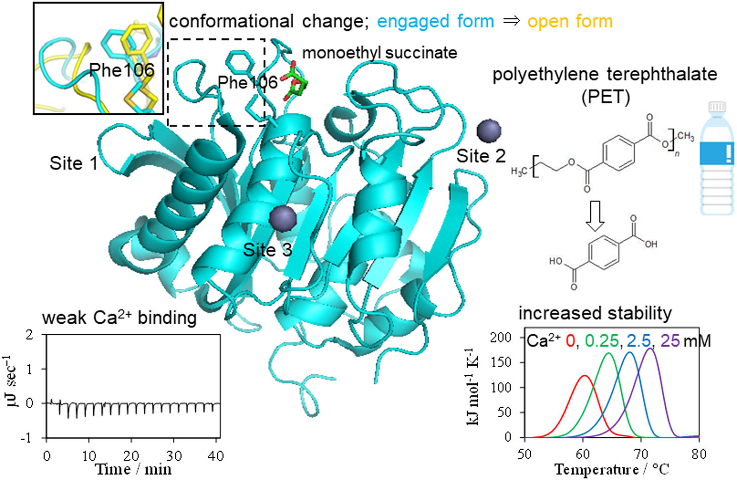
    A cutinase-like enzyme from Saccharomonospora viridis AHK190, Cut190, can depolymerize polyethylene terephthalate (PET). As high activity at approximately 70°C is required for PET depolymerization, structure-based protein engineering of Cut190 was carried out. Crystal structure information of the Cut190 mutants was used for protein engineering and for evaluating the molecular basis of activity and thermal stability. A variety of biophysical methods were employed to unveil the mechanisms underlying the unique features of Cut190, which included the regulation of its activity and thermal stability by Ca2+. Ca2+ association and dissociation can change the enzyme conformation to regulate catalytic activity. Weak metal-ion binding would be required for the naïve conformational change of Cut190, while maintaining its fluctuation, to “switch” the enzyme on and off. The activity of Cut190 is regulated by the weak Ca2+ binding to the specific site, Site 1, while thermal stability is mainly regulated by binding to another Site 2, where a disulfide bond could be introduced to increase the stability. Recent results on the structure-activity relationship of engineered Cut190 are reviewed, including the application for PET depolymerization by enzymes.

Regular Article
  • Izuru Kawamura, Hayato Seki, Seiya Tajima, Yoshiteru Makino, Arisu Shi ...
    Article type: Regular Article
    2021 Volume 18 Pages 177-185
    Published: 2021
    Released on J-STAGE: August 07, 2021
    Advance online publication: July 14, 2021
    JOURNAL OPEN ACCESS FULL-TEXT HTML
    Supplementary material

    Middle rhodopsin (MR) found from the archaeon Haloquadratum walsbyi is evolutionarily located between two different types of rhodopsins, bacteriorhodopsin (BR) and sensory rhodopsin II (SRII). Some isomers of the chromophore retinal and the photochemical reaction of MR are markedly different from those of BR and SRII. In this study, to obtain the structural information regarding its active center (i.e., retinal), we subjected MR embedded in lipid bilayers to solid-state magic-angle spinning nuclear magnetic resonance (NMR) spectroscopy. The analysis of the isotropic 13C chemical shifts of the retinal chromophore revealed the presence of three types of retinal configurations of dark-adapted MR: (13-trans, 15-anti (all-trans)), (13-cis, 15-syn), and 11-cis isomers. The higher field resonance of the 20-C methyl carbon in the all-trans retinal suggested that Trp182 in MR has an orientation that is different from that in other microbial rhodopsins, owing to the changes in steric hindrance associated with the 20-C methyl group in retinal. 13Cζ signals of Tyr185 in MR for all-trans and 13-cis, 15-syn isomers were discretely observed, representing the difference in the hydrogen bond strength of Tyr185. Further, 15N NMR analysis of the protonated Schiff base corresponding to the all-trans and 13-cis, 15-syn isomers in MR showed a strong electrostatic interaction with the counter ion. Therefore, the resulting structural information exhibited the property of stable retinal conformations of dark-adapted MR.

Review Article (Invited)
  • Kyoko Shinzawa-Itoh, Kazumasa Muramoto
    Article type: Review Article (Invited)
    2021 Volume 18 Pages 186-195
    Published: 2021
    Released on J-STAGE: August 19, 2021
    Advance online publication: July 16, 2021
    JOURNAL OPEN ACCESS FULL-TEXT HTML

    Cytochrome c oxidase (CcO), a terminal oxidase in the respiratory chain, catalyzes the reduction of O2 to water coupled with the proton pump across the membrane. Mitochondrial CcO exists in monomeric and dimeric forms, and as a monomer as part of the respiratory supercomplex, although the enzymatic reaction proceeds in the CcO monomer. Recent biochemical and crystallographic studies of monomeric and dimeric CcOs have revealed functional and structural differences among them. In solubilized mitochondrial membrane, the monomeric form is dominant, and a small amount of dimer is observed. The activity of the monomeric CcO is higher than that of the dimer, suggesting that the monomer is the active form. In the structure of monomeric CcO, a hydrogen bond network of water molecules is formed at the entrance of the proton transfer K-pathway, and in dimeric CcO, this network is altered by a cholate molecule binding between monomers. The specific binding of the cholate molecule at the dimer interface suggests that the binding of physiological ligands similar in size or shape to cholate could also trigger dimer formation as a physiological standby form. Because the dimer interface also contains weak interactions of nonspecifically bound lipid molecules, hydrophobic interactions between the transmembrane helices, and a Met–Met interaction between the extramembrane regions, these interactions could support the stabilization of the standby form. Structural analyses also suggest that hydrophobic interactions of cardiolipins bound to the trans­membrane surface of CcO are involved in forming the supercomplex.

Regular Article
  • Kenji Mishima, Mitsuo Shoji, Yasufumi Umena, Mauro Boero, Yasuteru Shi ...
    Article type: Regular Article
    2021 Volume 18 Pages 196-214
    Published: 2021
    Released on J-STAGE: September 01, 2021
    Advance online publication: July 30, 2021
    JOURNAL OPEN ACCESS FULL-TEXT HTML
    Supplementary material

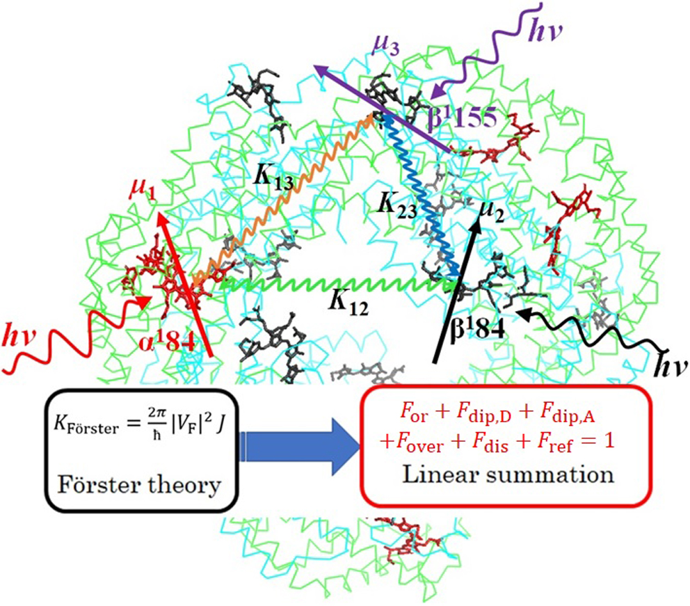
    In the present study, we provide a reformulation of the theory originally proposed by Förster which allows for simple and convenient formulas useful to estimate the relative contributions of transition dipole moments of a donor and acceptor (chemical factors), their orientation factors (intermolecular structural factors), intermolecular center-to-center distances (intermolecular structural factors), spectral overlaps of absorption and emission spectra (photophysical factors), and refractive index (material factor) to the excitation energy transfer (EET) rate constant. To benchmark their validity, we focused on the EET occurring in C-phycocyanin (C-PC) chromophores. To this aim, we resorted to quantum chemistry calculations to get optimized molecular structures of the C-PC chromophores within the density functional theory (DFT) framework. The absorption and emission spectra, as well as transition dipole moments, were computed by using the time-dependent DFT (TDDFT). Our method was applied to several types of C-PCs showing that the EET rates are determined by an interplay of their specific physical, chemical, and geometrical features. These results show that our formulas can become a useful tool for a reliable estimation of the relative contributions of the factors regulating the EET transfer rate.

Note
  • Daisuke Kuroda, Kouhei Tsumoto
    Article type: Note
    2021 Volume 18 Pages 215-222
    Published: 2021
    Released on J-STAGE: September 16, 2021
    Advance online publication: August 21, 2021
    JOURNAL OPEN ACCESS FULL-TEXT HTML
    Supplementary material

    Severe acute respiratory syndrome coronavirus 2 (SARS-CoV-2), which causes the coronavirus disease 2019 (COVID-19), spread rapidly around the globe. The main protease encoded by SARS-CoV-2 is essential for processing of the polyproteins translated from the viral RNA genome, making this protein a potential drug target. A recently reported mutation in the protease, P108S, may be responsible for milder symptoms observed in COVID-19 patients in Tokyo. Starting from a crystal structure of the SARS-CoV-2 main protease in the dimeric form, we performed triplicate 5.0-μs molecular dynamics simulations of the wild-type and P108S mutant. Our computational results suggest a link between the mutation P108S and dynamics of the catalytic sites in the main protease: The catalytic dyad become considerably inaccessible to substrates in the P108S mutant. Our results also demonstrate the potential of molecular dynamics simulations to complement experimental techniques and other computational methods, such as protein design calculations, which predict effects of mutations based on static crystal structures. Further studies are certainly necessary to quantitively understand the relationships between the P108S mutation and physical properties of the main protease, but the results of our study will immediately inform development of new protease inhibitors.

Editorial
Commentary and Perspective
Review Article (Invited)
  • Atsushi Hijikata, Clara Shionyu, Setsu Nakae, Masafumi Shionyu, Motono ...
    Article type: Review Article
    2021 Volume 18 Pages 226-240
    Published: 2021
    Released on J-STAGE: October 20, 2021
    Advance online publication: October 05, 2021
    JOURNAL OPEN ACCESS FULL-TEXT HTML

    More than one and half years have passed, as of August 2021, since the COVID-19 caused by the novel coronavirus named SARS-CoV-2 emerged in 2019. While the recent success of vaccine developments likely reduces the severe cases, there is still a strong requirement of safety and effective therapeutic drugs for overcoming the unprecedented situation. Here we review the recent progress and the status of the drug discovery against COVID-19 with emphasizing a structure-based perspective. Structural data regarding the SARS-CoV-2 proteome has been rapidly accumulated in the Protein Data Bank, and up to 68% of the total amino acid residues encoded in the genome were covered by the structural data. Despite a global effort of in silico and in vitro screenings for drug repurposing, there is only a limited number of drugs had been successfully authorized by drug regulation organizations. Although many approved drugs and natural compounds, which exhibited antiviral activity in vitro, were considered potential drugs against COVID-19, a further multidisciplinary investigation is required for understanding the mechanisms underlying the antiviral effects of the drugs.

Commentary and Perspective
Review Article (Invited)
  • Chan-Gi Pack
    Article type: Review Article (Invited)
    2021 Volume 18 Pages 244-253
    Published: 2021
    Released on J-STAGE: October 26, 2021
    Advance online publication: October 15, 2021
    JOURNAL OPEN ACCESS FULL-TEXT HTML

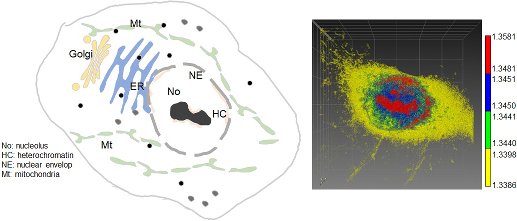
    The cell is three-dimensionally and dynamically organized into cellular compartments, including the endoplasmic reticulum, mitochondria, vesicles, and nucleus, which have high relative molecular density. The structure and functions of these compartments and organelles may be deduced from the diffusion and interaction of related biomolecules. Among these cellular components, various protein molecules can freely access the nucleolus or mitotic chromosome through Brownian diffusion, even though they have a densely packed structure. However, physicochemical properties of the nucleolus and chromosomes, such as molecular density and volume, are not yet fully understood under changing cellular conditions. Many studies have been conducted based on high-resolution imaging and analysis techniques using fluorescence. However, there are limitations in imaging only fluorescently labeled molecules, and cytotoxicity occurs during three-dimensional imaging. Alternatively, the recently developed label-free three-dimensional optical diffraction tomography (ODT) imaging technique can divide various organelles in cells into volumes and analyze them by refractive index, although specific molecules cannot be observed. A previous study established an analytical method that provides comprehensive insights into the physical properties of the nucleolus and mitotic chromosome by utilizing the advantages of ODT and fluorescence techniques, such as fluorescence correlation spectroscopy and confocal laser scanning microscopy. This review article summarizes a recent study and discusses the future aspects of the ODT for cellular compartments.

  • Takuma Sugi, Hiroshi Ito, Ken H. Nagai
    Article type: Review Article (Invited)
    2021 Volume 18 Pages 254-262
    Published: 2021
    Released on J-STAGE: November 27, 2021
    Advance online publication: October 20, 2021
    JOURNAL OPEN ACCESS FULL-TEXT HTML

    Active matter refers to systems composed of elements that are self-propelled by the dissipation of energy, in which dynamical patterns emerge, as is the case of flocks of birds and schools of fish. Some researchers in active matter physics seek to identify unified descriptions of such collective motions through interdisciplinary approaches by biologists and physicists. Through such collaborations, experimental studies pertaining to active matter physics have been developing recently, which allow us to verify the proposed mathematical models. Here, we review collective pattern formations and behaviors of animals from the perspective of active matter physics.

Commentary and Perspective
Review Article (Invited)
  • Katsumasa Irie
    Article type: Review Article (Invited)
    2021 Volume 18 Pages 274-283
    Published: 2021
    Released on J-STAGE: December 04, 2021
    Advance online publication: November 19, 2021
    JOURNAL OPEN ACCESS FULL-TEXT HTML

    Prokaryotic channels play an important role in the structural biology of ion channels. At the end of the 20th century, the first structure of a prokaryotic ion channel was revealed. Subsequently, the reporting of structures of various prokaryotic ion channels have provided fundamental insights into the structure of ion channels of higher organisms. Voltage-dependent Ca2+ channels (Cavs) are indispensable for coupling action potentials with Ca2+ signaling. Similar to other proteins, Cavs were predicted to have a prokaryotic counterpart; however, it has taken more than 20 years for one to be identified. The homotetrameric channel obtained from Meiothermus ruber generates the calcium ion specific current, so it is named as CavMr. Its selectivity filter contains a smaller number of negatively charged residues than mutant Cavs generated from other prokaryotic channels. CavMr belonged to a different cluster of phylogenetic trees than canonical prokaryotic cation channels. The glycine residue of the CavMr selectivity filter is a determinant for calcium selectivity. This glycine residue is conserved among eukaryotic Cavs, suggesting that there is a universal mechanism for calcium selectivity. A family of homotetrameric channels has also been identified from eukaryotic unicellular algae, and the investigation of these channels can help to understand the mechanism for ion selection that is conserved from prokaryotes to eukaryotes.

Commentary and Perspective
Editorial
Regular Article
  • Takayoshi Tsubo
    Article type: Regular Article
    2021 Volume 18 Pages 290-304
    Published: 2021
    Released on J-STAGE: December 18, 2021
    Advance online publication: December 02, 2021
    JOURNAL OPEN ACCESS FULL-TEXT HTML

    Successful synaptic integration is said to require that multiple excitatory postsynaptic potentials (EPSPs) occur almost simultaneously over a short period of time, so that they overlap and increase. However, if brain function is based on a chain of successful synaptic integrations, then constraints on the spacing of multiple EPSP generation must be released to allow for a higher probability of successful synaptic integration. This paper demonstrates that Ca2+ ions retained in spines after EPSP generation polarize spine neck fluid and dendritic fluid as a dielectric medium, that polarization is transmitted through dendrites to the cell body (soma), that polarization is enhanced by the addition of polarization from each spine, and that I propose that synaptic integration is successful when the membrane potential, as determined by the enhanced polarization and membrane capacitance, reaches the threshold of voltage-gated Na+ channels. Furthermore, the approach taken in this study suggests that a single neuron can integrate synapses for many combinations of synaptic inputs, that successful synaptic integration depends on spine neck capacitance and spine head size, and that spines farther from the soma are able to contribute to successful synaptic integration, and led to the elucidation of a number of important issues, including the fact that inhibitory post-synapses on dendrites suppress s effectively synaptic integration.

  • Hiroaki Hata, Duy Phuoc Tran, Mohamed Marzouk Sobeh, Akio Kitao
    Article type: Regular Article
    2021 Volume 18 Pages 305-316
    Published: 2021
    Released on J-STAGE: December 22, 2021
    Advance online publication: December 04, 2021
    JOURNAL OPEN ACCESS FULL-TEXT HTML
    Supplementary material

    We recently proposed a computational procedure to simulate the dissociation of protein/ligand complexes using the dissociation Parallel Cascade Selection Molecular Dynamics simulation (dPaCS-MD) method and to analyze the generated trajectories using the Markov state model (MSM). This procedure, called dPaCS-MD/MSM, enables calculation of the dissociation free energy profile and the standard binding free energy. To examine whether this method can reproduce experimentally determined binding free energies for a variety of systems, we used it to investigate the dissociation of three protein/ligand complexes: trypsin/benzamine, FKBP/FK506, and adenosine A2A receptor/T4E. First, dPaCS-MD generated multiple dissociation pathways within a reasonable computational time for all the complexes, although the complexes differed significantly in the size of the molecules and in intermolecular interactions. Subsequent MSM analyses produced free energy profiles for the dissociations, which provided insights into how each ligand dissociates from the protein. The standard binding free energies obtained by dPaCS-MD/MSM are in good agreement with experimental values for all the complexes. We conclude that dPaCS-MD/MSM can accurately calculate the binding free energies of these complexes.

Review Article (Invited)
  • Takashi Kikukawa
    Article type: Review Article (Invited)
    2021 Volume 18 Pages 317-326
    Published: 2021
    Released on J-STAGE: January 08, 2022
    Advance online publication: December 22, 2021
    JOURNAL OPEN ACCESS FULL-TEXT HTML
    Supplementary material

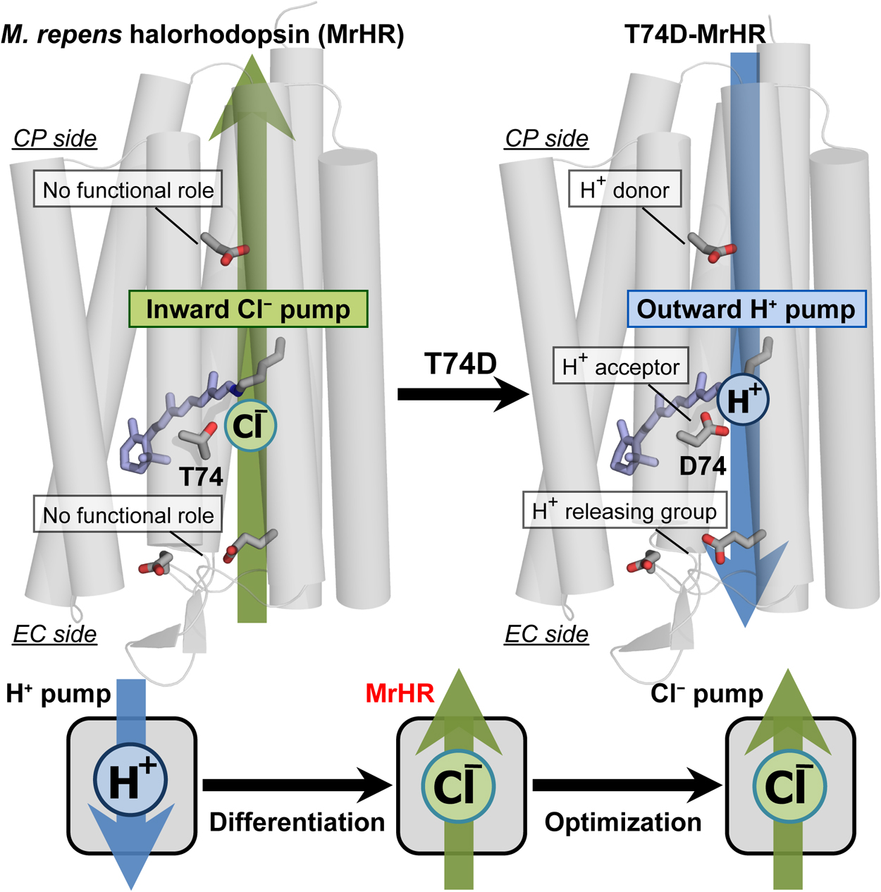
    Microbial rhodopsin is a ubiquitous membrane protein in unicellular microorganisms. Similar to animal rhodopsin, this protein consists of seven transmembrane helices and the chromophore retinal. However, unlike animal rhodopsin, microbial rhodopsin acts as not only a photosignal receptor but also a light-activated ion transporter and light-switchable enzyme. In this article, the third Cl pump microbial rhodopsin will be introduced. The physiological importance of Cl pumps has not been clarified. Despite this, their mechanisms, especially that of the first Cl pump halorhodopsin (HR), have been studied to characterize them as model proteins for membrane anion transporters. The third Cl pump defines a phylogenetic cluster distinct from other microbial rhodopsins. However, this Cl pump conserves characteristic residues for not only the Cl pump HR but also the H+ pump bacteriorhodopsin (BR). Reflecting close similarity to BR, the third Cl pump begins to pump H+ outwardly after single amino acid replacement. This mutation activates several residues that have no roles in the original Cl pump function but act as important H+ relay residues in the H+ pump mutant. Thus, the third Cl pump might be the model protein for functional differentiation because this rhodopsin seems to be the Cl pump occurring immediately after functional differentiation from the BR-type H+ pump.

feedback
Top