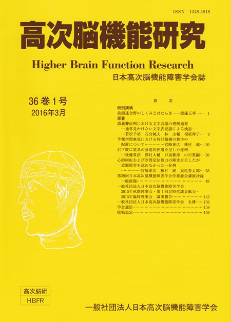
Volume 36, Issue 1
Displaying 1-50 of 53 articles from this issue
Special lecture
-
2016 Volume 36 Issue 1 Pages 1-8
Published: March 31, 2016
Released on J-STAGE: April 03, 2017
Download PDF (938K)
Original article
-
2016 Volume 36 Issue 1 Pages 9-19
Published: March 31, 2016
Released on J-STAGE: April 03, 2017
Download PDF (742K) -
2016 Volume 36 Issue 1 Pages 20-29
Published: March 31, 2016
Released on J-STAGE: April 03, 2017
Download PDF (604K) -
2016 Volume 36 Issue 1 Pages 30-37
Published: March 31, 2016
Released on J-STAGE: April 03, 2017
Download PDF (698K) -
2016 Volume 36 Issue 1 Pages 38-48
Published: March 31, 2016
Released on J-STAGE: April 03, 2017
Download PDF (1181K)
-
2016 Volume 36 Issue 1 Pages 49-51
Published: 2016
Released on J-STAGE: April 03, 2017
Download PDF (1076K) -
2016 Volume 36 Issue 1 Pages 51-53
Published: 2016
Released on J-STAGE: April 03, 2017
Download PDF (1080K) -
2016 Volume 36 Issue 1 Pages 53-55
Published: 2016
Released on J-STAGE: April 03, 2017
Download PDF (1080K) -
2016 Volume 36 Issue 1 Pages 55-57
Published: 2016
Released on J-STAGE: April 03, 2017
Download PDF (1082K) -
2016 Volume 36 Issue 1 Pages 57-59
Published: 2016
Released on J-STAGE: April 03, 2017
Download PDF (1081K) -
2016 Volume 36 Issue 1 Pages 59-61
Published: 2016
Released on J-STAGE: April 03, 2017
Download PDF (1079K) -
2016 Volume 36 Issue 1 Pages 61-62
Published: 2016
Released on J-STAGE: April 03, 2017
Download PDF (1072K) -
2016 Volume 36 Issue 1 Pages 62-64
Published: 2016
Released on J-STAGE: April 03, 2017
Download PDF (1080K) -
2016 Volume 36 Issue 1 Pages 64-66
Published: 2016
Released on J-STAGE: April 03, 2017
Download PDF (1081K) -
2016 Volume 36 Issue 1 Pages 66-69
Published: 2016
Released on J-STAGE: April 03, 2017
Download PDF (1087K) -
2016 Volume 36 Issue 1 Pages 69-70
Published: 2016
Released on J-STAGE: April 03, 2017
Download PDF (1073K) -
2016 Volume 36 Issue 1 Pages 70-73
Published: 2016
Released on J-STAGE: April 03, 2017
Download PDF (1084K) -
2016 Volume 36 Issue 1 Pages 73-75
Published: 2016
Released on J-STAGE: April 03, 2017
Download PDF (1081K) -
2016 Volume 36 Issue 1 Pages 75-78
Published: 2016
Released on J-STAGE: April 03, 2017
Download PDF (1088K) -
2016 Volume 36 Issue 1 Pages 78-80
Published: 2016
Released on J-STAGE: April 03, 2017
Download PDF (1080K) -
2016 Volume 36 Issue 1 Pages 80-81
Published: 2016
Released on J-STAGE: April 03, 2017
Download PDF (1073K) -
2016 Volume 36 Issue 1 Pages 82-84
Published: 2016
Released on J-STAGE: April 03, 2017
Download PDF (1081K) -
2016 Volume 36 Issue 1 Pages 84-86
Published: 2016
Released on J-STAGE: April 03, 2017
Download PDF (1080K) -
2016 Volume 36 Issue 1 Pages 86-89
Published: 2016
Released on J-STAGE: April 03, 2017
Download PDF (1087K) -
2016 Volume 36 Issue 1 Pages 89-91
Published: 2016
Released on J-STAGE: April 03, 2017
Download PDF (1080K) -
2016 Volume 36 Issue 1 Pages 91-92
Published: 2016
Released on J-STAGE: April 03, 2017
Download PDF (1074K) -
2016 Volume 36 Issue 1 Pages 92-94
Published: 2016
Released on J-STAGE: April 03, 2017
Download PDF (1081K) -
2016 Volume 36 Issue 1 Pages 94-96
Published: 2016
Released on J-STAGE: April 03, 2017
Download PDF (1080K) -
2016 Volume 36 Issue 1 Pages 96-98
Published: 2016
Released on J-STAGE: April 03, 2017
Download PDF (1081K) -
2016 Volume 36 Issue 1 Pages 98-100
Published: 2016
Released on J-STAGE: April 03, 2017
Download PDF (1083K) -
2016 Volume 36 Issue 1 Pages 100-101
Published: 2016
Released on J-STAGE: April 03, 2017
Download PDF (1075K) -
2016 Volume 36 Issue 1 Pages 102-104
Published: 2016
Released on J-STAGE: April 03, 2017
Download PDF (1080K) -
2016 Volume 36 Issue 1 Pages 104-105
Published: 2016
Released on J-STAGE: April 03, 2017
Download PDF (1073K) -
2016 Volume 36 Issue 1 Pages 105-107
Published: 2016
Released on J-STAGE: April 03, 2017
Download PDF (1081K) -
2016 Volume 36 Issue 1 Pages 107-109
Published: 2016
Released on J-STAGE: April 03, 2017
Download PDF (1077K) -
2016 Volume 36 Issue 1 Pages 109-110
Published: 2016
Released on J-STAGE: April 03, 2017
Download PDF (1075K) -
2016 Volume 36 Issue 1 Pages 110-111
Published: 2016
Released on J-STAGE: April 03, 2017
Download PDF (1074K) -
2016 Volume 36 Issue 1 Pages 111-113
Published: 2016
Released on J-STAGE: April 03, 2017
Download PDF (1082K) -
2016 Volume 36 Issue 1 Pages 113-115
Published: 2016
Released on J-STAGE: April 03, 2017
Download PDF (1081K) -
2016 Volume 36 Issue 1 Pages 115-116
Published: 2016
Released on J-STAGE: April 03, 2017
Download PDF (1073K) -
2016 Volume 36 Issue 1 Pages 116-118
Published: 2016
Released on J-STAGE: April 03, 2017
Download PDF (1082K) -
2016 Volume 36 Issue 1 Pages 118-119
Published: 2016
Released on J-STAGE: April 03, 2017
Download PDF (1074K) -
2016 Volume 36 Issue 1 Pages 119-121
Published: 2016
Released on J-STAGE: April 03, 2017
Download PDF (1079K) -
2016 Volume 36 Issue 1 Pages 121-123
Published: 2016
Released on J-STAGE: April 03, 2017
Download PDF (1080K) -
2016 Volume 36 Issue 1 Pages 123-124
Published: 2016
Released on J-STAGE: April 03, 2017
Download PDF (1073K) -
2016 Volume 36 Issue 1 Pages 124-126
Published: 2016
Released on J-STAGE: April 03, 2017
Download PDF (1082K) -
2016 Volume 36 Issue 1 Pages 126-128
Published: 2016
Released on J-STAGE: April 03, 2017
Download PDF (1080K) -
2016 Volume 36 Issue 1 Pages 128-130
Published: 2016
Released on J-STAGE: April 03, 2017
Download PDF (1080K) -
2016 Volume 36 Issue 1 Pages 130-132
Published: 2016
Released on J-STAGE: April 03, 2017
Download PDF (1079K) -
2016 Volume 36 Issue 1 Pages 132-135
Published: 2016
Released on J-STAGE: April 03, 2017
Download PDF (1087K)
