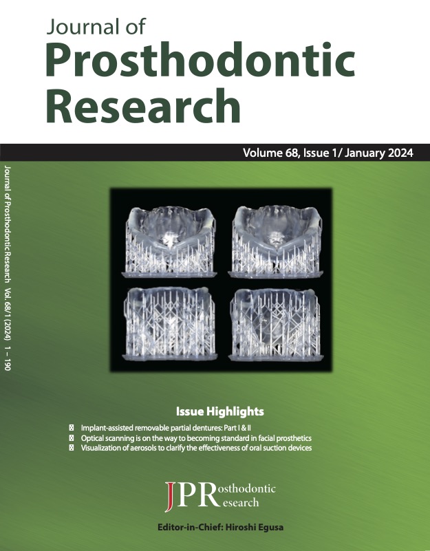
Journal of Prosthodontic Research
Online ISSN : 1883-9207
Print ISSN : 1883-1958
ISSN-L : 1883-1958
Volume 68, Issue 1
Displaying 1-22 of 22 articles from this issue
Editorial
Review articles
  • János König, Kata Kelemen, László Márk Czumbel, Bence Szabó, Gábor Var ...
    2024 Volume 68 Issue 1 Pages 1-11
    Published: 2024
    Released on J-STAGE: January 16, 2024
    Advance online publication: June 08, 2023
    JOURNAL OPEN ACCESS
    Supplementary material

    Purpose To assess the accuracy of scanning technologies for constructing facial prostheses on human faces.

    Study selection Our systematic search was performed on five databases. Studies reporting on human volunteers (P) whose faces were scanned with a scanning technology were eligible. The anthropometrical interlandmark distances (ILDs) were used as indicators of accuracy; the ILDs are measured on the virtual models (I) and directly on the faces (C). The virtual models deviated from their true values (O). Studies reporting the measurements on patients with or without facial deformities were included, but cadavers or inanimate objects were reasons for exclusion. We performed a mean difference (MD) / standardized MD analysis with a random effect model. The difficulties regarding the scanning procedure mentioned in the articles were also assessed.

    Results We found 3723 records after duplicate removal. Twenty five articles were eligible for the qualitative review, and ten articles were included in the quantitative synthesis. Eight different ILDs were compared in MD analyses. The differences were between -0.54-0.43 mm. We also performed a regional three-dimensional analysis to compare scanning technologies in each major region. No significant differences were found in any of the regions and axes. The most mentioned difficulties were artifacts due to motion or blinking.

    Conclusions The results suggest no systematic skew in linear dimensions neither between direct caliper measurements nor between measurements on the scanned models, scanning technologies, or facial regions.

    Download PDF (3706K)
  • Kata Kelemen, János König, Márk Czumbel, Bence Szabó, Péter Hegyi, Gáb ...
    2024 Volume 68 Issue 1 Pages 12-19
    Published: 2024
    Released on J-STAGE: January 16, 2024
    Advance online publication: June 08, 2023
    JOURNAL OPEN ACCESS
    Supplementary material

    Purpose Temporomandibular disorders (TMDs) are frequent stomatological disorders. However, their treatment is controversial. Therefore, we compared the efficacy of combination therapy (splint therapy along with physiotherapy, manual therapy, and counseling) with physiotherapy, manual therapy, and counseling alone. The extent of mouth opening and pain perception were the outcomes.

    Study selection Systematic searches for English publications were performed using four major literature databases (Cochrane Library, EMBASE, PubMed, and Web of Science). We included randomized controlled trials. We calculated mean differences with 95% confidence interval (CI) for pain perception and maximum mouth opening (MMO) for the two groups. The Hartung-Knapp adjustment was used for cases comprising at least five studies.

    Results Six articles were included in the pain perception category, and four were reviewed for MMO at baseline. Four articles assessed pain perception, and two assessed MMO at 1 month. Five articles were analyzed upon comparing pain perception at baseline and 1-month follow-up. The mean difference was -2.54 [95% CI: -3.38; to -1.70] in the intervention group and -2.33 [95% CI: -4.06; to -0.61] in the control group. Two articles were analyzed upon comparing MMO at baseline and 1-month follow-up. The mean difference in the intervention group was 3.69 [95% CI: -0.34; 7.72], whereas that in the control group was 3.62 [95% CI: -3.43; 10.67].

    Conclusions Both therapies can be used in the management of myogenic TMD. Due to the marginal differences between the baseline and 1-month values, our results could not confirm the efficacy of combination therapy.

    Download PDF (1315K)
  • Shinichiro Kuroshima, Yoshikazu Ohta, Yusuke Uto, Farah A. Al-Omari, M ...
    2023 Volume 68 Issue 1 Pages 20-39
    Published: 2023
    Released on J-STAGE: January 16, 2024
    Advance online publication: May 11, 2023
    JOURNAL OPEN ACCESS

    Purpose: This scoping review aimed to systematically map research regarding implant-assisted removable partial dentures (IARPDs), and identify existing gaps in knowledge.

    Study selection: Two reviewers independently conducted a search of the MEDLINE-PubMed and Scopus databases according to the Preferred Reporting Items for Systematic Review and Meta-Analyses (PRISMA) extension for Scoping Review and included articles published in English up to August 31, 2022, including human studies, reviews, and in vitro studies. Expert opinions, animal studies, and clinical studies involving complete overdentures were excluded, and ten aspects for establishing the treatment strategy for IARPDs were examined.

    Results: One hundred and twelve articles were chosen. There were two treatment modalities: IARPDs retained by implant- and tooth-supported surveyed single crowns (SCs) or fixed partial dentures (FPDs). In IARPDs retained by tooth-supported surveyed SCs or FPDs, the survival rate of dental implants for IARPDs was relatively higher with a wide range of marginal bone loss and many complications, but with improved functional performance, oral health-related quality of life, and patient satisfaction. There were limited data on survival or success rates and designs of IARPDs, attachment selections, length and diameter, inclination, placement sites, and loading protocols of implants, regardless of prosthetic types. There was limited information on maxillary IARPDs except for survival rates of implants.

    Conclusions: Although IARPDs could become a useful treatment strategy, there is limited scientific consensus with gaps in knowledge about their use. Additional well-designed clinical and in vitro studies are necessary to scientifically establish IARPDs as definitive prostheses in implant dentistry.

    Download PDF (1550K)
  • Shinichiro Kuroshima, Muneteru Sasaki, Farah A. Al-Omari, Yusuke Uto, ...
    2023 Volume 68 Issue 1 Pages 40-49
    Published: 2023
    Released on J-STAGE: January 16, 2024
    Advance online publication: May 20, 2023
    JOURNAL OPEN ACCESS

    Purpose: This systematic review aimed to evaluate the effects of implant placement sites on the biomechanical behavior of implant-assisted removable partial dentures (IARPDs) using finite element analysis (FEA).

    Study selection: Two reviewers independently conducted manual searches of the PubMed, Scopus, and ProQuest databases for articles investigating implant location in IARPDs using FEA, according to the 2020 Systematic Reviews and Meta-analyses statement. Studies published in English up to August 1, 2022, were included in the analysis based on the critical question.

    Results: Seven articles meeting the inclusion criteria were systematically reviewed. Six studies investigated mandibular Kennedy Class I and one study investigated mandibular Kennedy Class II. Implant placement reduced the displacement and stress distribution of the IARPD components, including dental implants and abutment teeth, regardless of the Kennedy Class type and dental implant placement site. Most of the included studies showed that, based on the biomechanical behavior, the molar region, rather than the premolar region, is the preferred implant placement site. None of the selected studies investigated the maxillary Kennedy Class I and II.

    Conclusions: Based on the FEA regarding mandibular IARPDs, we concluded that implant placement in both the premolar and molar regions improves the biomechanical behaviors of IARPD components, regardless of the Kennedy Class. Implant placement in the molar region results in more suitable biomechanical behaviors compared with implant placement in the premolar region in Kennedy Class I. No conclusion was reached for Kennedy Class II due to the lack of relevant studies.

    Download PDF (1559K)
  • Abdulaziz A. AlHelal
    2023 Volume 68 Issue 1 Pages 50-62
    Published: 2023
    Released on J-STAGE: January 16, 2024
    Advance online publication: June 07, 2023
    JOURNAL OPEN ACCESS

    Purpose: This study performed a systematic review to assess the biomechanical behavior of all-ceramic endowcrowns fabricated using computer-aided design and computer-aided manufacturing (CAD/CAM) for the restoration of endodontically treated teeth.

    Study selection: Three electronic databases (PubMed, Web of Science, and Scopus) were searched by operators specializing in health sciences databases searches to answer the following focused question based on the Population, Intervention, Comparison, Outcome (PICO) format: “Whether all-ceramic CAD/CAM endocrowns (Intervention) used to restore human endodontically treated teeth (Population) exhibit superior fracture resistance (Outcome) compared to non-CAD/CAM all-ceramic or non-ceramic endocrowns (Comparison).” The methodological quality assessment was performed using previously conducted systematic reviews of in vitro studies. The outcomes were expressed as the mean ± the standard deviation (SD).

    Results: Seventeen in vitro studies were included. The materials used in these studies were lithium disilicate glass-ceramic, polymer-infiltrated ceramic, zirconia-reinforced lithium silicate glass-ceramic, resin/hybrid nanoceramics, zirconia-reinforced lithium silicate ceramics, and feldspathic ceramic. The fracture resistance of endocrowns using different ceramics varied as follows: (i) IPS e.max CAD (2863.62 ± 51.47 N), (ii) Vita Enamic (1952 ± 378 N), (iii) Vita Suprinity (1859 ± 588 N), (iv) Cerasmart (1981 ± 169.5 N), (v) LAVA Ultimate (2484 ± 464 N), (vi) Celtra Duo (1618.30 ± 585.00 N), and (vii) Cerec Blocs (236.29 ± 32.12 N).

    Conclusions: CAD/CAM all-ceramic endocrowns can withstand occlusal forces in the posterior region. All-ceramic endocrowns improve the fracture strength of endodontically treated teeth. Lithium disilicate crowns were commonly and successfully used in the included studies. More in vitro investigations that implement uniformity in material and measurement approaches are required to strengthen the evidence currently available in the literature regarding the durability of all-ceramic endocrowns.

    Download PDF (720K)
  • Mengze Zhang, Dominic King Lun Ho, George Pelekos, Melissa Rachel Fok
    2023 Volume 68 Issue 1 Pages 63-77
    Published: 2023
    Released on J-STAGE: January 16, 2024
    Advance online publication: June 13, 2023
    JOURNAL OPEN ACCESS
    Supplementary material

    Purpose: To investigate survival rates and technical and biological complications of one-piece screw-retained hybrid abutments in implant-supported single crowns (SCs).

    Study selection: An electronic search was performed on five databases for clinical studies involving implant-supported single hybrid abutment crowns constructed using titanium-base (Ti base) abutments, with at least 12 months of follow-up. The RoB 2, Robins-I, and JBI tools were used to assess the risk of bias for the different study types. Success, survival, and complication rates were calculated, and a meta-analysis was performed to obtain a pooled estimate. Peri-implant health parameters were extracted and analyzed.

    Results: 22 records (20 studies) were included in this analysis. Direct comparisons between screw-retained hybrid abutment SCs and cemented SCs showed no significant differences in the 1-year survival and success rates. For SCs using a hybrid abutment crown design, their 1-year survival rate was 100% (95% CI: 100%-100%, I2 = 0.0%, P = 0.984), and a success rate of 99% (95% CI: 97%-100%, I2 = 50.3%, P = 0.023) was calculated. No confounding variables significantly affected the estimates. The individual technical complication rate was low at 1-year follow-up. The estimated incidence of all types of complications in hybrid abutment SCs is less than 1%.

    Conclusions: Within the limitations of this study, implant-supported SCs using a hybrid abutment crown design showed favorable short-term clinical outcomes. Additional well-designed clinical trials with at least a 5-year observation period are required to confirm their long-term clinical performance.

    Download PDF (2800K)
Original articles
  • Paula Pontevedra, Carlos Lopez-Suarez, Veronica Rodriguez, Celia Tobar ...
    2024 Volume 68 Issue 1 Pages 78-84
    Published: 2024
    Released on J-STAGE: January 16, 2024
    Advance online publication: March 28, 2023
    JOURNAL OPEN ACCESS

    Purpose To evaluate and compare the survival, success rates, and biological and technical complications of three-unit posterior monolithic and veneered zirconia and metal-ceramic (MC) posterior fixed partial dentures (FPDs) fabricated using a digital workflow and computer-aided design and computer-aided manufacturing (CAD/CAM) over a 5-year follow-up.

    Methods Ninety patients in need of three-unit posterior FPDs were randomized to receive monolithic zirconia (MZ), veneered zirconia (VZ), and MC restorations (n = 30 each). Teeth preparations were scanned using an intraoral scanner, and restorations were milled and cemented with resin cement. Clinical performance and periodontal parameters were assessed at baseline and yearly up to 5 years after insertion. Data analysis was performed using the Kaplan-Meier method, Friedman test, and Wilcoxon signed-rank test with Bonferroni correction and Mann-Whitney U test.

    Results The 5-year survival rates of the MZ, VZ, and MC FPDs were 87%, 97%, and 100%, respectively (P = 0.04). Most complications were biological in nature. Only one MZ FPD fractured 58 months after placement. All the restorations were assessed as satisfactory at each recall. Differences were found in the gingival index score over time in the VZ and MC groups. The margin index remained stable throughout the follow-up period in both zirconia groups.

    Conclusions The results of this study suggest that using a digital workflow to fabricate posterior FPDs is an adequate treatment option and that monolithic zirconia could be a viable alternative to metal-ceramic or veneered zirconia. However, further long-term studies are necessary to provide stronger evidence in patients with bruxism.

    Download PDF (1335K)
  • Jun Watanabe, Yoko Iwamatsu-Kobayashi, Kenji Kikuchi, Tomonari Kajita, ...
    2023 Volume 68 Issue 1 Pages 85-91
    Published: 2023
    Released on J-STAGE: January 16, 2024
    Advance online publication: February 22, 2023
    JOURNAL OPEN ACCESS
    Supplementary material

    Purpose: The hazards of aerosols generated during dental treatments are poorly understood. This study aimed to establish visualization methods, discover conditions for droplets/aerosols generated in simulating dental treatments and identify the conditions for effective suction methods.

    Methods: The spreading area was evaluated via image analysis of the droplets/aerosols generated by a dental air turbine on a mannequin using a light emitting diode (LED) light source and high-speed camera. The effects of different bur types and treatment sites, reduction effect of intra-oral suction (IOS) and extra-oral suction (EOS) devices, and effect of EOS installation conditions were evaluated.

    Results: Regarding the bur types, a bud-shaped bur on the air turbine generated the most droplets/aerosols compared with round-shaped, round end-tapered, or needle-tapered burs. Regarding the treatment site, the area of droplets/aerosols produced by an air turbine from the palatal plane of the anterior maxillary teeth was significantly higher. The generated droplet/aerosol area was reduced by 92.1% by using IOS alone and 97.8% by combining IOS and EOS. EOS most effectively aspirated droplets/aerosols when placed close (10 cm) to the mouth in the vertical direction (0°).

    Conclusions: The droplets/aerosols generated by an air turbine could be visualized using an LED light and a high-speed camera in simulating dental treatments. The bur shape and position of the dental air turbine considerably influenced droplet/aerosol diffusion. The combined use of IOS and EOS at a proper position (close and perpendicular to the mouth) facilitated effective diffusion prevention to protect the dental-care environment.

    Download PDF (2445K)
  • Saki Mikami, Taihiko Yamaguchi, Mebae Takahashi, Ai Kudo, Miku Saito, ...
    2024 Volume 68 Issue 1 Pages 92-99
    Published: 2024
    Released on J-STAGE: January 16, 2024
    Advance online publication: March 31, 2023
    JOURNAL OPEN ACCESS

    Purpose This study aimed to measure masseter muscle activity throughout the day in outpatients suspected of having awake bruxism (AB) and/or sleep bruxism (SB) and examine the relationship between AB and SB by comparing muscle activity during daytime wakefulness and nighttime sleep.

    Methods Fifty outpatients with suspected SB and/or AB participated in this study. A single-channel wearable electromyogram (EMG) device was used for EMG recording. The selected EMG bursts were divided into bursts during sleep (S-bursts) and bursts during awake state (A-bursts). The number of bursts per hour, average burst duration, and ratio of burst peak value to maximum voluntary contraction were calculated for both the S- and A-bursts. These values of the S- and A-bursts were then compared, and the correlations between them were analyzed. Additionally, the ratios of phasic and tonic bursts in the S- and A-bursts were compared.

    Results The number of bursts per hour was significantly higher for A-bursts than for S-bursts. No significant correlation was found between the numbers of S- and A-bursts. The ratio of phasic bursts was large and that of tonic bursts was small in both the S- and A-bursts. A comparison of the S- and A-bursts showed that the S-bursts had a significantly lower ratio of phasic bursts and higher ratio of tonic bursts than the A-bursts.

    Conclusions The number of masseteric EMG bursts during wakefulness did not show any association with that during sleep. It became clear that sustained muscle activity was not dominant in AB.

    Download PDF (1807K)
  • Anne Garling, Anna Krummel, Matthias Kern
    2023 Volume 68 Issue 1 Pages 100-104
    Published: 2023
    Released on J-STAGE: January 16, 2024
    Advance online publication: May 20, 2023
    JOURNAL OPEN ACCESS
    Supplementary material

    Purpose: This clinical study aimed to evaluate the outcomes of resin-bonded attachments (RBAs) for precision-retained removable dental prostheses (RDPs) after at least two years of clinical maintenance.

    Methods: Since December 1998, 205 RBAs (44 bonded to posterior teeth, 161 to anterior teeth) have been inserted in 123 patients (62 females and 61 males; mean age, 63.6 ± 9.6 years) who were recalled annually. The abutment teeth underwent a minimally invasive preparation limited to the enamel. RBAs were cast in a cobalt-chromium alloy with a minimum thickness of 0.5 mm and adhesively luted with a luting composite resin (Panavia 21 Ex or Panavia V5, Kuraray, Japan). We evaluated caries activity, plaque index, periodontal condition, and tooth vitality. The Kaplan-Meier survival curves were used to account for the reasons for failure.

    Results: The mean observation time of RBAs until the last recall visit was 84.5 ± 51.3 months (range, 3.6–270.6). During the observation period, 33 RBAs debonded in 27 patients (16.1%). The 10-year success rate according to the Kaplan-Meier analysis was 58.4%, which dropped to 46.2% after 15 years of observation if debonding was considered a failure. If rebonded RBAs were regarded as surviving, the 10- and 15-year survival rates would be 68.3% and 61%, respectively.

    Conclusions: The use of RBAs for precision-retained RDPs appears to be a promising alternative to conventionally retained RDPs. As reported in the literature, the survival rate and frequency of complications were comparable with those of conventional crown-retained attachments for RDPs.

    Download PDF (1181K)
  • Ece Ucar Başol, Pekka Kalevi Vallittu, Lippo Veli Juhana Lassila, Isil ...
    2023 Volume 68 Issue 1 Pages 105-113
    Published: 2023
    Released on J-STAGE: January 16, 2024
    Advance online publication: May 11, 2023
    JOURNAL OPEN ACCESS

    Purpose: The aim of this study is to evaluate the mechanical and adhesive properties of three different resin cements with bioactive glass (BAG) incorporated in two different ratios.

    Methods: BAG was added to different resin cements (3M Rely-X Ultimate, GC Link Ace, and GC Link Force) in different ratios (5% and 10% by weight). The three-point flexural strength, microhardness, and bond strength properties were evaluated. The fracture types of the groups were then analyzed using a stereo microscope. The data were analyzed using a multifactorial analysis of variance and Tukey’s post-hoc tests (α < 0.05).

    Results: The addition of BAG reduced the flexural strength of the resin cements (P < 0.05).The effect of BAG addition on the Vickers microhardness value was significantly different for each cement group (P < 0.05). In addition, with the exception of the GC link force group (10% BAG addition), the BAG addition decreased the bond strength of cements to dentin in all the groups (P = 0.171).

    Conclusions: The results of this study confirmed that different resin cements comprising different ratios of BAG exhibited different flexural strength, hardness, and bond-strength properties. Since the bond strength values increased with the addition of 10% BAG in the GC Link Force cement group, the effects of different BAG compositions could be worth investigating in future studies.

    Download PDF (1644K)
  • Sahaprom Namano, Manabu Kanazawa, Awutsadaporn Katheng, Bui Ngoc Huyen ...
    2024 Volume 68 Issue 1 Pages 114-121
    Published: 2024
    Released on J-STAGE: January 16, 2024
    Advance online publication: April 06, 2023
    JOURNAL OPEN ACCESS

    Purpose Additive manufacturing has revolutionized the fabrication of complete dentures. However, this process involves support structure, which is a construction part that holds the specimen during printing, and may prove to be disadvantageous. Therefore, this in vitro study compared the effect of support structure reduction on various volume and area distributions of a 3D-printed denture base to determine optimal parameters based on accuracy.

    Methods A complete maxillary denture base construction file was used as reference. Twenty denture bases were 3D printed under four conditions (total n=80): no support structure reduction (control), palatal support structure reduction (Condition P), border support structure reduction (Condition B), and palatal and border support structure reduction (Condition PB). Printing time and resin consumption were also recorded. The intaglio surface trueness and precision of all acquired data were exported to a 3D analysis software, and the dimensional changes to the denture base were analyzed using the root-mean-square estimate (RMSE) to assess geometric accuracy and generate color map patterns. Nonparametric Kruskal-Wallis and Steel-Dwass tests (α=0.05) analyzed the accumulated data.

    Results Control had the lowest RMSE values for trueness and precision. Nevertheless, it demonstrated a significantly lower RMSE than that of Condition B (P=0.02) in precision. Owing to negative deviation at the palatal region, Conditions P and PB had higher retention than Control and Condition B regarding the color map pattern.

    Conclusions Within the limitations of this study, the reduction of palatal and border support structures showed optimal accuracy with resource and cost savings.

    Download PDF (1804K)
  • Akio Fusayama, Tomoaki Mameno, Masahiro Wada, Kazuhiro Murakami, Toshi ...
    2023 Volume 68 Issue 1 Pages 122-131
    Published: 2023
    Released on J-STAGE: January 16, 2024
    Advance online publication: May 17, 2023
    JOURNAL OPEN ACCESS
    Supplementary material

    Purpose: To evaluate the reproducibility and reliability of a novel electromyogram (EMG) device with a flexible sheet sensor for measuring muscle activity related to mastication and swallowing.

    Methods: We developed a new EMG device made of elastic sheet electrodes to measure the masseter and digastric muscle activities for evaluating mastication and swallowing. To examine the measurement reproducibility of the new EMG device, masseter muscle activity was analyzed using the intraclass correlation coefficient (ICC). Further, we measured the maximum amplitude, duration, integrated value, and signal-to-noise ratio (SNR) using the new EMG device and conventional EMG devices and evaluated the reliability using ICC and Bland−Altman analysis.

    Results: We confirmed high ICC (1,1) and ICC (2,1) scores (0.92 and 0.88, respectively) while measuring the reproducibility of the new EMG device. When compared to the active electrode EMG device, we found a high correlation for the maximum amplitude (0.90), duration (0.99), integrated values (0.90), and SNR (0.75), with no observation of significant fixed errors. Moreover, the regression coefficient was not significant for any of the evaluation items and no proportional error was observed. Compared with the passive electrode EMG device, the maximum amplitude and duration were highly correlated (0.73 and 0.89). In addition, the SNR exhibited a significant fixed error. In contrast, the regression coefficient was not significant for any of the evaluation items and no proportional error was observed.

    Conclusions: Our results suggest that the new EMG device can be used to reliably and reproducibly evaluate muscle activity during mastication and swallowing.

    Download PDF (2376K)
  • Suguru Kimoto, Yasuhiko Kawai, Asako Suzuki, Yasuyo Koide, Hisatomo Ko ...
    2023 Volume 68 Issue 1 Pages 132-138
    Published: 2023
    Released on J-STAGE: January 16, 2024
    Advance online publication: June 13, 2023
    JOURNAL OPEN ACCESS

    Purpose: The purpose of this study is to investigate the effects of denture adhesives on masticatory performance via a 10-center, parallel, randomized, controlled trial of complete denture wearers in Japan.

    Methods: The trial was conducted between September 2013 and October 2016. The inclusion criteria were complete edentulism, willingness to undergo new complete denture treatment, and willingness to return for recall treatment. The exclusion criteria were age 90 years or older, presence of severe systemic illness, inability to understand the questionnaires, wearing metal base complete dentures, denture adhesive user, wearing prosthetics for maxillofacial defects, wearing complete dentures with tissue conditioners, and severe xerostomia. Randomization of the powder-type denture adhesive (powder), cream-type denture adhesive (cream), and control (saline) groups was performed using a sealed envelope system. Masticatory performance was measured using color-changeable chewing gum. Intervention blinding was not feasible.

    Results: Sixty-seven control, 69 powder, and 64 cream participants are analyzed using the intention-to-treat principle. The participants in all groups show significantly improved masticatory performance at post-intervention (paired t-test with Bonferroni correction P < 0.0001). However, no significant difference in masticatory performance is detected among the three groups (one-way analysis of variance). A significant negative correlation between pre- and post-changes in masticatory performance and intraoral condition scores is observed (Pearson’s correlation coefficient, P < 0.0001).

    Conclusions: Although denture adhesives improved the masticatory performance of complete denture wearers, their clinical effects are comparable to those of saline solution. The use of denture adhesives is more effective in complete denture wearers with unsatisfactory intraoral conditions.

    Download PDF (844K)
  • Takuya Mino, Yoko Kurosaki, Kana Tokumoto, Koji Izumi, Hiroshi Mitsumu ...
    2023 Volume 68 Issue 1 Pages 139-146
    Published: 2023
    Released on J-STAGE: January 16, 2024
    Advance online publication: May 20, 2023
    JOURNAL OPEN ACCESS

    Purpose: To compare the accuracy of the tooth morphology fusion (TMF) digital technique and customized impression transfer coping (conventional) technique when transferring the morphology of a provisional crown to a definitive screw-retained implant-supported crown.

    Methods: Six cases of partial edentulism (one anterior and five posterior) treated with oral implant placement in our clinic for the loss of three or fewer teeth in the maxilla or mandible between April 2017 and September 2018 were included. After implant placement and re-entry surgery, provisional restorations were made and adjusted to obtain the ideal morphology. Two definitive restorations were constructed by transferring the complete morphology of the provisional restorations, including the subgingival contour, using the TMF digital and conventional techniques. Three sets of surface morphological data were obtained using a desktop scanner. The three-dimensional total discrepancy volume (TDV) between the provisional restoration (reference) and the two definitive restorations was digitally measured by overlapping the surface data of the stone cast using the Boolean operation. Each TDV ratio (%) was calculated by dividing the TDV by the volume of provisional restoration. The median TDV ratios for TMF and conventional techniques were compared using the Wilcoxon signed-rank test.

    Results: The median TDV ratio between provisional and definitive restorations constructed using the TMF digital technique (8.05%) was significantly lower than that obtained using the conventional technique (13.56%, P < 0.05).

    Conclusions: In this preliminary intervention study, the TMF digital technique was more accurate than the conventional technique for the transfer of morphology from provisional to definitive prosthesis.

    Download PDF (2221K)
  • H. Lamia Elif Mutlu-Sağesen, E. Ayça Sağesen, Mutlu Özcan
    2024 Volume 68 Issue 1 Pages 147-155
    Published: 2024
    Released on J-STAGE: January 16, 2024
    Advance online publication: April 27, 2023
    JOURNAL OPEN ACCESS
    Supplementary material

    Purpose With an increase in patients' aesthetic demands and advancements in dental technologies, tooth-colored materials have grown in popularity. This study aimed to statistically analyze the scientific output of zirconia.

    Methods Articles published between 1980 and 2021 were downloaded from the Web of Science database and analyzed using various statistical/bibliometric methods. Correlations were evaluated using the Spearman's coefficient. Time-series forecasting was used to predict the number of articles in the coming years.

    Results Of the 18773 recordings, 16703 (88.9%) were articles. China contributed the most to the literature (n=3345, 20%). The Chinese Academy of Sciences was the most active institution (n=666). Furthermore, Ceramics International was the journal that published the most articles (N=611). The Journal of Catalysis was the journal with the highest average number of citations per article (average number of citations, 81.4). A high level of significant correlation was found between the number of articles produced by different countries on zirconia and gross domestic product (r=0.742, P<0.001).

    Conclusions It is expected that zirconia research will continue to increase parallel with the increase in aesthetic expectations. Recent trends include dental implants, resin cement, surface roughness, shear bond strength, monolithic zirconia, osseointegration, flexural strength, aging, geochemistry, zircon U-Pb dating, detrital zircon, adhesion, computer-aided design-computer-aided manufacturing, bond strength, adsorption, titanium, spark plasma sintering, corrosion, SEM, zirconium dioxide, surface modification, XRD, finite-element analysis, and yttria-stabilized zirconia. Clinicians and scientists interested in zirconia can refer to this comprehensive article as a useful resource for the relevant global and multidisciplinary outcomes.

    Download PDF (2132K)
  • Sebastián Correa, Loreto Matamala, Juan Pablo González, Mónica de la F ...
    2023 Volume 68 Issue 1 Pages 156-165
    Published: 2023
    Released on J-STAGE: January 16, 2024
    Advance online publication: May 20, 2023
    JOURNAL OPEN ACCESS

    Purpose: This study aimed to synthesize heat-cured poly(methyl methacrylate) (PMMA) acrylic formulated with copper nanoparticles (nCu) for producing dentures with antimicrobial properties and ability to prevent denture stomatitis (DS).

    Methods: nCu/PMMA nanocomposites were prepared through in situ formation of nCu into methyl methacrylate (MMA). The fabricated material was characterized using scanning electron microscopy, spectroscopy (energy-dispersive X-ray, attenuated total reflectance–Fourier-transform infrared, and X-ray photoelectron spectroscopy), X-ray diffraction analysis, and mechanical flexural tests (ISO 20795-1:2008). Antimicrobial activity against Candida albicans and oral bacteria was determined. MTS assay (ISO 10993-5:2009) and copper release experiments were conducted to assess cytotoxicity. In the clinical trial, participants wearing nCu/PMMA (n=25) and PMMA (n=25) dentures were compared; specifically, DS incidence and severity and Candida species proliferation were assessed for 12 months. Data were analyzed using analysis of variance with Tukey’s post hoc test (α=0.05).

    Results: nCu/PMMA nanocomposite loaded with 0.045% nCu exhibited the maximum antimicrobial activity against C. albicans and other oral bacteria without producing cytotoxicity in the wearer. nCu/PMMA dentures retained their mechanical and aesthetic properties as well as inhibited the growth of Candida species on both denture surface and patient palate. DS incidence and severity were lower in the nCu/PMMA denture group than in the PMMA denture group.

    Conclusions: PMMA acrylic produced with copper nanotechnology is antimicrobial, biocompatible, and aesthetic and can reduce DS incidence. Thus, this material may act as a novel preventive alternative for oral infections associated with denture use.

    Download PDF (4964K)
  • Kazuya Asami, Masanori Fujisawa, Konatsu Saito-Murakami, Shoko Miura, ...
    2023 Volume 68 Issue 1 Pages 166-171
    Published: 2023
    Released on J-STAGE: January 16, 2024
    Advance online publication: June 07, 2023
    JOURNAL OPEN ACCESS

    Purpose: No consensus exists regarding the examination methods and assessment criteria for awake bruxism (AB). In this study, masticatory muscle activity measured using electromyography (EMG) was recorded simultaneously with the ecological momentary assessment (EMA) of bruxism events. Data were collected to identify distinctive EMG parameters in AB.

    Methods: A total of 104 individuals were classified into the bruxism (BR) or control (CO) groups based on clinical findings. The participants recorded EMA on a tablet device under continuous EMG recording using a data log-type wireless EMG device. For EMA recording, a warning was randomly provided three times each hour for five hours. A receiver operating characteristic (ROC) curve was obtained based on EMA and EMG events. Maximum voluntary contraction (MVC) at the time of maximum bite force was set at 100%. The muscle activity was assessed as a relative value.

    Results: Based on discriminant analysis, analysis of participants who had four or more positive clenching EMA responses was appropriate. The assessed parameters for the EMG cutoff value determination procedure, which combined EMG and EMA, resulted in a cutoff value that enabled the distinction between the BR and CO groups. The area under the ROC curve was 0.77 and the cut-off value was 3.2 events/hour under the EMG of 20% MVC with a duration of ≥1 s.

    Conclusions: This is the first study to report a combined analysis of the EMA and EMG. These results suggest its effectiveness as a cutoff value for AB screening.

    Download PDF (1192K)
  • Alessandro Pozzi, Enrico Agliardi, Fabrizio Lio, Katalin Nagy, Alessan ...
    2023 Volume 68 Issue 1 Pages 172-180
    Published: 2023
    Released on J-STAGE: January 16, 2024
    Advance online publication: August 11, 2023
    JOURNAL OPEN ACCESS

    Purpose: To assess and compare the accuracies of intraoral scanners (IOS) and stereophotogrammetry (SPG) devices for complete-arch digital implant impressions.

    Methods: A 4-analog model was digitized using a desk scanner to obtain a reference file. Thirty test scans were conducted using the investigated IOS device, while an additional 30 scans were performed using the SPG device. Using the best-fit algorithm, the resulting 60 test files were aligned with the reference file. Linear (ΔX, ΔY, and ΔZ-axis) and angular deviations (ΔANGLE) were evaluated. Three-dimensional (3D) deviation was calculated based on the Euclidean distance (ΔEUC). The analysis was stratified according to the scanning device and implant position. Fisher’s F and t-tests were used to compare the variances and expected values of the two scanning systems.

    Results: IOS expressed a higher 3D (ΔEUC) mean deviation than SPG (52.8 µm vs. 33.4 µm, P < 0.0001), with extreme measurements up to 181.9 µm. A significantly higher standard deviation (SD) was associated with IOS (37.1 µm vs. 17.7 µm, P < 0.0001). Considering angular deviations, the IOS showed slightly higher angular mean deviations (ΔANGLE) than the SPG (0.28° vs. 0.24°, P = 0.0022), with extreme measurements of up to 0.73°. The SPG SD values were significantly lower than the IOS SD values (0.14° vs. 0.04°, P < 0.0001).

    Conclusions: The SPG showed significantly higher 3D and angular accuracies for complete arch implant impressions, with consistent repeatability. IOS scanning revealed significantly higher extreme deviations exceeding the acceptable threshold value. Despite study limitations, SPG appears more feasible than IOS for complete-arch digital implant impressions.

    Download PDF (1827K)
Technical report
  • Tamaki Hada, Yuriko Komagamine, Manabu Kanazawa, Shunsuke Minakuchi
    2024 Volume 68 Issue 1 Pages 181-185
    Published: 2024
    Released on J-STAGE: January 16, 2024
    Advance online publication: March 12, 2023
    JOURNAL OPEN ACCESS

    Purpose This technical procedure report explains the fabrication protocol for a newly developed 4D-printed sports mouthguard (MG) based on 4D-printing technology.

    Methods An intraoral scanner was used to scan a maxillary arch model. A two-layer sports MG was designed based on the scanned model using computer-aided design software and output in a standard tessellation language file format. Two types of filament materials were used for the MG material: a thermoplastic shape memory polyurethane elastomer with a unique glass transition temperature for the external layer and a thermoplastic elastomer for the internal layer. Both MGs were printed using a fused deposition modeling 3D printer and assembled using adhesives after trimming the support material. To confirm the shape-memory performance of the fabricated 4D-printed MG, a deviation analysis was performed by superimposing the internal surface data of the fabricated MG and the MG whose shape was recovered. The distance between the data obtained by deviation analysis was calculated, and the root mean square error value (mm) was determined.

    Conclusions The 4D-printing technology simplifies the complex processes required with conventional methods. It also overcomes the issues of conventional and 3D-printed MGs, such as the reduced fitting accuracy caused by deformation, because this technology employs shape memory materials.

    Download PDF (1502K)
Case report
  • Tao Zhang, Xiaoxiao Cai
    2023 Volume 68 Issue 1 Pages 186-190
    Published: 2023
    Released on J-STAGE: January 16, 2024
    Advance online publication: May 24, 2023
    JOURNAL OPEN ACCESS

    Patient: A 67-year-old woman presented with severe periodontitis-induced terminal dentition and proclined maxillary incisor. Three-dimensional facial esthetics-driven computer-assisted virtual tooth rearrangement was performed for implant-supported full-arch reconstruction. The digital workflow combines facial and spiral computed tomography (CT) scans to generate a virtual patient for three-dimensional (3D) facial analysis and obtain a visual treatment objective (VTO)-based lateral esthetic preview for virtual teeth rearrangement. Subsequently, this printed interim denture performed well in functionalization and esthetics, acted as a transitional removable denture, radiological template, and implant-supported interim denture, and guided the design of the final restoration.

    Discussion: Conventional methods for lateral esthetic preview, such as traditional wax rim try-in, confront difficulties in the treatment of terminal dentition, especially in the presence of proclined maxillary incisors. However, currently available software that aids information fusion and facial analysis can accurately predict soft-to-hard tissue movement and efficiently guide virtual tooth rearrangement for implant-supported full-arch reconstruction.

    Conclusions: The use of VTO-based lateral esthetic preview for implant-supported reconstruction improves pre- and postoperative information transfer accuracy and doctor-patient communication efficiency.

    Download PDF (3090K)
feedback
Top