NMC Case Report Journal
Online ISSN : 2188-4226
ISSN-L : 2188-4226
Current issue
Displaying 1-50 of 81 articles from this issue
CASE REPORT
  • Keisuke OHMAE, Motoyuki IWASAKI, Izumi KOYANAGI, Miki FUJIMURA
    2025Volume 12 Pages 1-5
    Published: December 31, 2025
    Released on J-STAGE: January 28, 2025
    JOURNAL OPEN ACCESS FULL-TEXT HTML

    Split cord malformation (SCM) is a rare congenital anomaly characterised by the division of the spinal cord into 2 distinct hemicords, often accompanied by bony or cartilaginous septum. We experienced a surgical case diagnosed as SCM with tethered cord syndrome (TCS) due to spinal lipoma. A 54-year-old female presented with a history of gradually progressive paraparesis and bladder dysfunction for a few months. Her magnetic resonance imaging revealed low-positioned conus medullaris below the level of S3 due to spinal lipoma and accompanied by other small multiple lipomas and a split cord below the level of L4/L5 without bony separation. The final diagnosis was SCM type II with TCS. Under careful intraoperative inspection, fibrous connecting strings were arising from the dorsal interface of the split cord to the dorsal arachnoid membrane as one of the responsible tethering structures. Circumferential meticulous dissection was performed around the split cord, followed by standard dissection of tethered conus owing to lipoma at the caudal end. Her symptoms were alleviated immediately after surgery. Although the surgical procedure has not been established because of its rarity, dissection around the split cord should precede the usual untethering of the caudal spinal cord.

  • Yutaro KANDA, Kozaburo MIZUTANI, Masashi KUMON, Saori SOEDA, Kosuke SU ...
    2025Volume 12 Pages 7-13
    Published: December 31, 2025
    Released on J-STAGE: January 28, 2025
    JOURNAL OPEN ACCESS FULL-TEXT HTML

    We report a rare case of an elite para-athlete with lumbar disk herniation whose pain generator was difficult to diagnose and treat. A 28-year-old woman with paraplegia below the T10 level felt pain in her low back and left leg during a match. Magnetic resonance imaging revealed a lateral lumbar disk herniation and a high-intensity zone on the left side at the L5-L6 level. Because the pain disappeared following L5 selective nerve block, we performed transforaminal full-endoscopic discectomy alone. Despite the disappearance of leg pain after surgery, her low back pain persisted. We performed thermal annuloplasty because reproducible pain and subsequent temporary pain relief by discography and discoblock after the initial surgery indicated discogenic pain. Her suffering from back pain gradually reduced. She returned to competition 2 months after the second surgery without intensive rehabilitation due to difficulty in performing core exercises for abdominal muscles. During the return match, she experienced a relapse of pain in the low back and left leg, which was caused by a recurrence of disk herniation. We performed a full-endoscopic discectomy with thermal annuloplasty again. Her clinical symptoms were immediately relieved. We enhanced her thoracic spine flexibility to prevent subsequent recurrence. Finally, she returned to international competition 2 months after the third surgery. Close attention to para-athletes is required to achieve an accurate diagnosis of pain generators and prevent recurrence due to their distinctive disorders. Thermal annuloplasty and rehabilitating thoracic movement can be an excellent option for para-athletes with discogenic low back pain.

  • Dai UZUKI, Yuhei ITO, Megumi KOIWAI, Tsuyoshi ICHIKAWA, Kazuhiro TASAK ...
    2025Volume 12 Pages 15-20
    Published: December 31, 2025
    Released on J-STAGE: January 28, 2025
    JOURNAL OPEN ACCESS FULL-TEXT HTML

    Myxoid meningioma is an extremely rare subtype of metaplastic meningioma, with only 17 cases reported in the literature. We present the case of an 85-year-old male who was diagnosed with multiple brain tumors following a magnetic resonance imaging scan that was performed because of progressive cognitive decline and aphasia for over 1 month. The tumors were located in the left anterior cranial fossa, left middle cranial fossa, and along the left sphenoid ridge. Gross total resection was performed on the tumors in the sphenoid ridge and middle cranial fossa. Histopathological examination confirmed the diagnosis of myxoid meningioma, although necrosis and increased cellular density suggested the possibility of an atypical meningioma. This is the first reported case of multiple lesions in a myxoid meningioma. Although myxoid meningiomas are classified as World Health Organization grade 1, their potential for rapid growth and clinically aggressive behavior warrant careful attention.

  • Hidekazu NAKATA, Nao SHIMOOKA, Takahiro UNO, Hanako SASAKI, Shiromaru ...
    2025Volume 12 Pages 21-26
    Published: December 31, 2025
    Released on J-STAGE: February 07, 2025
    JOURNAL OPEN ACCESS FULL-TEXT HTML

    The importance of traumatic dural venous sinus injury lies in the probability of massive blood loss at the time of trauma or emergency operation resulting in a high mortality rate during the perioperative period. We present 2 case reports highlighting the successful outcome of patients with head trauma and cerebral venous sinus injury treated with gauze packing. Hemorrhage was successfully halted in 2 patients, and all survived their operations. Both of them were discharged from the hospital and had an improved Glasgow Coma Scale score at the time of the last follow-up. There were no postoperative intracranial or wound infections. Intracranial packing to tamponade severe intracranial hemorrhage can be a lifesaving neurosurgical maneuver.

  • Nobuto HIRAI, Ryuichiro KAJIKAWA, Yusuke NISHIKAWA, Motoki NAKAMURA, E ...
    2025Volume 12 Pages 27-31
    Published: December 31, 2025
    Released on J-STAGE: February 07, 2025
    JOURNAL OPEN ACCESS FULL-TEXT HTML

    An aberrant course of the carotid artery can cause dysphagia by displacing the pharynx, but no definitive treatment strategy has been established for this condition. We report a case in which swallowing discomfort was improved by surgical transposition of a carotid artery following a highly medial course. A 79-year-old man presented with worsening swallowing discomfort over the preceding year. Computed tomography angiography of the head and neck showed tortuous right common and internal carotid arteries running through the retropharyngeal space. Videoendoscopic and videofluorographic examinations revealed intact swallowing function and the carotid artery compressing the right pharyngeal wall. Since the cause of swallowing discomfort was suspected to be the tortuous courses of the right common and internal carotid arteries, we performed surgery to change the course of the carotid artery. The surgical procedure moved the common and internal carotid arteries from the retropharyngeal space. The carotid artery was retracted anterolaterally with the carotid sheath, which was fixed to the sternocleidomastoid muscle to maintain the corrected course. Symptoms showed immediate improvement postoperatively. Our strategy appears effective as a method of treating swallowing discomfort due to tortuosity of the carotid arteries.

  • Kousuke SONO, Toshiyuki ENOMOTO, Hiromasa KOBAYASHI, Koichiro TAKEMOTO ...
    2025Volume 12 Pages 33-39
    Published: December 31, 2025
    Released on J-STAGE: February 07, 2025
    JOURNAL OPEN ACCESS FULL-TEXT HTML

    Intracranial mesenchymal tumor FET::cyclic adenosine monophosphate response element-binding fusion-positive is a soft tissue tumor with an extremely rare intracranial occurrence. Histological diagnosis is sometimes difficult, requiring confirmation of characteristic fusion genes. The patient was a 45-year-old male who presented with a chief complaint of pain and an abnormal sensation in the right trigeminal nerve area, in whom a neoplastic lesion in Meckel's cave was noted. The preoperative diagnoses included meningioma and schwannoma. The tumor was extirpated as much as possible, and the tumor tissue showed a high proliferative potential with rhabdoid features, raising the suspicion of a rhabdoid meningioma. However, immunostaining was positive for desmin and cluster of differentiation 99, suggesting the possibility of angiomatoid fibrous histiocytoma. For diagnosis, Ewing sarcoma breakpoint region1 gene-cyclic adenosine monophosphate response element-binding 1 fusion and Ewing sarcoma breakpoint region1 gene-activating transcription factor 1 fusion were examined at our institution, but were undetectable and did not lead to a diagnosis. Genomic profiling with FoundationOne CDx (Foundation Medicine, Cambridge, MA, USA) confirmed Ewing sarcoma breakpoint region1 gene-cyclic adenosine monophosphate response element modulator fusion and a diagnosis of intracranial mesenchymal tumor FET::cyclic adenosine monophosphate response element-binding fusion-positive was made. Diagnosis of intracranial mesenchymal tumor FET::cyclic adenosine monophosphate response element-binding fusion-positive requires both histological examination and confirmation of the fusion gene. Genomic profiling using the FoundationOne CDx is also useful when the fusion gene cannot be sufficiently confirmed at an individual's institution.

  • Yoshiki FUJIKAWA, Takahiro FUJISHIRO, Hideki TANABE, Toshihiro TAKAMI, ...
    2025Volume 12 Pages 41-46
    Published: December 31, 2025
    Released on J-STAGE: February 07, 2025
    JOURNAL OPEN ACCESS FULL-TEXT HTML

    Spinal subdural hematoma is a rare condition whereas intracranial chronic subdural hematoma is well-recognized and documented in clinical settings. Despite various theories that have been proposed, the exact pathogenesis of spinal subdural hematoma remains to be elucidated. Herein, we report a rare case of spinal subdural hematoma with a co-existing intracranial chronic subdural hematoma and deduce its etiology using histopathological findings. A 76-year-old Japanese man with slight hemiparesis due to intracranial chronic subdural hematoma underwent burr hole surgery with successful drainage of the hematoma. The patient could not walk unassisted after the surgery. Spinal magnetic resonance imaging was performed, revealing a subdural hematoma extending from the T11 to S2 levels. The patient subsequently underwent the evacuation of spinal subdural hematoma 12 days after initial surgery. Intraoperatively, we identified a dark liquefied hematoma with capsule formation. Histological examination of the resected capsule revealed loose fibrovascular tissues comprising capillaries, collagen fibers, a small number of macrophages, and hemosiderin deposits. These findings were very similar to the pathology of intracranial chronic subdural hematoma so the pathogenesis of spinal subdural hematoma in this case was considered identical to that of subacute or chronic subdural hematoma. The patient was transferred to a rehabilitation hospital, and 4 months after the lumbar surgery, no recurrence was observed in the magnetic resonance imaging examination. Subacute or chronic spinal subdural hematoma may result from various pathogeneses that clinicians should consider for correct diagnosis and appropriate management. Our case provides important insights into the pathogenesis of subacute or chronic spinal subdural hematoma.

  • Shogo WAKITA, Kentaro HORIGUCHI, Shigeki NAKANO, Yoshinori HIGUCHI
    2025Volume 12 Pages 47-51
    Published: December 31, 2025
    Released on J-STAGE: February 07, 2025
    JOURNAL OPEN ACCESS FULL-TEXT HTML

    Gamma knife surgery is useful for treating cerebral arteriovenous malformations. However, some radiation-induced long-term complications have been reported. One of these is a chronic expanding hematoma. We present a case of chronic expanding hematoma, successfully treated with endoscopic resection. The patient, a woman in her 30s, experienced a cerebral hemorrhage 17 years ago associated with an arteriovenous malformation in the corpus callosum. The lesion was completely embolized with n-butyl-2-cyanoacrylate embolization twice, followed 2 years later by gamma knife surgery. The patient presented to the emergency room with headache and left hemiplegia. A computed tomography scan showed hemorrhagic changes consistent with the cystic lesion and worsening edematous changes around it. An endoscopic tumor resection (interhemispheric approach) was performed, and most of the lesion was removed. Pathology did not detect any neoplastic lesions, and a diagnosis of chronic expanding hematoma was performed based on the presence of abnormal vascular neoplasia. The postoperative course was uneventful, and the headache promptly resolved. The pathophysiology of chronic expanding hematoma involves slow and progressive hematoma expansion due to repeated local hemorrhage, causing intense cerebral edema around the lesion. Surgical removal is effective, and edematous changes and neurological symptoms can be quickly relieved after surgery. Endoscopic surgery is particularly effective for deep lesions owing to its ability to manipulate within a narrow surgical field. In this case, the lesion was removed with minimal invasiveness and no complications, leading to early symptom relief and resolution of the surrounding brain edema changes.

  • Shunsuke NAKAMURA, Hiroya SHIMAUCHI-OHTAKI, Fumiaki HONDA, Yuta GOTO, ...
    2025Volume 12 Pages 53-58
    Published: December 31, 2025
    Released on J-STAGE: February 07, 2025
    JOURNAL OPEN ACCESS FULL-TEXT HTML

    Central nervous system germ cell tumors are rare and account for 2% to 3% of all central nervous system tumors in Japan. Here, we report an extremely rare case of a primary spinal intramedullary mixed germ cell tumor. A 33-year-old man presented with a chief report of dysuria and numbness in the right lower extremity. Magnetic resonance imaging revealed a mass lesion on the left side of the intramedullary spinal cord at the Th10-11 vertebral body level with hyperintensity on T2-weighted images, isointensity on T1-weighted images, and uniform contrast in gadolinium. Cerebrospinal fluid examination revealed a few atypical cells. Although tumor removal using the posterior median sulcus approach was attempted, only a biopsy was performed because intraoperative rapid pathology suggested a possible diagnosis of germinoma. Permanent pathology revealed a mixed germ cell tumor (mainly comprising a germinoma with a yolk sac tumor). Postoperatively, cerebrospinal irradiation and 8 courses of the carboplatin and etoposide regimen were administered. No recurrence or new lesions were observed on magnetic resonance imaging at 94 months postoperatively. Our extensive literature search found only 4 cases of a mixed germ cell tumor of primary intramedullary origin in the spinal cord. Most spinal germ cell tumors described in the literature are either germinomas or mature teratomas; however, mixed germ cell tumors can also occur, albeit infrequently. Although additional cases need to be accumulated, our case suggests that yolk sac elements in spinal mixed germ cell tumors might not be directly associated with poor life expectancy.

TECHNICAL NOTE
  • Ryohei SAITO, Takeshi MIKAMI, Yukinori AKIYAMA, Katsuya KOMATSU, Yasuh ...
    2025Volume 12 Pages 59-64
    Published: December 31, 2025
    Released on J-STAGE: March 07, 2025
    JOURNAL OPEN ACCESS FULL-TEXT HTML
    Supplementary material

    The conventional transcranial approach for treating lower basilar artery aneurysms is challenging because the deep and narrow surgical field limits surgical maneuvering for clipping. We report a case of a 77-year-old female who presented with right hemiparesis caused by a partially thrombosed giant aneurysm in her lower basilar artery. We performed neck clipping and thrombus debulking using a combined transcranial and endonasal approach. This approach enabled us to secure the parent artery from the endonasal side, which is difficult to achieve with the transcranial approach alone. It also allowed for early thrombus removal, thereby widening the surgical field on the transcranial side. Additionally it compensated for the transcranial blind spot by observing the clip tip from the endonasal side. After surgery, the patient required 3 months of rehabilitation for residual right hemiparesis before being discharged. Follow-up imaging revealed a reduction in the size of the partially thrombosed aneurysm. To our knowledge, this is the first report of a combined transcranial and endonasal approach for the treatment of a basilar artery aneurysm. For lower basilar artery aneurysms, this combined approach may offer advantages in achieving safe parent artery control, thrombus debulking, and neck clipping.

CASE REPORT
  • Hana KIKUCHI, Shin-Ichiro OSAWA, Kazuo KAKINUMA, Shoko OTA, Kazuto KAT ...
    2025Volume 12 Pages 65-71
    Published: December 31, 2025
    Released on J-STAGE: March 07, 2025
    JOURNAL OPEN ACCESS FULL-TEXT HTML
    Supplementary material

    Hippocampectomy is effective for drug-resistant mesial temporal lobe epilepsy with hippocampal sclerosis. However, multiple studies have reported high risks associated with hippocampectomy in patients with mesial temporal lobe epilepsy without hippocampal sclerosis on magnetic resonance imaging and in those with preserved memory function. Verbal memory and language functions are believed to coexist in the same hemisphere. We present a case of left mesial temporal lobe epilepsy with atypical memory function lateralization revealed by super-selective infusion of propofol to the intracranial artery (super-selective Wada test). A 24-year-old right-handed man with drug-resistant focal impaired awareness seizures was diagnosed with left mesial temporal lobe epilepsy without hippocampal sclerosis, but he showed preserved verbal intelligence quotient and memory, suggesting a high risk of severe memory decline after hippocampectomy. We performed super-selective Wada test to the posterior cerebral artery to assess the lateralization of verbal and visual memory separately, and to the middle cerebral artery to assess language function. The results revealed right-sided dominance for both verbal and visual memory, although the language was left-dominant. Hippocampectomy was performed and resulted in freedom from seizures. Memory assessments 1 year postoperatively showed no decline in all subtests. In patients with drug-resistant epilepsy exhibiting atypical neuropsychological profiles, the memory-dominant, and language-dominant hemispheres may not align; detailed evaluations of function lateralization are necessary for tailored treatment.

  • Derya KARATAS, Saygi UYGUR, Irmak Tekeli BARUT, Hamide SAYAR, Ahmet DA ...
    2025Volume 12 Pages 73-78
    Published: December 31, 2025
    Released on J-STAGE: March 07, 2025
    JOURNAL OPEN ACCESS FULL-TEXT HTML

    Infant-type hemispheric glioma is a subtype of pediatric high-grade glioma that is seen among those under the age of 3-5 years and congenital infant-type hemispheric gliomas are another subtype which is seen among those under 6 months of age. The molecular features and overall survival of infant-type hemispheric gliomas are distinct from those of pediatric and adult high-grade gliomas.

    The present study reports a fraternal twin case of a giant hemispheric infant-type hemispheric glioma diagnosed at the age of 2 months and treated with gross total resection. Even though the Ki-67 proliferative index of the tumor was 70%, she has been surviving for 4 years. After molecular analysis, the first case of infant-type hemispheric glioma that had concurrent echinoderm microtubule-associated protein-like 4::anaplastic lymphoma kinase gene fusion and an A269V point mutation on exon 7 in epidermal growth factor receptor gene was identified.

    Among echinoderm microtubule-associated protein-like 4::anaplastic lymphoma kinase gene fusion cases, the present study presents a rare twin case of giant infant-type hemispheric glioma that has a long recurrence-free survival without radiation or targeted therapy. In contrast, the neurocognitive test results of this case at the age of 4 years corresponded to 12-15 months of age.

  • Yumi KONDO, Yasuo SASAGAWA, Sho TAMAI, Mitsutoshi NAKADA
    2025Volume 12 Pages 79-84
    Published: December 31, 2025
    Released on J-STAGE: March 07, 2025
    JOURNAL OPEN ACCESS FULL-TEXT HTML

    Herein, we present 3 cases of Rathke's cleft cysts that shrank spontaneously within a short period. In addition, we present a literature review, discussing the characteristics and of action mechanisms of rapid Rathke's cleft cyst regression. Case 1 was that in a 17-year-old boy diagnosed with an Rathke's cleft cyst on the basis of magnetic resonance imaging. The Rathke's cleft cysts had expanded to compress the optical chiasm. Therefore, surgical intervention was planned. However, without any medical treatment, magnetic resonance imaging performed 3 months later revealed a spontaneous reduction in size, and surgery was canceled. Case 2 was in a 10-year-old boy who presented with a severe headache. Surgery was planned for a sellar cystic lesion; however, magnetic resonance imaging performed 2 months later revealed spontaneous shrinkage without any treatment. The patient's symptoms disappeared, and the surgery was canceled. Case 3 was in a 25-year-old man who presented with a headache. Magnetic resonance imaging at presentation revealed a cystic lesion in the sella turcica, but repeat magnetic resonance imaging 4 months later revealed spontaneous shrinkage. Fifty-nine cases of spontaneously shrinking Rathke's cleft cysts, initially characterized by severe headache (54.2%), have been reported; of these, 57% of cases were in patients younger than 39 years. Furthermore, 64% of patients showed evidence of cyst reduction on magnetic resonance imaging within 6 months. Type 1-weighted magnetic resonance imaging scans showed isointensity or high signal intensity in 91% of cases. In conclusion, spontaneous shrinkage of Rathke's cleft cysts can occur within a few months. Type 1-weighted images are characterized by high signal intensity, and episodic headaches may indicate shrinkage. The possibility of short-term regression of Rathke's cleft cyst should be considered before scheduled surgery.

  • Sota WAKAHARA, Joji INAMASU, Hiroaki FUKUMOTO, Mizuto SATO, Takahiro M ...
    2025Volume 12 Pages 85-90
    Published: December 31, 2025
    Released on J-STAGE: April 01, 2025
    JOURNAL OPEN ACCESS FULL-TEXT HTML

    A male patient in his 50s had a head-on collision while driving. Prehospital emergency services recorded pulseless electrical activity on an electrocardiogram, and chest compressions were initiated. Before hospital arrival, return of spontaneous circulation was achieved after 17-min resuscitation during transport. His Glasgow Coma Scale score was 6, with unequal-size pupils unresponsive to light stimuli. A head computed tomography scan revealed a left acute subdural hematoma with a marked midline shift, and computed tomography of the cervical spine showed that the atlanto-occipital and atlanto-axial joint spaces were significantly widened. Initially, an emergency decompressive craniectomy for hematoma evacuation was performed, followed by posterior cervical fixation surgery in the subacute phase. After brain surgery, neurocritical care management was implemented for brain protection. Cervical spine magnetic resonance imaging revealed multiple ligament injuries at the craniovertebral junction, which confirmed the diagnosis of atlanto-occipital and atlanto-axial dislocation. On day 9, posterior fixation from the occiput to the fourth cervical vertebrae was performed. Subsequently, he was transferred to a rehabilitation hospital on day 45. No neurological sequelae were noted except for the neck rotation limitations due to the fixation surgery, and he could return to his previous job. Although craniovertebral junction ligamentous injuries are rare, they may coexist with severe traumatic brain injury. A careful reading of preoperative images focusing on the inter-joint space is important to detect craniovertebral junction ligamentous injuries in patients with traumatic brain injury inflicted with high-energy trauma.

  • Sae YAMANAKA, Tomoko HANADA, Takuichiro HIGASHI, Manaka MATSUNAGA, Chi ...
    2025Volume 12 Pages 91-95
    Published: December 31, 2025
    Released on J-STAGE: April 01, 2025
    JOURNAL OPEN ACCESS FULL-TEXT HTML

    Wilson's disease is an autosomal recessive disorder of copper metabolism. A current unresolved issue is the worsening of neurological symptoms during the initial treatment phase, particularly with chelation therapy. This phenomenon, termed "early neurological worsening," is attributed to the rapid mobilization and redistribution of copper during treatment initiation. We report the case of a 10-year-old boy, with neuro-hepatic Wilson's disease who developed treatment-refractory generalized dystonia, which improved with intrathecal baclofen therapy. The patient experienced walking discomfort 5 months before referral to our hospital, with rapid progression to dysphagia and a 3 kg weight loss. Initially, he presented with dystonia, including foot inversion. Wilson's disease was diagnosed based on physiological, clinical, and imaging findings, with confirmation of a homozygous mutation in the ATP7B gene. The patient was treated with trientine hydrochloride, followed by zinc monotherapy. Despite appropriate chelation therapy, dystonia progressed to severe axial torsion involving the trunk. His condition deteriorated to status dystonicus, with high-grade fever, elevated creatine phosphokinase levels, and dehydration, requiring midazolam sedation. These symptoms were attributed to "early neurological worsening." A trial of intrathecal baclofen injection provided symptom relief, leading to the implantation of a baclofen pump, which significantly reduced the status dystonicus. At discharge, the patient had a modified Rankin Scale score of 5. Three years later, although wheelchair-dependent, his oral intake and speech are progressively improving with training. This is the first reported case of status dystonicus in Wilson's disease successfully treated with intrathecal baclofen, highlighting its potential as a viable treatment option for Wilson's disease-associated debilitating dystonia.

  • Motoki ISHIDA, Naoki SHIMOYA, Hiromi SHIBATA, Takayuki OHNO, Takashi I ...
    2025Volume 12 Pages 97-101
    Published: December 31, 2025
    Released on J-STAGE: April 01, 2025
    JOURNAL OPEN ACCESS FULL-TEXT HTML

    Cerebral amyloid angiopathy is a vascular disorder characterized by amyloid β deposition in leptomeningeal and cortical blood vessels. Rarely, amyloid β accumulation induces inflammatory responses, classified as cerebral amyloid angiopathy-related inflammation or amyloid β-related angiitis, with the latter being extremely rare. We report a case of a 74-year-old woman presenting with visual field disturbance, occipital pain, speech impairment, agraphia, and acalculia. Imaging revealed subarachnoid hemorrhage without an identifiable source on cerebral angiography. Magnetic resonance imaging demonstrated abnormal enhancement in the dura and pia mater of the left temporal and parietal lobes. Brain biopsy confirmed amyloid β-related angiitis, showing histopathological findings of amyloid deposition with granulomatous inflammation. Steroid pulse therapy led to marked clinical and radiological improvement. This case highlights the importance of considering amyloid β-related angiitis in the differential diagnosis of angiographically negative subarachnoid hemorrhage. Early recognition and immunosuppressive therapy are essential to reduce neurological deficits, with brain biopsy providing a definitive diagnosis.

  • Tetsuo OTA, Daisu ABE, Hiroshi SHINTAKU, Motoki INAJI, Yoji TANAKA, Ta ...
    2025Volume 12 Pages 103-108
    Published: December 31, 2025
    Released on J-STAGE: April 01, 2025
    JOURNAL OPEN ACCESS FULL-TEXT HTML

    Capillary malformations in the central nervous system, especially in the cauda equina, are rare. Although the typical symptoms are lower back pain and motor weakness in the lower extremities, we report here a rare case in which the initial symptom of a capillary malformation in the cauda equina was normal-pressure hydrocephalus. A 67-year-old woman was admitted to our hospital for close examination of hydrocephalus. Imaging studies showed no abnormalities other than an 8-mm intradural extramedullary tumor at the lumbar 2 level. Given lumbar puncture revealed a high protein level and the pressure was within the normal range, we assumed the hydrocephalus was caused by a schwannoma in the cauda equina. Within 7 days after admission, the patient's consciousness level decreased rapidly. Therefore, we immediately placed a ventriculoperitoneal shunt. Nine months after admission, the tumor increased from 8 mm to 12 mm in diameter, and tumor resection was performed. Because intraoperative neuromonitoring revealed that the tumor arose from a motor nerve, and the appearance of the tumor was not that of a typical schwannoma, we decided to remove the tumor subcapsularly. The pathological diagnosis was a capillary malformation. No neurological deficits developed in the patient postoperatively, and the high cerebrospinal fluid protein level normalized after the tumor resection. Although hydrocephalus caused by a capillary malformation in the cauda equina is relatively rare, it should be differentiated, and the strategy for its resection should be considered using the information obtained from preoperative neurological symptoms and intraoperative neuromonitoring.

  • Takanori FUKUNAGA, Shingo TOYOTA, Tomoaki MURAKAMI, Shuki OKUHARA, Ter ...
    2025Volume 12 Pages 109-114
    Published: December 31, 2025
    Released on J-STAGE: April 01, 2025
    JOURNAL OPEN ACCESS FULL-TEXT HTML

    Cavernous malformation, also known as cavernoma or cavernous hemangioma, is a benign vascular malformation characterized by abnormal proliferation of capillaries that lack intervening neural tissue. Spinal intradural extramedullary cavernous malformations are exceedingly rare and, therefore, are often misdiagnosed. Here, we report a rare case of thoracic intradural extramedullary cavernous malformation that mimicked meningioma on preoperative imaging. A 77-year-old male patient presented with a two-month history of myelopathy, progressive paresthesia in the lower extremities, and difficulty walking, noted during an outpatient clinic visit. Spinal magnetic resonance imaging detected an intradural extramedullary mass lesion on the dorsal side at the Thoracic (Th) 10 vertebral level, compressing the thoracic cord. Preoperative imaging revealed a uniform T1- and T2-weighted intensity signal, suggesting meningioma. Intraoperatively, the mass appeared reddish with multiple abnormal vessels and adhered firmly to the thoracic cord. The lesion was completely resected, and postoperative pathological examination confirmed the diagnosis of cavernous malformation. The patient's preoperative symptoms improved postoperatively. Thoracic intradural extramedullary cavernous malformations are extremely rare and typically present with heterogeneous intensity signals on magnetic resonance imaging. Additionally, they can cause subarachnoid hemorrhage, emphasizing the importance of considering them in the preoperative differential diagnosis, even though imaging characteristics may be atypical. Surgical gross total resection should be considered to prevent future hemorrhage and neurological deterioration.

  • Tomohito HISHIKAWA, Satoshi MURAI, Masafumi HIRAMATSU, Jun HARUMA, Yuk ...
    2025Volume 12 Pages 115-119
    Published: December 31, 2025
    Released on J-STAGE: April 01, 2025
    JOURNAL OPEN ACCESS FULL-TEXT HTML

    The aim of this single-center, single-arm study was to evaluate the safety of adenosine-assisted clipping surgery for unruptured cerebral aneurysms. Five patients underwent aneurysmal clipping during adenosine-induced hypotension at ≤60 mmHg. The mean age of patients was 63.4±8.5 years, and the mean aneurysm size was 5.3±1.1 mm. The prevalence of patients with modified Rankin Scale scores of zero 30 days after surgery was 100%. The degree of aneurysm obliteration was complete in 4 patients and residual dome in 1 patient. The mean total dosage of adenosine was 37.4±18.8 mg. The mean duration of systolic blood pressure at ≤60 mmHg was 64.2±28.3 secs. No patients exhibited paroxysmal atrial fibrillation within 24 hours after adenosine administration or elevation of high-sensitivity cardiac troponin T on postoperative day 1. There was no reduction in either motor-evoked or somatosensory-evoked potential amplitude during surgery. Adenosine-induced hypotension is a safe procedure in clipping surgery for unruptured cerebral aneurysms.

  • Jiro AKIMOTO, Hirokazu FUKUHARA, Yuta NAKAMURA, Yamato YOSHIOKA, Sho O ...
    2025Volume 12 Pages 121-125
    Published: December 31, 2025
    Released on J-STAGE: April 01, 2025
    JOURNAL OPEN ACCESS FULL-TEXT HTML

    Chronic spinal subdural hematoma is an extremely rare condition. We recently encountered a case of symptomatic thoracolumbar chronic spinal subdural hematoma in an older patient caused by a fall. The patient was a man in his 80s with a history of cerebral infarction, who was receiving oral antiplatelet therapy. He was hospitalized for conservative treatment for a brain contusion and mild acute subdural hematoma, and was discharged home after 6 days. However, 9 days after the injury, the patient developed back pain, weakness in both lower limbs, and urinary incontinence and was brought to our hospital. A computed tomography scan on admission displayed a high-density area in the thoracolumbar spinal canal, and magnetic resonance imaging 2 weeks after the fall displayed a spinal subdural hematoma from 8th thoracic to sacral 2nd, with a hyperintensity signal on T1weighted image and T2 weighted image and partial low intensity on T2* imaging. On day 22 after the injury, lumbar drainage was performed, and a motor-oil-like hematoma was aspirated. A total of 330 mL of hematoma content was drained for 3 days. Immediately after treatment, the patient's back pain and lower limb weakness improved, and imaging confirmed the disappearance of the spinal subdural hematoma. Most reported cases to date of chronic spinal subdural hematoma were treated with invasive laminectomy for hematoma removal. In the present case, the authors suspected this condition from the late subacute stage of onset and were able to cure the patient with minimum invasive lumbar drainage after diagnosis of liquefaction of the hematoma by magnetic resonance imaging.

  • Takafumi TANEI, Satoshi MAESAWA, Yusuke NISHIMURA, Yoshitaka NAGASHIMA ...
    2025Volume 12 Pages 127-132
    Published: December 31, 2025
    Released on J-STAGE: April 01, 2025
    JOURNAL OPEN ACCESS FULL-TEXT HTML

    Sacroiliac joint dysfunction is one of the causes of lower back pain, and although it has characteristic pain locations and aggravating factors, it is difficult to diagnose and is often overlooked. A case of relief of typical pain symptoms due to sacroiliac joint dysfunction by spinal cord stimulation is presented. A 60-year-old woman presented with severe chronic pain in the left lumbar, lower buttock, iliac, and groin areas that worsened even when sitting for short periods, as well as numbness in the right lower extremity. The patient had chronic lower back pain since experiencing acute lumbosacral sprains in her 20s and 40s, and her symptoms worsened without any trigger in her 60s. Standard imaging examinations showed no lesions that could be causing the pain, and blood tests showed no inflammation or other abnormalities. Although pharmacological treatment did not provide sufficient analgesia, sacroiliac joint block provided a significant analgesic effect, leading to a definitive diagnosis of sacroiliac joint dysfunction. A spinal cord stimulation trial was performed using percutaneous 8-contact leads placed at the thoracic vertebra 8-11 level, and pain relief was confirmed. One month later, 2 new percutaneous 16-contact leads and an implantable pulse generator were implanted simultaneously. One month after implantation, the visual analog scale and the quick inventory of depression symptomatology scores decreased dramatically from 83 to 8 and from 16 to 4, respectively. In addition, the numbness of the right lower extremity disappeared. These analgesic effects were sustained for 12 months.

  • Kazuhiro TOUHARA, Takamune ACHIHA, Shingo TOYOTA, Shuki OKUHARA, Motoh ...
    2025Volume 12 Pages 133-138
    Published: December 31, 2025
    Released on J-STAGE: April 11, 2025
    JOURNAL OPEN ACCESS FULL-TEXT HTML

    A 24-year-old woman, who was diagnosed with cardiac sarcoidosis as an adolescent, was brought to the emergency room with right hemiparesis and impaired consciousness 21 days after giving birth to her second child by cesarean section. Brain magnetic resonance imaging revealed high diffusion-weighted signal changes in the left insular cortex and temporal lobe. Magnetic resonance angiography revealed occlusion of the left internal carotid artery. She was treated with alteplase administration and mechanical thrombectomy, resulting in the improvement of neurological symptoms. Subsequent examination revealed a ventricular aneurysm caused by cardiac sarcoidosis, leading to the diagnosis of cardioembolic stroke. It was also assumed that the patient's postpartum period caused increased coagulability, which contributed to the ischemic stroke. It is important to recognize that abnormal cardiac function and morphology due to cardiac sarcoidosis and increased coagulability during the postpartum period may contribute to ischemic stroke.

  • Daisuke TANIKAWA, Mitsuaki SHIRAHATA, Kokyo SAKURADA, Masayoshi FUKUOK ...
    2025Volume 12 Pages 139-146
    Published: December 31, 2025
    Released on J-STAGE: April 11, 2025
    JOURNAL OPEN ACCESS FULL-TEXT HTML

    One of the most serious complications of cranial radiotherapy is the development of radiation-induced glioma, which is estimated to occur in 1%-4% of patients who have received cranial irradiation and has a worse prognosis than sporadic glioblastoma. Although comprehensive genetic analysis has recently uncovered the molecular characteristics of radiation-induced glioma, the full picture remains unclear due to its rarity. A 45-year-old man presented with generalized seizures caused by multiple brain tumors involving the right frontal lobe, thalamus, and brainstem. The patient had a history of whole-brain radiotherapy for recurrent Burkitt's lymphoma at the age of 12. He underwent craniotomy, and the histological diagnosis revealed a high-grade glioma with isocitrate dehydrogenase-wildtype, which was presumed to be a radiation-induced glioma that developed 33 years after whole-brain irradiation. Next-generation sequencing identified a CDKN2A/B deletion, as well as coamplification of several receptor tyrosine kinases-encoding genes, including PDGFRA, KIT, and KDR, all of which are located at 4q12. Amplification of this region is broadly observed across cancers and is associated with poor prognosis in sporadic glioblastoma. Nevertheless, the patient received chemoradiotherapy with temozolomide, followed by temozolomide maintenance therapy, resulting in a complete response of all lesions. Although radiation-induced gliomas are generally difficult to treat, our patient unexpectedly responded well to conventional chemoradiotherapy despite the coamplification of multiple receptor tyrosine kinases-encoding genes, which is typically suggestive of an aggressive phenotype. Our case indicates that some radiation-induced gliomas may have distinct molecular characteristics influencing the therapeutic response, which differ from those of sporadic glioblastomas.

  • Takashi MIZUTANI, Kento TAKEBAYASHI, Yasushi OSHIMA, Hiroki IWAI, Hiro ...
    2025Volume 12 Pages 147-152
    Published: December 31, 2025
    Released on J-STAGE: April 11, 2025
    JOURNAL OPEN ACCESS FULL-TEXT HTML
    Supplementary material

    Surgical treatment of thoracic disc herniation is challenging for spinal surgeons because of the kyphotic structure of the thoracic spine and the obstruction caused by the ribs and lungs during the lateral approach. In particular, highly migrating thoracic disc herniation requires the removal of surrounding structures, including the ribs, pedicles, and vertebral body. We present a case in which an upward-migrating T11/12 thoracic disc herniation was safely removed using full-endoscopic spine surgery. The patient was a 63-year-old man with sudden-onset leg pain and muscle weakness. A physical examination revealed thoracic myelopathy. Magnetic resonance imaging and computed tomography showed an upward-migrating T11/12 thoracic disc herniation without ossification or calcification. Given the severe compression of the spinal cord, a full endoscopic discectomy was performed via the posterolateral approach to relieve his myelopathy. In this study, we demonstrate surgical techniques for removing upward-migrating thoracic disc herniation using full-endoscopic spine surgery. Two important surgical techniques are emphasized: 1) Removal of the inferior and superior articular processes using a high-speed drill along the articular surface at an early stage of surgery. 2) Removal of a small caudal part of the upper vertebral body to insert forceps between the vertebral body and the posterior longitudinal ligament at a later stage of surgery. Our technique is a rapid and minimally invasive method for managing upward-migrating thoracic disc herniation without ossification.

  • Ryosuke IKEMACHI, Yusuke TOMITA, Yoshihiro OTANI, Joji ISHIDA, Kentaro ...
    2025Volume 12 Pages 153-158
    Published: December 31, 2025
    Released on J-STAGE: April 11, 2025
    JOURNAL OPEN ACCESS FULL-TEXT HTML

    We report a case of adult-onset gangliocytoma in the parietal lobe. A 54-year-old woman presented with sensory disturbance in her right upper limb. A computed tomography scan revealed a cystic and calcified lesion in the left parietal lobe. She underwent a left parietal craniotomy with gross total resection, and the pathological diagnosis was confirmed as gangliocytoma. Gangliocytomas are very rare tumors of the central nervous system, predominantly affecting children and young adults, and are often located in the temporal lobe. Reports of gangliocytomas developing after middle age are uncommon. To assess the epidemiology of gangliocytoma, we utilized data from the Surveillance, Epidemiology, and End Results database. From January 1, 2000, to December 31, 2021, a total of 74 cases were identified, 18 of which were in patients over 50 years of age. While 27 patients had tumors in the temporal lobe, the most frequent site, others had tumors in different locations. Notably, there were no patients over 50 with gangliocytoma in the parietal lobe in the Surveillance, Epidemiology, and End Results registry. These findings suggest that in older patients, although gangliocytomas located outside the temporal lobe are rare, they are kept in mind as one of the differential diagnoses.

  • Naoki NISHIZAWA, Masao UMEGAKI, Takahiro MATSUHASHI, Shigenori TAKETSU ...
    2025Volume 12 Pages 159-165
    Published: December 31, 2025
    Released on J-STAGE: April 11, 2025
    JOURNAL OPEN ACCESS FULL-TEXT HTML

    Dysphagia involving the occipital bone is a critical complication after posterior fusion surgery. However, to our knowledge, dysphagia after posterior fixation of the cervical spine C1-2 has not been reported. Herein, we report a patient with dysphagia after posterior fusion of the cervical spine C1-2. The patient was a man in his 80s. He was referred to our department with a chief complaint of dexterity difficulty. After a thorough examination, we diagnosed subluxation of the atlantoaxial vertebrae and performed C1-2 posterior fusion surgery. Dysphagia appeared the day after surgery, which was thought to be caused by the change in cervical spine alignment after fixation surgery. The patient's dysphagia improved with continued rehabilitation without surgical treatment. The narrowest oropharyngeal airway space, occipital and external acoustic meatus to axis angle, pharyngeal inlet angle, and S-line were examined. We found that our patient had a low range of motion of the O-C1 joint and that compensation via mobility of the O-C1 joint was insufficient to compensate for the decrease in the O-C2 angle after C1-2 fixation. Preoperative evaluation of the O-C1 joint range of motion and increasing O-C2a from preoperative levels may be important for preventing dysphagia onset after posterior fixation of the cervical spine C1-2.

  • Youhei TAKEUCHI, Masahiro YOSHIDA, Yasuo NISHIJIMA, Kuniyasu NIIZUMA, ...
    2025Volume 12 Pages 167-173
    Published: December 31, 2025
    Released on J-STAGE: April 25, 2025
    JOURNAL OPEN ACCESS FULL-TEXT HTML

    Partially thrombosed and dissecting giant peripheral aneurysms present significant challenges for treatment with both direct surgical and endovascular approaches. We report a case of partially thrombosed, dissecting giant aneurysm in the peripheral segment of the middle cerebral artery treated with straightforward endovascular parent artery occlusion without saccular packing. A 30-year-old male presented with sudden loss of consciousness and subarachnoid hemorrhage and was transferred to our hospital. On admission, his level of consciousness was assessed as Japan Coma Scale 100 and Glasgow Coma Scale 8 (E1V2M5), with severe paralysis of the right upper and lower limbs. Computed tomography and magnetic resonance imaging demonstrated a partially thrombosed middle cerebral artery aneurysm with a maximum diameter of 31 mm at the M2 non-branching segment of the left middle cerebral artery mid-trunk. Cerebral angiography indicated potential collateral circulation, no retrograde opacification of the aneurysm, and absence of perforating branches near the lesion, so we selected endovascular parent artery occlusion that targeted only the proximal portion of the aneurysm. Postoperative care focused on managing intracranial pressure and overall systemic management. The patient recovered without significant infarction and was discharged home with a modified Rankin Scale score of 1 4 months after the onset. In general, peripheral aneurysms are less likely to cause perforator infarcts, and larger aneurysms are more tolerant of parent artery occlusion. Therefore, simple endovascular parent artery occlusion targeting only the proximal portion of the aneurysm offers both anatomical and pathophysiological advantages and provides a viable option when direct surgery is challenging.

  • Takeru HIRATA, Yudai MIYAMA, Takahiro OTA
    2025Volume 12 Pages 175-179
    Published: December 31, 2025
    Released on J-STAGE: April 25, 2025
    JOURNAL OPEN ACCESS FULL-TEXT HTML

    Variations in the posterior inferior cerebellar artery are well-documented. However, bilateral posterior inferior cerebellar artery originating from the posterior meningeal artery is extremely rare, and no such cases have been previously reported. Anastomosis between the anterior inferior cerebellar artery and posterior inferior cerebellar artery is a rare arterial anomaly that may arise due to remnants of the primitive lateral basilovertebral anastomosis during embryonic development.

    A 61-year-old male presented with headache, vomiting, and left lower extremity ataxia. Brain magnetic resonance imaging revealed an acute infarction in the left medulla, and magnetic resonance angiography suggested a dissection of the left vertebral artery. Digital subtraction angiography revealed the posterior meningeal artery originating from the extracranial vertebral artery, supplying the bilateral posterior inferior cerebellar artery hemispheric branches. The original posterior inferior cerebellar artery from the distal vertebral artery supplied only the cerebellar vermis. Additionally, an anterior inferior cerebellar artery-posterior inferior cerebellar artery anastomosis was observed on the left side.

    This case highlights two rare arterial anomalies: bilateral posterior inferior cerebellar artery hemispheric branches originating from the posterior meningeal artery and an anterior inferior cerebellar artery-posterior inferior cerebellar artery anastomosis, offering insights into the embryologic development and unique vascular structures involved.

  • Masafumi YOSHIDA, Koki MORIYOSHI, Kento DOI, Yukihiro YAMAO, Natsue KI ...
    2025Volume 12 Pages 181-188
    Published: December 31, 2025
    Released on J-STAGE: April 25, 2025
    JOURNAL OPEN ACCESS FULL-TEXT HTML

    Intracranial solitary fibrous tumor is a rare tumor accounting for 0.4% of intracranial tumors, with a high local recurrence rate and a tendency toward metastasis outside the central nervous system. We experienced a case of distant metastasis to the soft tissues and the kidney without local recurrence after gross total resection of the primary intracranial tumor. A 58-year-old male with cognitive impairment and right hemianopsia had a tumor of 5 cm maximum diameter in the left occipitotemporal convexity. Magnetic resonance imaging examinations showed a tumor with a slightly high signal on T1-weighted images, a low signal on T2-weighted images, and uniform and prominent contrast-enhanced images. Intratumoral flow voids were markedly observed. A head computed tomography performed 3 years earlier showed no mass lesions in the same region. Following tumor embolization, a gross total tumor resection was performed. The pathological diagnosis was grade-1 solitary fibrous tumor according to the 2021 World Health Organization central nervous system 5 criteria. A total of 45 Gy of postoperative local radiation therapy was administered. The patient recovered from his cognitive impairment and his right hemianopsia also improved. Although subsequent imaging studies showed no local recurrence, over 8 years after surgery, distant metastases were found in the subcutaneous soft tissue of the medial right femur and the left kidney, all of which were surgically removed. In a literature review, we identified 213 cases of intracranial solitary fibrous tumor having distant metastases with or without local recurrence from 18 reports and found that ours was the ninth case of distant metastasis despite gross total resection without local recurrence.

  • Takahiro UENO, Shuhei MORITA, Seiichiro EGUCHI, Kenta MASUI, Takakazu ...
    2025Volume 12 Pages 189-195
    Published: December 31, 2025
    Released on J-STAGE: May 20, 2025
    JOURNAL OPEN ACCESS FULL-TEXT HTML

    A 79-year-old man presented with progressive ptosis, diplopia, and headaches, which led to the diagnosis of a rare primary malignant melanoma of the sphenoid sinus. Imaging revealed extensive bone destruction and aggressive tumor behavior. The rarity of primary malignant melanoma in the sphenoid sinus complicates early detection. Despite undergoing surgical decompression, immunotherapy, and radiation therapy, the tumor rapidly recurred, necessitating supportive care. This case highlights the need for neurosurgeons to include primary malignant melanoma in the differential diagnosis of skull base tumors, particularly in the sphenoid sinus, due to its potential for aggressive local invasion and significant impact on critical neurovascular structures. Early recognition and intervention are crucial for managing this rare and aggressive malignancy effectively.

  • Miyuki OGANE, Iwao YAMAKAMI, Shunsuke KUBOTA, Toru SERIZAWA, Yoshinori ...
    2025Volume 12 Pages 197-201
    Published: December 31, 2025
    Released on J-STAGE: May 20, 2025
    JOURNAL OPEN ACCESS FULL-TEXT HTML

    Hydrocephalus following Gamma Knife surgery for vestibular schwannoma is typically treated with cerebrospinal fluid diversion. However, additional cerebrospinal fluid diversion (shunt placement) causes a lifelong risk of shunt malfunction and infection. We report two cases of vestibular schwannoma in which the hydrocephalus with progressive tumor growth after Gamma Knife surgery was treated by an initial tumor removal, resolving hydrocephalus without cerebrospinal fluid diversion and causing long-term tumor control.

    Patient 1 underwent Gamma Knife surgery for a 22-mm tumor vestibular schwannoma of Koos grade III and developed symptomatic hydrocephalus with progressive tumor growth. Tumor removal at 17 months after Gamma Knife surgery resolved the hydrocephalus without tumor recurrence 8 years after surgery. Patient 2 underwent Gamma Knife surgery for an 18-mm tumor vestibular schwannoma of Koos grade IV and developed rapid tumor growth and symptomatic hydrocephalus 2 years after Gamma Knife surgery. Patient 2 underwent subtotal tumor removal at 40 months after Gamma Knife surgery resolving hydrocephalus without residual tumor progression at 14 years after Gamma Knife surgery.

    Subtotal tumor removal may be a primary treatment option in patients with vestibular schwannoma treated with Gamma Knife surgery and developing hydrocephalus with tumor progression. This might help avoid cerebrospinal fluid diversion with long-term tumor control.

  • Asuka SASAO, Ayumu YAMAOKA, Yukinori AKIYAMA, Yusuke KIMURA, Katsuya K ...
    2025Volume 12 Pages 203-208
    Published: December 31, 2025
    Released on J-STAGE: May 20, 2025
    JOURNAL OPEN ACCESS FULL-TEXT HTML

    Rheumatoid meningitis is a rare central neurological complication associated with rheumatoid arthritis. We report an unusual case of rheumatoid meningitis presenting with clinical and radiological findings resembling subarachnoid hemorrhage, with no history of rheumatoid arthritis diagnosis and negative serum rheumatoid factor. A woman in her fifties presented with a severe headache and loss of consciousness. Magnetic resonance imaging suggested subarachnoid hemorrhage involving the interhemispheric fissure and adjacent bilateral cerebral sulci. Cerebral angiography did not reveal any vascular abnormalities, and an initial diagnosis of subarachnoid hemorrhage of unknown etiology was made. Follow-up magnetic resonance imaging failed to identify a source of bleeding, evidence of hematoma resolution, or new vascular lesions. Although she had a family history of rheumatoid arthritis, she had no formal rheumatoid arthritis diagnosis, and serum rheumatoid factor tests were negative. Contrast-enhanced magnetic resonance imaging showed leptomeningeal thickening along the falx cerebri, raising suspicion of an inflammatory condition such as meningitis. Her symptoms progressively worsened, and on day 16, a meningeal biopsy was performed. Histopathological examination confirmed meningitis and subsequent blood tests revealed positive anticyclic citrullinated peptide antibodies. A comprehensive evaluation ultimately led to the diagnosis of rheumatoid meningitis. This case highlights that rheumatoid meningitis can mimic the clinical and imaging findings of subarachnoid hemorrhage, even in patients without a prior rheumatoid arthritis diagnosis or with negative serum rheumatoid factor. Furthermore, rheumatoid meningitis may show progressive deterioration, emphasizing the importance of early meningeal biopsy during the acute phase for accurate diagnosis and improved prognosis. Clinicians should closely monitor changes in clinical and radiological findings and consider early biopsy in such cases.

  • Yousuke TOMITA, Yoshiaki ADACHI, Nobuo TAMESA, Isao DATE
    2025Volume 12 Pages 209-213
    Published: December 31, 2025
    Released on J-STAGE: June 04, 2025
    JOURNAL OPEN ACCESS FULL-TEXT HTML

    Congenital hydrocephalus manifesting in old age is exceptionally rare. We present a case of hydrocephalus in an older woman, where congenital hydrocephalus was suggested following a thalamic hemorrhage. A woman in her 90s gradually developed gait and cognitive impairments. Eight months before her admission, she suffered a thalamic hemorrhage, after which she became bedridden and was transferred to our hospital following impaired consciousness from asphyxiation. Upon admission, her Japan Coma Scale score was 200. A head computed tomography scan revealed significant ventricular enlargement. A tap test was conducted to diagnose normal pressure hydrocephalus, resulting in a slight improvement in her Japan Coma Scale score to 20. Consequently, a ventriculoperitoneal shunt was performed. Two weeks after the ventriculoperitoneal shunt, her consciousness improved to a Japan Coma Scale score of 2, and she resumed oral intake. Two months after the surgery, head magnetic resonance imaging revealed tetraventriculomegaly, an open aqueduct, a wide foramen of Magendie, and a large cisterna magna. These findings raised the suspicion of previously undetected congenital hydrocephalus, exacerbated by panventriculomegaly with a wide foramen of Magendie and a large cisterna magna, revealed after the thalamic hemorrhage. At the final evaluation, she required only minimal assistance with oral intake, showing significant improvement not only compared with her condition before the ventriculoperitoneal shunt procedure but also to her state before the thalamic hemorrhage.

  • Yuki WATABE, Kyoka NISHITA, Kaoru TOMIKAWA, Koshi SEKI, Shisei YOSHIDA ...
    2025Volume 12 Pages 215-219
    Published: December 31, 2025
    Released on J-STAGE: June 04, 2025
    JOURNAL OPEN ACCESS FULL-TEXT HTML

    Spontaneous spinal epidural hematoma is a clinical entity as a hematoma occurring in the spinal epidural space with unknown etiology. It is known to sometimes show spontaneous regression, and recurrence is rare and repeating even rarer. We had an experience of repeated recurrent spontaneous spinal epidural hematoma, eventually leading to surgery.

    A 25-year-old man, a habitual exerciser of strength training, had a sudden onset of cervical back pain during sleep the same night after training. Radiological examinations revealed a cervical epidural hematoma but no vascular anomalies, and the patient's symptoms and the hematoma resolved rapidly and spontaneously in a day. Within 2.5 years, he had another three more similar hemorrhagic episodes with rapid and spontaneous resolution. On the 5th episode, the patient underwent surgical treatment with removal of the epidural tissue and packing of the epidural space. The pathological diagnosis was only the normal connective tissue and veins. He has had no recurrence in the 1.5 years after surgery with the same strength training as before.

    The appropriate timing and method of treatment for recurrent spontaneous spinal epidural hematoma with rapid resolution is still under debate, but surgery should be considered if there are multiple recurrences. It is important to collapse the epidural space with packing to prevent further recurrence.

  • Shunsuke KUMAGAI, Ryosuke OGURA, Kohei SHIBUYA, Jotaro ON, Shoji SAITO ...
    2025Volume 12 Pages 221-226
    Published: December 31, 2025
    Released on J-STAGE: June 04, 2025
    JOURNAL OPEN ACCESS FULL-TEXT HTML

    We report a case of syringomyelia in a 55-year-old man with a unique obstruction of Magendie's foramen. Spinal magnetic resonance imaging revealed a large syrinx extending from C1 to C3, with intermittent syringomyelia extending down to Th11. While the obstruction was not clearly evident on T2-weighted imaging, three-dimensional constructive interference in steady-state imaging demonstrated a thickened membranous tissue blocking the cerebrospinal fluid outlet, leading to syrinx formation.

    Surgical intervention, involving the resection of the thickened membrane to open the foramen of Magendie, resulted in considerable improvement in the syringomyelia and neurological symptoms. Histopathological examination revealed gliomesenchymal tissue, suggesting an embryonic origin of the obstruction. This case highlights the importance of detailed imaging, particularly three-dimensional constructive interference in steady-state sequence, in diagnosing foramen of Magendie obstruction and the potential for successful surgical treatment in selected cases. Histopathological examination is crucial for differentiating gliomesenchymal tissue from adhesive arachnoiditis.

  • Yasutaka HORIUCHI, Ryuta YASUDA, Takeshi OKADA, Masanori TSUJI, Seiji ...
    2025Volume 12 Pages 227-232
    Published: December 31, 2025
    Released on J-STAGE: June 04, 2025
    JOURNAL OPEN ACCESS FULL-TEXT HTML

    A man in his 60s was referred to our hospital due to a large basilar artery aneurysm incidentally detected 12 years before. Cerebral angiography revealed the basilar artery aneurysm and another 2 aneurysms at the left middle cerebral artery. Coil embolization was performed for the basilar artery aneurysm, and the middle cerebral artery aneurysms were to be monitored because of their small size. The middle cerebral artery aneurysms had enlarged on magnetic resonance imaging over 8 years, and therefore we recommended surgery. Clipping was performed 4 years after the recommendation and intraoperative findings revealed that there were 3 aneurysms at the left middle cerebral artery, one of which closest to the M2 superior trunk harbored an entirely sclerosed dome wherein no blood inflow was observed. Rather than simple growth of the 2 middle cerebral artery aneurysms initially found 12 years before, it appeared that one of the 2 middle cerebral artery aneurysms had spontaneously occluded, and another had been newly formed and enlarged. Spontaneous occlusion of small, unruptured saccular, intracranial aneurysms is rare, and just 10 cases have been reported in the literature. To the best of our knowledge, this is the first report in which the spontaneous occlusion of such an aneurysm was directly observed during microscopic surgery.

TECHNICAL NOTE
  • Shoto YAMADA, Ayumu YAMAOKA, Kanae HASHIMOTO, Katsuya KOMATSU, Sangnyo ...
    2025Volume 12 Pages 233-239
    Published: December 31, 2025
    Released on J-STAGE: June 11, 2025
    JOURNAL OPEN ACCESS FULL-TEXT HTML

    The monitoring of intraoperative optic nerve protection using visual evoked potential has been increasingly used in neurosurgical procedures. Although visual evoked potential is a valuable tool, its application is often hindered by anesthetic limitations and challenges related to measurement and recording environments. This case study indicates the successful use of visual evoked potential monitoring during long-term anesthesia maintenance and in artifact-prone environments for the treatment of a ruptured cerebral arteriovenous malformation. We report the case of a woman in her 30s who underwent surgical treatment for a ruptured cerebral arteriovenous malformation in the right occipital lobe, adjacent to the optic radiation. Visual evoked potential monitoring was essential during both transarterial embolization and subsequent microsurgical resection. During transarterial embolization, the implementation of improved measurement environments, localized light stimulation, and optimized recording conditions facilitated stable visual evoked potential monitoring despite the high levels of environmental noise in the angiography suite. Post-embolization, deep sedation was required to mitigate the risk of postoperative bleeding, causing an 18-hr duration of anesthesia before microsurgical resection. Despite the prolonged anesthesia, visual evoked potential monitoring was successfully maintained by carefully managing anesthesia depth. This case shows that visual evoked potential monitoring can be reliable and reproducible during extended periods of anesthesia and in challenging, artifact-rich environments. These findings underscore the feasibility of using visual evoked potential in complex neurosurgical cases, even under less-than-ideal conditions.

CASE REPORT
  • Eiji ITO, Kiyoshi SAITO, Masazumi FUJII, Shigenori TAKEBAYASHI, Lushun ...
    2025Volume 12 Pages 241-247
    Published: December 31, 2025
    Released on J-STAGE: June 11, 2025
    JOURNAL OPEN ACCESS FULL-TEXT HTML

    Neurofibromatosis 2-related schwannomatosis is a rare genetic disorder marked by the development of multiple central nervous system tumors and, occasionally, non-hydrocephalic intracranial hypertension. This report details two cases of papilledema in patients with neurofibromatosis 2-related schwannomatosis, characterized by normal ventricular morphology on Magnetic resonance imaging and refractory to conservative treatment. Both patients-a 23-year-old and a 31-year-old female-demonstrated complete resolution of papilledema and restoration of visual function following ventriculoperitoneal shunt placement. These cases underscore the critical role of early ventriculoperitoneal surgical intervention in mitigating vision loss and emphasize the necessity for rigorous clinical surveillance in this unique patient population.

  • Yusuke OKAMURA, Hiroaki NAGASHIMA, Kazuhiro TANAKA, Norio CHIHARA, Ken ...
    2025Volume 12 Pages 249-254
    Published: December 31, 2025
    Released on J-STAGE: June 11, 2025
    JOURNAL OPEN ACCESS FULL-TEXT HTML

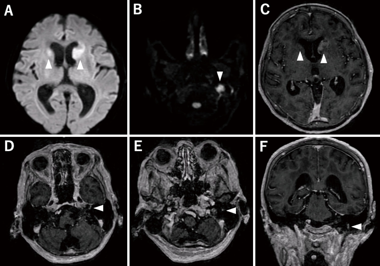
    Neurolymphomatosis is a rare lymphoma infiltrating cranial nerves, peripheral nerves, and spinal nerve roots. We present a unique case of neurolymphomatosis involving the central nervous system, initially resembling polyneuropathy in Sjögren's syndrome. A 79-year-old woman experienced dry eye symptoms for 2 years before presenting with left facial nerve palsy and lumbar pain to her previous physician. Brain magnetic resonance imaging revealed high signal intensity on diffusion-weighted imaging solely in the left peripheral facial nerve and lower cranial nerves. Initially diagnosed with Sjögren's syndrome-associated polyneuritis, she underwent treatment with high-dose steroid pulse therapy and intravenous immunoglobulin. Despite treatment, her symptoms progressed, leading to referral to our hospital with multiple peripheral nerve palsies and severe lumbar pain. Subsequent brain magnetic resonance imaging demonstrated gadolinium enhancement in the bilateral caudate nucleus heads and the left facial and lower cranial nerves. Stereotactic brain tumor biopsy-confirmed diffuse large B-cell lymphoma upon pathological analysis. Treatment with rituximab, high-dose methotrexate significantly improved her symptoms, with no recurrence observed for 12 months. These findings underscore the diagnosis of neurolymphomatosis involving the central nervous system, initially masquerading as Sjögren's syndrome. Neurolymphomatosis should be considered in cases of refractory Sjögren's syndrome presenting with multiple nerve palsies.

  • Yoshihiro OKADA, Yusuke TOMITA, Hisakazu NISHIMORI, Hiroki UENO, Kanji ...
    2025Volume 12 Pages 255-260
    Published: December 31, 2025
    Released on J-STAGE: June 11, 2025
    JOURNAL OPEN ACCESS FULL-TEXT HTML

    Methotrexate-related lymphoproliferative disorder is a rare but serious complication of methotrexate treatment. Recurrent methotrexate-related lymphoproliferative disorder in the central nervous system is uncommon. An 86-year-old woman, who had been on methotrexate for 3 years to treat rheumatoid arthritis, was admitted with left hemiplegia and sensory disturbance. Brain magnetic resonance imaging revealed a right basal ganglia lesion that was hyperintense on diffusion-weighted imaging and showed patchy enhancement. Methotrexate-related lymphoproliferative disorder was suspected, and methotrexate was discontinued, which led to improvement in her symptoms and partial remission. Twenty-eight months after methotrexate cessation, the patient presented with reduced activity and left hemiparesis. Brain magnetic resonance imaging showed homogeneously enhancing lesions in the right frontal lobe, basal ganglia, and brainstem. A craniotomy for biopsy of the right frontal lobe lesion confirmed the diagnosis of methotrexate-related lymphoproliferative disorder. Whole-brain radiation (40 Gy in 20 fractions) was administered without chemotherapy due to her advanced age and poor performance status. Her symptoms improved, and she was transferred to a rehabilitation hospital. Methotrexate-related lymphoproliferative disorder of the central nervous system can recur spontaneously, even without resumption of methotrexate, making long-term follow-up necessary after methotrexate cessation.

  • Yoshiyuki TAKAISHI, Hiroto KAJIMOTO, Hirotomo TANAKA, Masahiro SUGIHAR ...
    2025Volume 12 Pages 261-265
    Published: December 31, 2025
    Released on J-STAGE: June 11, 2025
    JOURNAL OPEN ACCESS FULL-TEXT HTML

    Low back pain during pregnancy is common. However, lumbar disc herniation in pregnancy is rare, and when severe, it can cause lower extremity pain that resists conservative treatment or lead to cauda equina syndrome. In this report, we describe a case of severe lower back pain that did not improve with conservative treatment, necessitating surgery at 12 weeks of gestation. Additionally, we reviewed the literature. A 35-year-old woman, 7 weeks pregnant, presented to our outpatient clinic with right lower extremity pain. Magnetic resonance imaging revealed a herniated disc at the right L4/5. Acetaminophen, a pregnancy-safe analgesic was prescribed. Despite initiating treatment, the pain persisted, and the patient was hospitalized for bed rest. After one month of conservative treatment with no relief, a repeat magnetic resonance imaging showed an enlarged herniated disc. After consulting with the patient and her family, surgery was performed at Kobe University Hospital, which offers comprehensive neurosurgery, obstetrics and gynecology, and anesthesiology services. At 12 weeks of gestation, the patient underwent a microscopic discectomy in the prone position under general anesthesia. Fluoroscopy for surgical level confirmation was minimized. The surgery lasted approximately 1 hour, and the right lower extremity pain was alleviated. Five months later, the patient delivered a healthy child. We report a case of surgery for a herniated disc during pregnancy, along with a review of the literature.

  • Kosuke NAKAJO, Hiroki MORISAKO, Tsutomu ICHINOSE, Takeo GOTO
    2025Volume 12 Pages 267-273
    Published: December 31, 2025
    Released on J-STAGE: June 11, 2025
    JOURNAL OPEN ACCESS FULL-TEXT HTML

    The trigone of the lateral ventricle is deep and surrounded by eloquent gyri and subcortical fibers. Resection of intraventricular trigone tumors has therefore been challenging, and the optimal surgical approach to the trigone of the lateral ventricle remains controversial. Three patients with large intraventricular trigone meningioma (≥4 cm in diameter) underwent surgical excision using a combined transtemporal and high-parietal approach at Osaka City University Hospital between July 2016 and January 2021. Clinical and imaging studies, as well as surgical complications, were retrospectively reviewed based on medical records from our institution. We also reviewed 153 patients with intraventricular trigone meningioma from 11 reports in the literature and assessed pre- and postoperative symptoms. Gross total resection of the tumor was achieved in all cases. None of the patients showed deterioration of neurological symptoms at 3 months after tumor resection, although one patient experienced transient language dysfunction several weeks after surgery. No cases showed recurrence or required additional therapy. According to our literature review, postoperative visual field defects are more likely to persist than postoperative language dysfunction at 3 months postoperatively. In conclusion, combining the transtemporal and high-parietal approaches appears to be useful for treating large intraventricular trigone meningioma. Postoperative language dysfunction after resection of intraventricular trigone meningioma may tend to resolve more rapidly than postoperative visual field defect.

  • Ryota TAOMOTO, Mikiko AOKI, Toshiyuki ENOMOTO, Noriko HIRAO, Hiroshi A ...
    2025Volume 12 Pages 275-281
    Published: December 31, 2025
    Released on J-STAGE: June 30, 2025
    JOURNAL OPEN ACCESS FULL-TEXT HTML
    Supplementary material

    Primary histiocytic sarcoma of the central nervous system is an extremely rare malignancy, with only 43 cases reported to date. It often presents diagnostic challenges due to its rarity and similarities with other central nervous system tumors, particularly glioblastoma. We report a case of primary central nervous system histiocytic sarcoma in a 72-year-old woman, one of the older patients among the reported cases. The patient presented with gait disturbance and left hemiplegia. Initial imaging and intraoperative findings suggested a high-grade glioma. However, comprehensive pathological examination, including immunohistochemistry, electron microscopy, and genetic analyses, led to the diagnosis of histiocytic sarcoma. Notably, the tumor exhibited CDKN2A homozygous deletion and a unique methylation profile distinct from gliomas. Despite treatment with surgery, radiation, and chemotherapy, the patient experienced meningeal dissemination 8 months post-surgery and died 11 months after diagnosis. This case highlights the importance of comprehensive diagnostic evaluation in challenging central nervous system tumors and provides valuable insights into the molecular characteristics of central nervous system histiocytic sarcoma, particularly regarding CDKN2A deletion and methylation patterns. Awareness of this rare entity is crucial for accurate diagnosis and appropriate management of central nervous system tumors.

  • Kotaro KOHARA, Tatsuya MAEGAWA, Eitaro OKUMURA, Ryo HASHIMOTO, Motoo K ...
    2025Volume 12 Pages 283-288
    Published: December 31, 2025
    Released on J-STAGE: June 30, 2025
    JOURNAL OPEN ACCESS FULL-TEXT HTML

    Foramen magnum decompression is an established surgical method for the treatment of Chiari type 1 malformation with syringomyelia. However, in some cases, neurologic symptoms that improve only after foramen magnum decompression may deteriorate again, and it has been suspected that increased instability of the craniocervical junction may be a factor. We report a case of Chiari type 1 malformation accompanied by basilar invagination and syringomyelia in which atlantoaxial facet joint distraction and fixation was performed as a revision surgery for deteriorating neurologic symptoms after foramen magnum decompression. The patient was a 16-year-old boy with chief complaints of gait disturbance and repeated falls. He was diagnosed with Chiari type 1 malformation accompanied by basilar invagination and syringomyelia. The clivo-axial angle was narrow at 105.8°. Initially, only foramen magnum decompression was performed, and the symptoms were relieved after the procedure but re-exacerbated within 2 weeks. The condition was speculated to have worsened instability at the atlantoaxial segment and ventral medullary compression; then, atlantoaxial facet joint distraction and fixation was performed secondarily, and symptoms improved. There is no clear surgical standard for performing foramen magnum decompression, atlantoaxial facet joint distraction and fixation, or a combination of both for Chiari type 1 malformation. Foramen magnum decompression provides horizontal decompression at the craniocervical junction, and atlantoaxial facet joint distraction and fixation achieves vertical indirect decompression of the ventral medulla and stabilization of the atlantoaxial segment in case with basilar invagination. Our experience suggests that combined foramen magnum decompression and atlantoaxial facet joint distraction and fixation may be particularly beneficial in Chiari type 1 malformation cases with basilar invagination and narrow clivo-axial angle.

  • Ichiro MAEDA, Taichiro IMAHORI, Shigeru MIYAKE, Hiroki GOTO, Rikuo NIS ...
    2025Volume 12 Pages 289-294
    Published: December 31, 2025
    Released on J-STAGE: June 30, 2025
    JOURNAL OPEN ACCESS FULL-TEXT HTML

    Cerebral embolism following endoscopic injection sclerotherapy using N-butyl-2 cyanoacrylate for esophagogastric varices is a rare but serious complication. This condition occurs due to the migration of N-butyl-2 cyanoacrylate into the cerebral circulation, which has traditionally been attributed to paradoxical embolism via a patent foramen ovale. However, recent reports have suggested the possibility of alternative embolic pathways. We report the case of a 76-year-old woman who presented with acute gastric variceal bleeding and underwent endoscopic injection sclerotherapy using N-butyl-2 cyanoacrylate and Lipiodol. Shortly after the procedure, the patient developed global aphasia and right hemiparesis. Head computed tomography revealed scattered ultra-hyperdense materials and magnetic resonance imaging confirmed left middle cerebral artery M2 occlusion. Mechanical thrombectomy was performed, achieving successful recanalization and neurological improvement. Imaging findings and histopathological analysis of the retrieved thrombus strongly supported the diagnosis of N-butyl-2 cyanoacrylate embolism. Retrospective evaluation of preoperative contrast-enhanced computed tomography identified a portopulmonary venous anastomosis, a right-to-left shunt connecting gastric varices to the pulmonary veins. This anastomosis is a collateral pathway associated with portal hypertension. This case suggests that even in the absence of a patent foramen ovale, portopulmonary venous anastomosis can serve as a route for N-butyl-2 cyanoacrylate migration into the cerebral circulation. To the best of our knowledge, this is the first reported case of mechanical thrombectomy for N-butyl-2 cyanoacrylate cerebral embolism following endoscopic injection sclerotherapy. This case underscores the importance of early detection and risk assessment of such right-to-left shunting and highlights mechanical thrombectomy as a viable intervention for cases where thrombolysis is contraindicated.

  • Mayuko KUNII, Tomofumi TAKENAKA, Nobuto HIRAI, Yuhei HOSHIKUMA, Shogo ...
    2025Volume 12 Pages 295-301
    Published: December 31, 2025
    Released on J-STAGE: August 02, 2025
    JOURNAL OPEN ACCESS FULL-TEXT HTML

    Dural arteriovenous fistula can present with cerebral venous sinus thrombosis and retrograde leptomeningeal venous drainage. Given the associated intracranial hemorrhage risk, immediate retrograde leptomeningeal venous drainage obliteration is desirable. Herein, we report a case of dural arteriovenous fistula with acute cerebral venous sinus thrombosis and intracranial hemorrhage that was successfully managed using sinus thrombectomy alone. A 76-year-old woman was admitted to our hospital with a sudden headache. Radiological assessments showed a subarachnoid hemorrhage in the left cerebellopontine cistern, Borden type III (Cognard type III) dural arteriovenous fistula with cerebral venous sinus thrombosis in the left transverse-sigmoid sinus, and retrograde leptomeningeal venous drainage in the superficial middle cerebral vein, the vein of Labbé, and the petrosal vein. To prevent re-bleeding, we performed an acute thrombectomy of the transverse-sigmoid sinus, aiming to obliterate retrograde leptomeningeal venous drainage. Following the procedure, the dural arteriovenous fistula was downgraded to Borden type I (Cognard type IIa). In this case, a simple thrombectomy alone prevented the re-bleeding of a dural arteriovenous fistula without direct fistula access. Understanding the relative contributions of the fistula and cerebral venous sinus thrombosis to retrograde leptomeningeal venous drainage in transverse-sigmoid sinus dural arteriovenous fistula is essential for refining the treatment strategy. Sinus thrombectomy is a tentative treatment, and this strategy is the emergency procedure to reduce re-bleeding risk. However, it may be a viable approach to prevent re-bleeding by improving venous outflow in dural arteriovenous fistula cases complicated by acute cerebral venous sinus thrombosis and intracranial hemorrhage.

  • Yotaro OKAZAKI, Satoshi HORI, Ryosuke TAKAGI, Taishi NAKAMURA, Makoto ...
    2025Volume 12 Pages 303-308
    Published: December 31, 2025
    Released on J-STAGE: August 02, 2025
    JOURNAL OPEN ACCESS FULL-TEXT HTML

    Reversible cerebral vasoconstriction syndrome is characterized by severe headaches and diffuse, segmental constriction of cerebral arteries. We report a rare pediatric case of triptan-induced reversible cerebral vasoconstriction syndrome, complicated by both subarachnoid hemorrhage and cerebral infarction. A 10-year-old boy presented with persistent severe headaches initially suspected to be migraines, for which he was prescribed rizatriptan. Five days after starting rizatriptan, he developed impaired consciousness (Japan Coma Scale 10) and partial right-sided hemiparesis, prompting emergency hospitalization. A head computed tomography scan revealed subarachnoid hemorrhage in the left frontal cortical region. Cerebral angiography showed stenotic changes in the internal carotid artery, middle cerebral artery, anterior cerebral artery, and posterior cerebral artery. Suspecting triptan-induced reversible cerebral vasoconstriction syndrome, rizatriptan was discontinued, and verapamil treatment was initiated. Subsequent magnetic resonance imaging and magnetic resonance angiography revealed cerebral infarction in the right cerebellum, left middle cerebral artery territory, and right occipital lobe, with persistent stenotic changes from earlier findings. Edaravone was added to the treatment regimen. The patient's condition gradually improved, and follow-up magnetic resonance angiography showed almost complete resolution of the prior stenotic changes. His partial right-sided hemiparesis nearly resolved, and he was discharged home. Triptans should be used with caution, as they may precipitate reversible cerebral vasoconstriction syndrome or exacerbate cerebral vasoconstriction. Careful monitoring and vascular imaging are essential for patients presenting with symptoms following triptan administration.

  • Mayuko MORITSUBO, Takuya FURUTA, Yoshihiro NATORI, Yoshihiro OHISHI, Y ...
    2025Volume 12 Pages 309-315
    Published: December 31, 2025
    Released on J-STAGE: August 02, 2025
    JOURNAL OPEN ACCESS FULL-TEXT HTML

    Composite or collision tumors in the central nervous system can significantly impact disease progression and metastasis, potentially affecting treatment efficacy. Studying the mechanisms associated with these tumors can provide neuro-oncologists with insights into tumor diversity, progression, and aid in the development of novel treatments. We encountered an 84-year-old female with memory disturbance who presented with tumors consistent with wild-type isocitrate dehydrogenase high-grade glioma and low-grade B-cell lymphoma at the same site. Magnetic resonance imaging revealed a solid enhanced mass in the right frontal lobe. A pre-operative suspicion of primary central nervous system lymphoma led to a brain biopsy. Histologically, 2 types of lesions were observed; the first consisted of atypical glial cells with diffuse infiltration and mitoses, positive for glial fibrillary acidic protein and negative for anti-isocitrate dehydrogenase 1 (IDH1) -R132H, characterized by partial amplification of PDGFRA and homozygous deletion of CDKN2A. The second type consisted of small atypical lymphoid positive forCD20, showing immunoglobulin heavy chain (IgH) rearrangement, and minimal invasion of vessel walls while filling the perivascular space. Based on these findings, collision neoplasms of high-grade gliomas and marginal zone B-cell lymphomas were suspected. To our knowledge, this is the first reported co-existence of a glioma and intracranial lymphoma.

  • Yuto KATSUYAMA, Yasuhiro AIDA, Daisuke KITA, Fumihide ENKAKU, Tsuyoshi ...
    2025Volume 12 Pages 317-321
    Published: December 31, 2025
    Released on J-STAGE: August 02, 2025
    JOURNAL OPEN ACCESS FULL-TEXT HTML

    Iatrogenic cerebral amyloid angiopathy is a rare yet clinically relevant condition that may develop decades after cadaveric dura mater transplantation. We present the case of a 48-year-old man who experienced recurrent, bilateral, multilobar intracerebral hemorrhages approximately 40 years after childhood dura mater transplantation. Pathological examination confirmed amyloid β protein deposition around small cerebral blood vessels in the brain parenchyma. Genetic testing excluded mutations associated with hereditary cerebral amyloid angiopathy. According to the patient's operative record, at age 7 years, a left parietal bone tumor and the adjacent dura mater were removed, and a cadaveric dural patch was transplanted onto the intact brain surface. This case suggests that exogenous amyloid β protein can be transmitted through cadaveric dura mater, even when placed on intact brain tissue with preserved glymphatic and dural lymphatic drainage function, manifesting decades after transplantation.

  • Kensuke IKEDA, Keisuke MARUYAMA, Hiroki KAGIWATA, Atsushi YAMAMICHI, K ...
    2025Volume 12 Pages 323-329
    Published: December 31, 2025
    Released on J-STAGE: August 02, 2025
    JOURNAL OPEN ACCESS FULL-TEXT HTML

    Posterior reversible encephalopathy syndrome is a well-known condition that causes reversible vasogenic edema, mainly in the occipital lobe. However, no guideline for its diagnosis or treatment has been established to date. While many atypical cases have been reported in recent years, posterior reversible encephalopathy syndrome associated with cranioplasty has not yet been reported. A man in his 50s underwent right decompressive craniectomy for brain swelling secondary to ischemic stroke. He was transferred to another hospital for rehabilitation 9 months later and was again admitted for cranioplasty after 2 months. Because of sustained brain distension, right cranioplasty was performed simultaneously with ventriculoperitoneal shunting. No sudden change in blood pressure occurred during the perioperative period. However, generalized convulsions occurred postoperatively. Magnetic resonance imaging showed signal changes in the right frontal lobe, left basal ganglia, left thalamus, and right occipital lobe. When cranioplasty was performed simultaneously with ventriculoperitoneal shunting for a skull defect complicated by brain distension, intraoperative cerebrospinal fluid drainage and continuous cerebrospinal fluid drainage by shunts may have caused a sudden decrease in intracranial pressure and an increase in cerebral perfusion pressure, possibly leading to the development of posterior reversible encephalopathy syndrome.

feedback
Top