NMC Case Report Journal
Online ISSN : 2188-4226
ISSN-L : 2188-4226
Volume 10
Displaying 1-50 of 57 articles from this issue
CASE REPORT
  • Takahiro KUMAGAWA, Naoki OTANI, Yuzo KAKEI, Hiroshi NEGISHI, Takeshi S ...
    2023 Volume 10 Pages 1-7
    Published: December 31, 2023
    Released on J-STAGE: January 16, 2023
    JOURNAL OPEN ACCESS FULL-TEXT HTML

    Initial three-dimensional computed tomography and cerebral angiography fail to identify any aneurysm in 20% of cases of subarachnoid hemorrhage. Basilar artery (BA) perforator aneurysms are rare, and approximately 30%-60% were not identified by initial angiography. A 71-year-old male was transferred with a sudden onset of headache and loss of consciousness. Computed tomography demonstrated subarachnoid hemorrhage, but no ruptured aneurysm was detected. Repeat preoperative cerebral angiography indicated a bifurcation aneurysm of the circumflex branch of the superior cerebellar artery perforator, but microsurgical observation identified the BA perforator aneurysm. If the location of the BA perforator aneurysm cannot be clearly identified, as in this case, repeat angiography should be considered, and the treatment strategy should be decided based on a detailed consideration of the site of the aneurysm.

  • Kazuo KAKINUMA, Keisuke MORIHARA, Yoshiteru SHIMODA, Nobuko KAWAKAMI, ...
    2023 Volume 10 Pages 9-14
    Published: December 31, 2023
    Released on J-STAGE: February 09, 2023
    JOURNAL OPEN ACCESS FULL-TEXT HTML

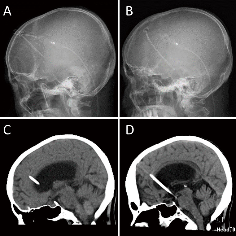
    Idiopathic normal-pressure hydrocephalus (iNPH) is a neurological disorder that typically presents with gait disturbance, cognitive impairment, and urinary incontinence. Although most patients respond to cerebrospinal-fluid shunting, some do not react well because of shunt failure. A 77-year-old female with iNPH underwent ventriculoperitoneal shunt implantation, and her gait impairment, cognitive dysfunction, and urge urinary incontinence improved. However, 3 years after shunting (at the age of 80), her symptoms gradually recurred for 3 months and she did not respond to shunt valve adjustment. Imaging studies revealed that the ventricular catheter detached from the shunt valve and migrated into the cranium. With immediate revision of the ventriculoperitoneal shunt, her gait disturbance, cognitive dysfunction, and urinary incontinence improved. When a patient whose symptoms have been relieved by cerebrospinal-fluid shunting experiences an exacerbation, it is important to suspect shunt failure, even if many years have passed since the surgery. Identifying the position of the catheter is crucial to determine the cause of shunt failure. Prompt shunt surgery for iNPH can be beneficial, even in elderly patients.

  • Takafumi TANEI, Satoshi MAESAWA, Yusuke NISHIMURA, Yoshitaka NAGASHIMA ...
    2023 Volume 10 Pages 15-20
    Published: December 31, 2023
    Released on J-STAGE: February 09, 2023
    JOURNAL OPEN ACCESS FULL-TEXT HTML

    Central poststroke pain is a chronic, intractable, central neuropathic pain. Spinal cord stimulation is a neuromodulation therapy for chronic neuropathic pain. The conventional stimulation method induces a sense of paresthesia. Fast-acting subperception therapy is one of the latest new stimulation methods without paresthesia. A case of achieving pain relief of central poststroke pain affecting both the arm and leg on one side by double-independent dual-lead spinal cord stimulation using fast-acting subperception therapy stimulation is presented. A 67-year-old woman had central poststroke pain due to a right thalamic hemorrhage. The numerical rating scale scores of the left arm and leg were 6 and 7, respectively. Using dual-lead stimulation at the Th 9-11 levels, a spinal cord stimulation trial was performed. Fast-acting subperception therapy stimulation achieved pain reduction in the left leg from 7 to 3. Therefore, a pulse generator was implanted, and the pain relief continued for 6 months. Then, two additional leads were implanted at the C 3-5 levels, and pain in the arm decreased from 6 to 4. Independent setting and adjustments of the dual-lead stimulation were required because the thresholds of paresthesia perception were significantly different. To achieve pain relief in both the arm and leg, double-independent dual-lead stimulation placed at cervical and thoracic levels is an effective treatment. Fast-acting subperception therapy stimulation may be effective for central poststroke pain, especially in cases where the paresthesia is perceived as uncomfortable or the conventional stimulation itself is ineffective.

  • Takayuki KAKIMOTO, Satoshi MURAI, Noboru KUSAKA, Fukiko BABA, Yohei IN ...
    2023 Volume 10 Pages 21-25
    Published: December 31, 2023
    Released on J-STAGE: February 23, 2023
    JOURNAL OPEN ACCESS FULL-TEXT HTML

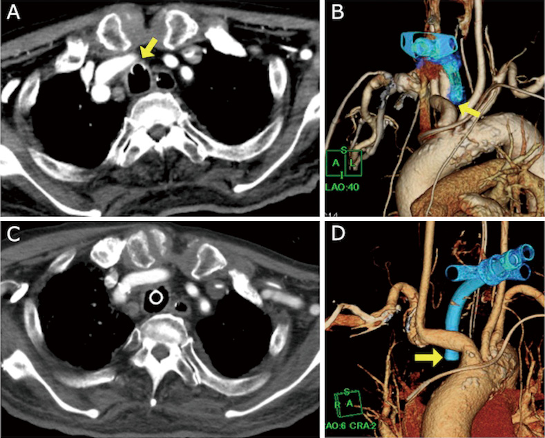
    A 78-year-old man underwent a tracheostomy after embolization for a dural arteriovenous fistula. Seventy days after tracheostomy, arterial bleeding appeared through the tracheal stoma. The bleeding stopped spontaneously. However, two days later, arterial bleeding reappeared, and he was diagnosed with a tracheo-innominate artery fistula (TIF). He then underwent urgent endovascular covered stent placement. After the procedure, there was no bleeding. TIF can be a fatal complication after tracheostomy and it is generally treated with open chest surgery. However, a successful endovascular treatment for TIF has recently been reported and may yield better results.

  • Takumi HOSHIMARU, Fugen TAKAGI, Yuichiro TSUJI, Ryokichi YAGI, Ryo HIR ...
    2023 Volume 10 Pages 27-32
    Published: December 31, 2023
    Released on J-STAGE: February 23, 2023
    JOURNAL OPEN ACCESS FULL-TEXT HTML

    Primary germ cell tumors of the central nervous system (CNS) typically occur in the neurohypophysis, hypothalamus, or pineal gland and rarely in the spinal cord. We report a case of a spinal intramedullary tumor, which was first detected on magnetic resonance imaging (MRI) 41 months after the initial symptoms, with a verified pathological diagnosis of germinoma. The initial symptom was an abnormal sensation in the left plantar region that gradually worsened, resulting in severe sensory disturbance, difficulty in standing, and even bladder rectal disturbance. Repeated MRI after the onset failed to provide an imaging diagnosis. The MRI was performed 41 months after the onset and revealed a previously undiagnosed, contrast-enhancing spinal intramedullary neoplastic lesion at the Th11-12 level. Gross total resection of the tumor was successfully performed, and the pathology confirmed the diagnosis of pure germinoma. Postoperative chemotherapy, followed by local radiation, was successfully administered. Among primary germinomas of the CNS, occult germinoma that lacks imaging findings suggestive of tumors in the early stages of onset and becomes apparent over time is often reported as a primary neurohypophyseal germinoma, particularly in adolescents presenting with diabetes insipidus. In the present case, the lesion appeared to correspond to a primary occult germinoma of the intramedullary spinal cord.

  • Hideki NAKAJIMA, Shigetoshi SHIMIZU, Takuro TSUCHIYA, Hidenori SUZUKI
    2023 Volume 10 Pages 33-39
    Published: December 31, 2023
    Released on J-STAGE: February 23, 2023
    JOURNAL OPEN ACCESS FULL-TEXT HTML

    Blood blister aneurysms (BBAs) or pseudoaneurysms (PAs) in the internal carotid artery (ICA) have fragile necks; therefore, conventional neck clipping is difficult. The standard treatment for BBAs or PAs is trapping with high or low flow bypass. However, there is no consensus on whether or not anterior clinoidectomy should be performed together. Two patients with ruptured ICA PA (anterior protrusion) or BBA (posterior protrusion) were presented to our hospital. Complete trapping was safely performed for both types of aneurysms via extradural anterior clinoidectomy and the extradural approach with dural incision. The advantages of the procedure are 1) safe proximal clipping, 2) early identification of the ICA C3 portion, 3) minimized frontal lobe retraction, 4) optic canal opening to allow mobility of the optic nerve, and 5) dural ring incision to allow mobility of the ICA.

  • Hirohisa YAJIMA, Satoshi KOIZUMI, Tomoki KANDA, Naoyuki SHONO, Taichi ...
    2023 Volume 10 Pages 41-45
    Published: December 31, 2023
    Released on J-STAGE: March 15, 2023
    JOURNAL OPEN ACCESS FULL-TEXT HTML

    Several studies have reported the coexistence of chronic subdural hematoma (CSDH) and dural arteriovenous fistula (DAVF); however, the association between these two entities remains unknown. A case of coexisting CSDH and DAVF that was successfully treated with burr hole surgery and middle meningeal artery (MMA) embolization is reported herein. We visualized the positional relationship between CSDH and DAVF by fusion three-dimensional computer graphics images reconstructed from multimodal imaging studies, which revealed that the shunt point of the DAVF was far from the burr hole and was in contact with the CSDH membrane at the center of the CSDH. Additionally, the chronological development of CSDH in the presence of DAVF and the complete disappearance of both DAVF and CSDH after MMA embolization were also demonstrated. This study suggests a possible association between recurrent CSDH and DAVF.

  • Mizuka IKEZAWA, Kenko MAEDA, Syuntaro TAKASU, Masaya TAKEMOTO, Masahir ...
    2023 Volume 10 Pages 47-50
    Published: December 31, 2023
    Released on J-STAGE: March 15, 2023
    JOURNAL OPEN ACCESS FULL-TEXT HTML

    The accessory meningeal artery (AMA) demonstrates various potential anastomoses with the external (ECA) and internal (ICA) carotid arteries. However, rarely does the AMA markedly dilate and compensate for ICA blood flow. A 52-year-old woman with nonspecific symptoms was diagnosed with multiple cerebral aneurysms and abnormal blood vessels observed on magnetic resonance angiography. Digital subtraction angiography revealed four aneurysms and anastomoses between the left AMA and inferolateral trunk (ILT). In addition, two sequential severe flexions were observed in the cervical portion of the left ICA. No ischemic lesions were detected on magnetic resonance imaging. In conclusion, we experienced a rare case in which the AMA-ILT anastomosis was highly developed. This case also presented with the unusual characteristics of an anomaly in the extracranial ICA and multiple aneurysms.

  • Yoshitaka TSUJIMOTO, Junya HANAKITA, Kazuhiro MIYASAKA, Manabu MINAMI, ...
    2023 Volume 10 Pages 51-54
    Published: December 31, 2023
    Released on J-STAGE: March 15, 2023
    JOURNAL OPEN ACCESS FULL-TEXT HTML

    We report a rare case presenting radiculopathy caused by unilateral lumbosacral facet abnormality resembling facet interlocking. The patient was a 57-year-old man with no medical and traumatic history. He began to exhibit numbness below his left ankle followed by pain at the left buttock with no obvious causes. He visited our hospital approximately 1 year after the onset of his symptom. Preoperative images revealed a left lumbosacral facet abnormality resembling facet interlocking. His left S1 nerve root was compressed by the dislocated left L5 inferior articular process and bone fragment. His symptom was consistent with left S1 radiculopathy without an obvious stenosis of the left L5 intervertebral foramen; thus, we performed partial facetectomy of the left L5/S1, posterior decompression of the S1 nerve root tunnel, and removal of bone fragment. After the operation, his symptom completely disappeared with satisfactory result. There are several types of congenital facet anomalies in the lumbosacral facet joint; however, congenital unilateral lumbosacral facet abnormality resembling facet interlocking described in this paper has not been reported. His clinical symptom was completely recovered after simple decompression surgery. In this paper, we report the interesting and unique findings of facet abnormality resembling facet interlocking.

  • Ryutaro MAKINO, Hitoshi YAMAHATA, Akari MACHIDA, Ayumi TANIGUCHI, Ryos ...
    2023 Volume 10 Pages 55-60
    Published: December 31, 2023
    Released on J-STAGE: March 15, 2023
    JOURNAL OPEN ACCESS FULL-TEXT HTML

    Internal carotid artery aplasia or hypoplasia above the cervical bifurcation is rare, occurring in less than 0.01% of the general population. Unilateral neurocristopathy complicated by unilateral internal carotid artery agenesis or hypogenesis has been reported, but bilateral internal carotid artery hypoplasia is rare and scarcely reported. Herein, we report a novel case of Treacher Collins syndrome complicated by bilateral internal carotid artery hypoplasia. A 94-year-old woman presented with complaints of headache and vomiting. Computed tomography revealed a subarachnoid hemorrhage and dysplasia of the bilateral zygoma, mandible, and external auditory meatus. The patient had severe hearing loss and visual impairment. Computed tomography angiography revealed bilateral internal carotid artery hypoplasia and multiple aneurysmal changes in the intracranial arteries. We diagnosed the patient with a ruptured anterior inferior cerebellar artery aneurysm and performed coil embolization. The patient's unique facial features were consistent with neurocristopathy, especially Treacher Collins syndrome. Developmental anomalies of neural crest cells can present as vascular abnormalities and craniofacial malformations. Special care is required for endovascular treatment and airway management in cases of neurocristopathy because of the specific craniofacial anomalies.

  • Yurie RAI, Takayuki HARA
    2023 Volume 10 Pages 61-66
    Published: December 31, 2023
    Released on J-STAGE: March 24, 2023
    JOURNAL OPEN ACCESS FULL-TEXT HTML

    Nilotinib, one of the tyrosine kinase inhibitors, has been used to treat chronic myeloid leukemia (CML) and Philadelphia chromosome-positive acute lymphoblastic leukemia (Ph+ ALL). Nilotinib-associated cerebral arterial occlusive disease, which is treated with medicine with/without bypass surgery or stenting, has been sporadically reported to occur. The mechanism of the nilotinib-associated cerebral disease has not been clarified and is still controversial. Here we present the case of a 39-year-old woman with Ph+ ALL treated with nilotinib, which led to symptomatic intracranial arterial stenosis. We performed high-flow bypass surgery and observed the arterial stenotic change in the stenotic portion intraoperatively, whose findings strongly supported the theory of atherosclerosis and seemed to be irreversible.

  • Juntaro FUJITA, Yusuke TOMITA, Koichi ICHIMURA, Rie YAMASAKI, Shohei N ...
    2023 Volume 10 Pages 67-73
    Published: December 31, 2023
    Released on J-STAGE: March 24, 2023
    JOURNAL OPEN ACCESS FULL-TEXT HTML

    Melanoma carries a high risk of brain metastasis. A small subset of metastatic melanomas, known as amelanotic melanomas, does not present black coloration, reflecting a lack of melanin pigmentation. Here, we report a case of B-Raf proto-oncogene (BRAF) V600E mutation associated with a metastatic brain tumor caused by the amelanotic melanoma. A 60-year-old man was transferred to our department following acute onsets of left upper limb paralysis and convulsion. In the brain imaging, multiple lesions in the right frontal lobe and left basal ganglia were detected, and the presence of an enlarged left axillary lymph node was revealed. Consequently, we removed the right frontal lesion and performed a biopsy of the left axillary lymph node. Histological analysis of both specimens indicated an amelanotic melanoma, and genetic testing revealed a BRAF V600E mutation. The residual intracranial lesions were treated with stereotactic radiotherapy and molecular-targeted therapy, with dabrafenib and trametinib as the systemic treatment. Based on the Response Evaluation Criteria in Solid Tumors, we determined that the patient achieved complete remission (CR) under uninterrupted molecular-targeted therapy over a period of 10 months. After the temporary withdrawal of dabrafenib and trametinib to avoid hepatic dysfunction, a new intracranial lesion appeared. CR of this lesion was achieved following reinstatement of the two drugs. These results suggest that, under limited conditions, molecular-targeted therapy can produce a sustained response against the intracranial metastasis of melanoma, and the therapy with reduced dose is still effective against a recurrent case after cessation of the therapy due to the toxicity.

  • Takayuki NAGASE, Joji ISHIDA, Susumu SASADA, Tatsuya SASAKI, Yoshihiro ...
    2023 Volume 10 Pages 75-80
    Published: December 31, 2023
    Released on J-STAGE: March 24, 2023
    JOURNAL OPEN ACCESS FULL-TEXT HTML

    Diffusely infiltrative midline gliomas are known to have a poor prognosis. The standard treatment for typical diffuse midline glioma in the pons is local radiotherapy as surgical resection is inappropriate. This case reports a brainstem glioma in which stereotactic biopsy and foramen magnum decompression were concomitantly performed to confirm the diagnosis and improve symptoms. A 23-year-old woman was referred to our department with a chief complaint of headache for six months. Magnetic resonance imaging (MRI) showed diffuse T2 hyperintense swelling of the brainstem with the pons as the main locus. Enlargement of the lateral ventricles was observed because of cerebrospinal fluid obstruction out of the posterior fossa. This was atypical for a diffuse midline glioma in terms of the longstanding slow progression of symptoms and patient age. Stereotactic biopsy was performed for diagnosis, and foramen magnum decompression (FMD) was concomitantly performed to treat the obstructive hydrocephalus. The histological diagnosis was astrocytoma, IDH-mutant. Post-surgery, the patient's symptoms were relieved, and she was discharged on the fifth day after surgery. The hydrocephalus was resolved, and the patient returned to normal life without any symptoms. The tumor size follow-up with MRI demonstrated no marked change for 12 months. Even though diffuse midline glioma is considered to have a poor prognosis, clinicians should contemplate if it is atypical. In atypical cases like the one described herein, surgical treatment may contribute to pathological diagnosis and symptom improvement.

  • Satoshi MIYAMOTO, Hisayuki HOSOO, Eiichi ISHIKAWA, Yuji MATSUMARU
    2023 Volume 10 Pages 81-85
    Published: December 31, 2023
    Released on J-STAGE: March 24, 2023
    JOURNAL OPEN ACCESS FULL-TEXT HTML
    Supplementary material

    Middle meningeal arteriovenous fistula (MMAVF) is a shunt between the middle meningeal artery and the vein surrounding the artery. We report an extremely rare case of spontaneous MMAVF; then, we evaluated the effectiveness of trans-arterial embolization for spontaneous MMAVF and the possible cause of spontaneous MMAVF.

    A 42-year-old man with tinnitus, a left temporal headache, and pain surrounding the left mandibular joint was diagnosed with MMAVF on digital subtraction angiography. Trans-arterial embolization with detachable coils was conducted, which resulted in a fistula closure and symptoms' diminishment. The cause of MMAVF was thought to be the rupture of the middle meningeal artery aneurysm.

    A middle meningeal artery aneurysm can be a cause of spontaneous MMAVF, and trans-arterial embolization might be an optimal treatment.

  • Masatoshi MORIMOTO, Shunsuke TAMAKI, Takayuki OGAWA, Shutaro FUJIMOTO, ...
    2023 Volume 10 Pages 87-92
    Published: December 31, 2023
    Released on J-STAGE: April 10, 2023
    JOURNAL OPEN ACCESS FULL-TEXT HTML
    Supplementary material

    Various approaches to lumbar interbody fusion have been described. The usefulness of full-endoscopic trans-Kambin's triangle lumbar interbody fusion has recently been reported. This technique has several advantages in patients with degenerative spondylolisthesis, including the ability to improve symptoms without decompression surgery. Moreover, given that the entire procedure is performed percutaneously, it can be performed without increasing the operation time or surgical invasiveness, even in obese patients. In this article, we discuss these advantages and illustrate them with representative cases.

  • Toshitsugu TERAKADO, Akibumi OMI, Yuji MATSUMARU, Eiichi ISHIKAWA
    2023 Volume 10 Pages 93-98
    Published: December 31, 2023
    Released on J-STAGE: April 10, 2023
    JOURNAL OPEN ACCESS FULL-TEXT HTML

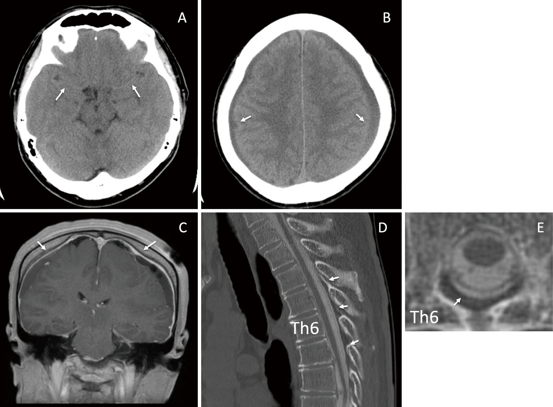
    Both chronic subdural hematoma (CSDH) and spontaneous intracranial hypotension (SIH) cause headaches. However, the etiologies are different: SIH headache is caused by decreased intracranial pressure (ICP), whereas CSDH headache results from increased ICP. Moreover, CSDH is treated by hematoma drainage, while SIH is treated by epidural blood patch (EBP). Treatment for the cases of combined SIH and CSDH is not well-established. Herein, we report two cases wherein ICP was monitored and safely controlled by EBP after hematoma drainage. Case 1: A 55-year-old man with progressive consciousness disturbance was diagnosed with bilateral CSDH. He underwent bilateral hematoma drainage; however, the headache became apparent during standing. We diagnosed SIH by diffuse pachymeningeal enhancement on brain MRI and epidural contrast medium leakage on CT myelography. Due to the re-enlargement of bilateral CSDH, we performed EBP after hematoma drainage and ICP monitor insertion. Finally, the headache and bilateral CSDH were resolved. Case 2: A 54-year-old man with persistent headache was diagnosed with bilateral CSDH. He underwent multiple hematoma drainage sessions. However, headache on standing persisted. We diagnosed SIH by diffuse pachymeningeal enhancement on brain MRI and epidural contrast medium leakage on CT myelography. Due to the re-enlargement of the left CSDH, we performed EBP after left hematoma drainage and ICP monitor insertion. Finally, the headache and bilateral CSDH were resolved. EBP after hematoma drainage and ICP monitoring was useful for SIH with bilateral CSDH. By monitoring ICP before EBP, the ICP was safely controlled and CSDH was resolved.

  • Isamu MIURA, Shiro HORISAWA, Takakazu KAWAMATA, Takaomi TAIRA
    2023 Volume 10 Pages 99-102
    Published: December 31, 2023
    Released on J-STAGE: April 10, 2023
    JOURNAL OPEN ACCESS FULL-TEXT HTML

    Cervical dystonia, characterized by the involuntary contraction of cervical muscles, is the most common form of adult dystonia. In a patient with intractable cervical dystonia, we carried out a myotomy of the left obliquus capitis inferior and selective peripheral denervation (SPD) of the posterior branches of the C3-C6 spinal nerves based on preoperative 18F-fluorodeoxyglucose (18F-FDG) positron emission tomography/computed tomography (PET/CT). The patient was a 65-year-old, right-handed man with an unremarkable medical history. His head rotated involuntarily to the left. Medication and botulinum toxin injections were ineffective, and surgical treatment was considered. 18F-FDG PET/CT imaging revealed FDG uptake in the left obliquus capitis inferior, right sternocleidomastoideus, and left splenius capitis. Myotomy of the left obliquus capitis inferior and SPD of the posterior branches of the C3-C6 spinal nerves was performed under general anesthesia. During the 6-month follow-up, the patient's Toronto Western Spasmodic Torticollis Rating Scale score improved from 35 to 9. This case shows that preoperative 18F-FDG PET/CT is effective in identifying dystonic muscles and determining the surgical strategy for cervical dystonia.

  • Fumihiro SAGO, Yusuke SAKAMOTO, Kenko MAEDA, Masaya TAKEMOTO, Jungsu C ...
    2023 Volume 10 Pages 103-108
    Published: December 31, 2023
    Released on J-STAGE: April 21, 2023
    JOURNAL OPEN ACCESS FULL-TEXT HTML

    In this study, we report a case of catastrophic propeller brain injury with large scalp defect treated with omental flap reconstruction.

    A 62-year-old man was accidentally caught in a powered paraglider propeller during maintenance. The rotor blades impacted the left part of his head. On arrival at the hospital, he presented with a Glasgow Coma Scale score of E4V1M4. On some areas on his head, skin was noticeably cut off, and the brain tissue out-slipped through an open skull fracture. Continuous bleeding from the superior sagittal sinus (SSS) and the brain surface was observed during emergency surgery. Massive bleeding from the SSS was controlled using a number of tenting sutures and hemostatic agents. We evacuated the crushed brain tissue and coagulated the severed middle cerebral arteries. Dural plasty using the deep fascia of the thigh was performed. The skin defect was closed using an artificial dermis. The administration of high-dose antibiotics has failed to prevent meningitis. Moreover, the severed skin edges and fasciae were necrotic. Plastic surgeons performed debridement and vacuum-assisted closure therapy to promote wound healing. Follow-up head computed tomography revealed hydrocephalus. Lumbar drainage was performed; however, sinking skin flap syndrome was observed. After removing the lumbar drainage, cerebrospinal fluid leakage occurred. We then performed cranioplasty with a titanium mesh and omental flap on day 31. After the surgery, perfect wound healing and infection control were achieved; however, severe disturbance of consciousness remained. The patient was transferred to a nursing home. Primary hemostasis and infection control are mandatory. An omental flap has been determined to be effective in controlling infection by covering the exposed brain tissue.

  • Hiroshi OKETANI, Katsumi HARIMAYA, Teruaki ONO, Kazushige TERADO, Sato ...
    2023 Volume 10 Pages 109-113
    Published: December 31, 2023
    Released on J-STAGE: April 21, 2023
    JOURNAL OPEN ACCESS FULL-TEXT HTML

    Lumbar canal stenosis (LCS) has been reported as a precipitating factor by which a tethered spinal cord, which is asymptomatic during childhood, develops into tethered cord syndrome (TCS) in adulthood. However, only a few reports on surgical strategies for such cases are available. A 64-year-old woman presented with unbearable pain in the left buttock and dorsal aspect of the thigh approximately 1 year ago. Magnetic resonance imaging showed cord tethering with a filar-type spinal lipoma and LCS due to the thickening of the ligamentum flavum at the L4-5 vertebral level. Five months after the decompressive laminectomy for the treatment of LCS, an untethering surgery was performed at the dural cul-de-sac at the S4 level. The severed end of the filum was elevated rostrally by 7 mm, and the pain subsided postoperatively. This case study shows that surgeries for both lesions should be indicated for adult-onset TCS triggered by LCS.

  • Takahiro YOKOYAMA, Tatsuya ISHIKAWA, Yosuke MOTEKI, Takayuki FUNATSU, ...
    2023 Volume 10 Pages 115-119
    Published: December 31, 2023
    Released on J-STAGE: April 21, 2023
    JOURNAL OPEN ACCESS FULL-TEXT HTML

    PulseRider (Cerenovus, Irvine, CA, USA) is a relatively novel device used for the treatment of wide-neck aneurysms with a coil-assisted effect. However, treatment options for recurrent aneurysms after PulseRider-assisted coil embolization remain controversial. Here we report a case of recurrent basilar tip aneurysm (BTA) treated with Enterprise 2 after PulseRider-assisted coil embolization. A woman in her 70s underwent coil embolization for a subarachnoid hemorrhage with ruptured BTA 16 years ago. Recurrence was detected at 6-year follow-up, and an additional coil embolization was performed. Nevertheless, gradual recurrence still occurred, and PulseRider-assisted coil embolization was performed without any complications 9 years after the second treatment. However, recurrence was detected once more at 6-month follow-up. Thus, stent-assisted coil embolization using Enterprise 2 (Cerenovus) through PulseRider was selected for angular remodeling. Enterprise 2 was deployed between the right P2 segment of the posterior cerebral artery (PCA) and basilar artery (BA) after an effective coil embolization, which achieved effective angular remodeling between the right PCA and BA. The patient's postoperative course was uneventful, and no recanalization was detected after half a year. Although PulseRider is effective for wide-neck aneurysm treatment, recurrence remains a possibility. Additional treatment using Enterprise 2 is safe and effective with the expectation of angular remodeling.

  • Eitaro OKUMURA, Hiroyuki ONUKI, Kunitoshi OTSUKA, Shigeki SUNAGA, Asas ...
    2023 Volume 10 Pages 121-124
    Published: December 31, 2023
    Released on J-STAGE: May 17, 2023
    JOURNAL OPEN ACCESS FULL-TEXT HTML

    We present a case of autoimmune-acquired factor XIII deficiency associated with systemic lupus erythematosus, which was diagnosed as a cause of repeated intracerebral hemorrhage. An intracerebral hemorrhage occurred in a 24-year-old female patient. Craniotomy was performed to remove the hematoma, but rebleeding occurred at the same site on days 2 and 11, respectively. Detailed blood tests revealed that factor XIII activity decreased. Although autoimmune-acquired factor XIII deficiency is a very rare disease, it can sometimes be fatal when intracerebral hemorrhage occurs. If there is repeated intracerebral hemorrhage, factor XIII activity should be confirmed.

  • Takashi IWAMA, Katsuhiro MIZUTANI, Hajime KUBO, Masahiro KATSUMATA, Ta ...
    2023 Volume 10 Pages 125-130
    Published: December 31, 2023
    Released on J-STAGE: May 17, 2023
    JOURNAL OPEN ACCESS FULL-TEXT HTML

    Patients with neurofibromatosis type 1 not only have characteristic skin findings but are also known to have vascular disorders due to vascular vulnerability. A 44-year-old man with previously undiagnosed neurofibromatosis type 1 was brought to the emergency room due to a sudden subcutaneous hematoma with no history of trauma. Angiography revealed extravasation from the parietal branch of the right superficial temporal artery, which was embolized with n-butyl-2-cyanoacrylate. However, the next day, the patient exhibited an increased subcutaneous hematoma, and new extravascular leakage was detected at the frontal branch of the superficial temporal artery, which was also embolized with n-butyl-2-cyanoacrylate. The patient had physical findings characteristic of neurofibromatosis type 1, such as café-au-lait spots, and was subsequently diagnosed with neurofibromatosis type 1. No obvious neurofibroma or any other subcutaneous lesion associated with neurofibromatosis type 1 was identified in the affected area. Massive idiopathic arterial bleeding in the scalp, although infrequent, can be fatal. Neurofibromatosis type 1 should be considered when a subcutaneous scalp hematoma is observed without a history of trauma, even if the facial skin structure appears normal. Neurofibromatosis type 1 is also known to have multiple sources of hemorrhage. Thus, it is important to repeatedly evaluate vascular structures via cerebral angiography, contrast-enhanced computed tomography, and magnetic resonance imaging, if necessary.

  • Takaharu KAWAJIRI, Hayato TAKEUCHI, Yoshinobu TAKAHASHI, Yuji SHIMURA, ...
    2023 Volume 10 Pages 131-137
    Published: December 31, 2023
    Released on J-STAGE: May 17, 2023
    JOURNAL OPEN ACCESS FULL-TEXT HTML

    Eating disorders caused by brain tumors are infrequently seen. Recent studies revealed that a neurocircuit from the nucleus tractus solitarius of the medulla oblongata to the hypothalamus participates in the control of appetite. Among brain tumors, those located in the brain stem, especially a solitary one in the medulla oblongata, are rare. Tumors in the brainstem are generally considered gliomas, and with the difficulty in reaching the lesion, treatment without histological confirmation is often performed. However, there are a few reported cases of medulla oblongata tumors other than gliomas. We describe a case of a 56-year-old man who presented with persistent anorexia. Magnetic resonance images revealed a solitary tumor in the medulla oblongata. After several examinations, craniotomy for the biopsy of the tumor using the cerebellomedullary fissure approach was carried out and primary central nervous system lymphoma (PCNSL) was histologically proven. The patient was treated with effective adjuvant therapy and was discharged home after he recovered from the symptoms. No tumor recurrence was recognized 24 months after surgery. A PCNSL arising only from the medulla oblongata is very rare, and anorexia can be an initial symptom of a tumor in the medulla oblongata. Surgical intervention is safely achieved and is a key to a better clinical outcome.

  • Taku SUGIYAMA, Miki FUJIMURA
    2023 Volume 10 Pages 139-143
    Published: December 31, 2023
    Released on J-STAGE: May 17, 2023
    JOURNAL OPEN ACCESS FULL-TEXT HTML

    During microvascular decompression (MVD) for trigeminal neuralgia (TGN), the mastoid emissary veins (MEV) are routinely sacrificed using the suboccipital retrosigmoid approach. The technical nuances when the MEV is an important collateral venous pathway for the obstructive internal jugular vein (IJV) have not been described thus far. Herein, we demonstrate for the first time a modified surgical technique for MVD to preserve the MEV. A 62-year-old man with a 10-year history of TGN refractory to carbamazepine was referred to our hospital for MVD. Preoperative imaging revealed that the superior cerebellar artery was the offending vessel. Computed tomography angiography also revealed that his contralateral IJV pathway was hypoplastic, and the ipsilateral pathway was severely stenosed by the external compression of the elongated styloid process and the transverse process of the first cervical vertebra. The ipsilateral MEV and the connecting occipital veins were enlarged as the sole collateral pathways of intracranial venous drainage. A modified MVD technique, including an upside-down L-shaped skin incision, layer-by-layer dissection of the occipital muscles, and denuding of the intraosseous part of the MEV, was used to cure the TGN with the preservation of the venous pathway. After surgery, the pain completely diminished without any complications. In conclusion, such technical modifications would be applicable in cases where the MEV needs to be preserved during posterior fossa surgery. Preoperative screening of the venous system is also recommended.

  • Naoya KIDANI, Yusuke INOUE, Shohei NISHIGAKI, Juntaro FUJITA, Yuta SOT ...
    2023 Volume 10 Pages 145-150
    Published: December 31, 2023
    Released on J-STAGE: May 17, 2023
    JOURNAL OPEN ACCESS FULL-TEXT HTML

    An appropriate therapeutic option for pial arteriovenous fistula (PAVF) can vary according to the angioarchtecture of the lesion. We present a case of adult infratentorial PAVF treated by transarterial coil embolization. A 26-year-old man was referred to our institution for an asymptomatic intracranial vascular lesion. Cerebral angiograms revealed PAVF fed by three arteries in the right cerebellomedullary cistern. The feeding arteries were accurately identified by three-dimensional rotational angiography and were successfully embolized using coils while normal arterial flow was preserved. This case report suggests that stepwise transarterial coil embolization can cure PAVF under detailed evaluation of its angioarchitecture.

  • Shota HORIIKE, Yasuhiro NAKAJIMA, Mamoru MATSUO, Akinori KAGEYAMA, Aya ...
    2023 Volume 10 Pages 157-162
    Published: December 31, 2023
    Released on J-STAGE: June 06, 2023
    JOURNAL OPEN ACCESS FULL-TEXT HTML

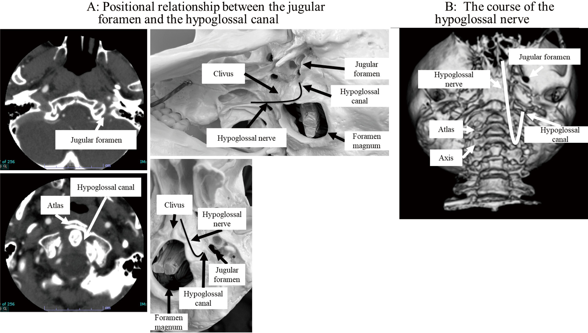
    A 68-year-old man presented with a Jefferson fracture leading to lower cranial nerve palsies affecting the ninth, tenth, and twelfth cranial nerves with a traumatic basilar impression. On the X day, the patient underwent occipitocervical posterior fixation surgery; the surgery was uneventful. However, just after the surgery, epipharyngeal palsy and airway obstruction occurred. Consequently, tracheostomy was needed. On the X+8 day, speech-language pathology (SLP) therapy was initiated for decannulation. On the X+21 day, the patient could clear all the checkpoints and was decannulated. On the X+36 day, the patient was discharged home and SLP therapy was continued. On the X+171 day, his SLP therapy was halted. However, the patient continued to complain that he could not speak as fast as before, and his quality of life remained compromised. Some studies reported that lower cranial nerve palsies affecting the ninth to the twelfth cranial nerve occur in conjunction with Jefferson fractures. Thus, SLP therapy is crucial for Jefferson fracture cases.

  • Masahiro NISHIHORI, Takashi IZUMI, Kazuhito TAKEUCHI, Shunsaku GOTO, F ...
    2023 Volume 10 Pages 163-168
    Published: December 31, 2023
    Released on J-STAGE: June 06, 2023
    JOURNAL OPEN ACCESS FULL-TEXT HTML

    A 61-year-old man presented with massive epistaxis, amaurosis, nausea, and severe headache. A detailed examination revealed a subarachnoid hemorrhage and prolactinoma. Angiography showed a small internal carotid artery pseudoaneurysm and inadequate collateral circulation; thus, uncomplicated coil embolization was performed. Considering the side effects of medication, such as cerebrospinal fluid rhinorrhea, the patient was followed up for asymptomatic prolactinoma without medication after discharge. At 40 months later, aneurysm recurrence was confirmed. Flow diverter device placement was performed, and the outcomes were excellent. In the present report, we described a rare case of a ruptured internal carotid artery aneurysm in an untreated prolactinoma and discussed the literature.

  • Haruhiko KIKUTA, Shinya JINGUJI, Taku SATO, Mudathir BAKHIT, Ryo HIRUT ...
    2023 Volume 10 Pages 169-175
    Published: December 31, 2023
    Released on J-STAGE: June 06, 2023
    JOURNAL OPEN ACCESS FULL-TEXT HTML

    Double or multiple pituitary adenomas expressing different types of transcription factors and collision tumors of pituitary adenomas and craniopharyngiomas are rare. In this report, we present a case of pituitary adenoma of two different cell populations, Pit-1 and SF-1, and an adenoma and craniopharyngioma collision tumor with coexisting Graves' disease. The patient had a 16-mm pituitary tumor with pituitary stalk calcification and optic chiasm compression but no visual dysfunction. Based on hormonal profile results, the tumor in the sella was considered a nonfunctioning pituitary adenoma; nevertheless, the pituitary stalk was invaded by a different lesion, which was later confirmed to be a craniopharyngioma. Using an endoscopic endonasal approach, the pituitary adenoma was removed; however, a small remnant remained medial to the right cavernous sinus. Because the pituitary stalk lesion was isolated from the pituitary adenoma, it was preserved to maintain pituitary function. Three years after the initial surgery, the patient suffered from Graves' disease and was treated with antithyroid medications. However, the intrasellar residual and pituitary stalk lesions gradually increased in size. A second surgery was performed, and the residual intrasellar and stalk lesions were completely removed. As per the initial and second histopathologies, the pituitary adenoma comprised different cell groups positive for thyroid-(TSH) and follicle-stimulating hormones, and each cell group was positive for Pit-1 and SF-1. The pituitary stalk lesion was an adamantinomatous craniopharyngioma. We believe that TSH-producing adenoma was involved in the development of Graves' disease or that treatment for Graves' disease increased TSH-producing adenoma.

  • Hayato YOKOYAMA, Masahiro NISHIHORI, Takashi IZUMI, Shunsaku GOTO, Mic ...
    2023 Volume 10 Pages 177-183
    Published: December 31, 2023
    Released on J-STAGE: June 26, 2023
    JOURNAL OPEN ACCESS FULL-TEXT HTML

    Intracranial aneurysms (IA) in infants are reportedly rare at 0.5% to 4.5% of all aneurysms. Furthermore, subarachnoid hemorrhage in infants younger than three months are even rarer as it has been reported in approximately 20 cases only till date.

    A 3-month-old infant with seizures and impaired consciousness was admitted to our hospital. Three-dimensional computed tomography angiography (3D-CTA) revealed a dissecting aneurysm with a maximum diameter of 13 mm in the right M2. Internal trapping using detachable coil were successfully performed, following which he was discharged without significant neurological deficit after one month of onset. Thus, we have reported a rare case of a large ruptured dissecting IA in a 3-month-old infant, in the right middle cerebral artery (MCA), successfully treated with an endovascular therapy, along with a literature review.

  • Kento TAKAHARA, Tomoru MIWA, Takashi IWAMA, Masahiro TODA
    2023 Volume 10 Pages 185-189
    Published: December 31, 2023
    Released on J-STAGE: June 26, 2023
    JOURNAL OPEN ACCESS FULL-TEXT HTML

    The occipital transtentorial approach (OTA), which is often applied for superior cerebellar lesions, has an inevitable risk of homonymous hemianopsia due to the retraction of the occipital lobe. The endoscopic approach provides increased visibility of the surgical field due to the wide-angled panoramic view and is minimally invasive in approaching deep brain lesions compared to the conventional microscopic approach. However, little is known regarding endoscopic OTA for the removal of cerebellar lesions. We experienced a case of a hemangioblastoma in the paramedian superior surface of the cerebellum that was successfully treated with endoscopic OTA combined with gravity retraction while avoiding postoperative visual dysfunction.

    A 48-year-old woman was diagnosed with a hemangioblastoma in the superior surface of the cerebellum. She underwent tumor removal with endoscopic OTA combined with gravity retraction of the occipital lobe instead of using brain retractors. The narrower space was sufficient for surgical manipulation with a panoramic view obtained by endoscopy. The simultaneous observation of the lesion with both an endoscope and a microscope revealed the superiority of infratentorial visualization with an endoscope. Gross total removal was achieved with no postoperative complications, including visual dysfunction.

    Endoscopic OTA may reduce the risk of postoperative visual dysfunction because of its minimally invasive nature, which is enhanced when combined with gravity retraction. Additionally, the panoramic view of the endoscope allows favorable visualization of an infratentorial lesion, which is otherwise hidden partly by the tentorium. The use of endoscopy is compatible with OTA, and endoscopic OTA could be an option for superior cerebellar lesions for avoiding visual dysfunction.

  • Shogo DOFUKU, Masayuki SATO, Takashi AOKA, Rika NAKAMURA, Kenta OHARA, ...
    2023 Volume 10 Pages 191-195
    Published: December 31, 2023
    Released on J-STAGE: June 26, 2023
    JOURNAL OPEN ACCESS FULL-TEXT HTML

    We report a rare case of isolated internal carotid artery occlusion complicated by central retinal artery occlusion that was successfully treated with mechanical thrombectomy for internal carotid artery occlusion. A 59-year-old man visited the emergency room because of right monocular blindness. Magnetic resonance imaging showed multiple acute small embolic infarctions in the right frontal lobe, and magnetic resonance angiography revealed right internal carotid artery occlusion without the associated occlusion of the circle of Willis, which indicates the patency of the anterior and middle cerebral arteries. An electrocardiogram showed atrial fibrillation. Therefore, we performed mechanical thrombectomy with a stent retriever under continuous manual aspiration with a balloon-guiding catheter and confirmed complete recanalization, anterograde flow in the right ophthalmic artery, and retinal brush. The procedure was completed without complications, and the patient noticed an improvement in visual acuity immediately after the procedure. When a patient with atrial fibrillation complains of monocular blindness, it is important to consider internal carotid artery occlusion due to cardioembolism, to perform an examination promptly, and to consider early treatment, including mechanical thrombectomy.

  • Tomoya YOKOYAMA, Shigeki SUNAGA, Hiroyuki ONUKI, Kunitoshi OTSUKA, Hir ...
    2023 Volume 10 Pages 197-202
    Published: December 31, 2023
    Released on J-STAGE: June 26, 2023
    JOURNAL OPEN ACCESS FULL-TEXT HTML

    We report a case of a 73-year-old man who developed nonconvulsive status epilepticus as a complication of cerebral hyperperfusion syndrome after carotid endarterectomy for carotid artery stenosis. On postoperative day 1, the patient experienced headaches and vomiting. Resting N-isopropyl-p-[123I] iodoamphetamine single-photon emission computed tomography showed increased cerebral blood flow to the entire right hemisphere, and the patient was diagnosed with cerebral hyperperfusion syndrome. He was treated with antihypertensive and antiseizure medications, sedated using propofol, intubated, and placed under mechanical ventilation. On postoperative day 3, computed tomography perfusion imaging showed a reduction in hyperperfusion, and propofol sedation was terminated on postoperative day 4. However, the patient exhibited prolonged impaired awareness and roving eye movements, and long-term video electroencephalographic monitoring revealed electrographic seizures. The patient was diagnosed with nonconvulsive status epilepticus. Propofol sedation was resumed, and the antiseizure medication dose was increased. Subsequently, the state of hyperperfusion in the right hemisphere diminished, and electroencephalographic findings improved, allowing sedation to be terminated on postoperative day 7. The findings from this case suggest that when clinical subtle symptoms, such as impaired awareness and roving eye movements, are observed during treatment of cerebral hyperperfusion syndrome, video electroencephalography should be performed to detect electrographic seizures.

  • Takafumi TANEI, Yusuke NISHIMURA, Yoshitaka NAGASHIMA, Motonori ISHII, ...
    2023 Volume 10 Pages 203-208
    Published: December 31, 2023
    Released on J-STAGE: July 13, 2023
    JOURNAL OPEN ACCESS FULL-TEXT HTML

    Hereditary neuropathy with liability to pressure palsies is an extremely rare genetic disorder; it is an autosomal dominant disorder with a high incidence of neuropathic and/or musculoskeletal pain. A case of achieving pain relief by spinal cord stimulation using differential target multiplexed stimulation for a 44-year-old female patient with hereditary neuropathy with liability to pressure palsies who was experiencing severe pain in her back, face, and all four limbs is presented. In her early teens, the initial symptoms were numbness and weakness of a limb after movement, which improved spontaneously. Transient pain in her back followed by systemic and persistent muscle weakness and pain developed. Deletion of the gene for peripheral myelin protein 22 was detected by peripheral nerve biopsy. The diagnosis of hereditary neuropathy with liability to pressure palsies was made in her early thirties. A spinal cord stimulation trial was performed because her severe pain continued despite administering many medications. Therefore, two spinal cord stimulation systems were implanted at the C3-5 and Th8-9 levels by two procedures. Pain in her back, arms, and legs decreased from 8 to 1, 5 to 1, and 6 to 2 on the numerical rating scale, respectively. Furthermore, opioid usage was tapered. The pain of hereditary neuropathy with liability to pressure palsies has a complicated pathogenesis and is resistant to pharmacological treatment. Spinal cord stimulation using differential target multiplexed stimulation may be a viable treatment option.

  • Natsumi TESHIMA, Kazuhito MATSUZAKI, Noriya ENOMOTO, Masaaki KORAI, Yo ...
    2023 Volume 10 Pages 209-213
    Published: December 31, 2023
    Released on J-STAGE: July 13, 2023
    JOURNAL OPEN ACCESS FULL-TEXT HTML

    Pituitary gamma knife surgery (GKS) is a treatment option for poststroke thalamic pain syndrome. Complications such as hypopituitarism, transient enuresis, and transient hyponatremia have been reported. However, cerebrospinal fluid (CSF) leakage has not yet been reported as a complication of pituitary GKS for poststroke thalamic pain syndrome. Herein, we report a case of delayed CSF rhinorrhea that developed 9 years after GKS for poststroke thalamic pain syndrome. A 64-year-old man presented to our hospital with bacterial meningitis and CSF rhinorrhea. Pituitary GKS for poststroke thalamic pain had been performed 9 years prior to his admission to our hospital. Computed tomography revealed pneumocephalus, fluid in the sphenoid and maxillary sinuses, and a partial bony defect of the sella turcica floor with communication between the paranasal and intracranial spaces. The CSF rhinorrhea resolved with bed rest and a lumbar CSF drain but recurred several days later. The patient underwent direct endoscopic surgical repair of the skull base. The sellar floor was covered with an autologous fascia graft harvested from the rectus sheath, and the sphenoid sinus was packed with abdominal fat grafts. The patient recovered, and the CSF rhinorrhea has not recurred for 2 years. Long-term follow-up is necessary after pituitary GKS, considering the complication of delayed CSF leakage.

  • Eisaku TERADA, Takeo NISHIDA, Yuya FUJITA, Yohei MAEDA, Masaki HAYAMA, ...
    2023 Volume 10 Pages 215-220
    Published: December 31, 2023
    Released on J-STAGE: July 13, 2023
    JOURNAL OPEN ACCESS FULL-TEXT HTML

    We report a case of invasive sphenoid sinus aspergillosis with progressive internal carotid artery (ICA) stenosis and contralateral carotid occlusion that was successfully treated with percutaneous transluminal angioplasty and stenting (PTAS). A 70-year-old man presented with right-sided visual disturbance, ptosis, and left hemiparesis. Magnetic resonance imaging of the head revealed a space-occupying lesion within the sphenoid sinus with infiltration of the bilateral cavernous sinuses, right ICA occlusion, and multiple watershed cerebral infarcts involving the right cerebral hemisphere. The patient was diagnosed with invasive sinus aspergillosis based on transnasal biopsy findings. Despite intensive antifungal therapy using voriconazole, rapidly progressive aspergillosis led to a new stenotic lesion in the left ICA, which increased the risk of bilateral cerebral hypoperfusion. We performed successful PTAS to prevent critical ischemic events. Finally, aspergillosis was controlled with voriconazole treatment, and the patient was discharged. He showed a favorable outcome, with a patent left ICA observed at a 3-year follow-up. PTAS may be feasible in patients with ICA stenosis and invasive sinus aspergillosis.

  • Ryuichi NODA, Atsuya AKABANE, Mariko KAWASHIMA, Masafumi SEGAWA, Sho T ...
    2023 Volume 10 Pages 221-226
    Published: December 31, 2023
    Released on J-STAGE: August 03, 2023
    JOURNAL OPEN ACCESS FULL-TEXT HTML

    Spontaneous regression of an arteriovenous malformation (AVM) is a rare condition observed in 0.3%-1.3% of patients with AVMs and is most likely caused by hemorrhagic events. The regression of an unruptured AVM is rarer than that of a ruptured AVM. Moreover, due to its low frequency of occurrence, the etiology and natural course of spontaneous regression of an AVM is still unclear. This is the first report presenting a case of a spontaneous regression of an unruptured AVM caused by a gradual drainer vein thrombosis that was suspected to result from hypercoagulability due to protein S deficiency.

  • Tomoki KIMURA, Yoshikazu ARAI, Shintaro YAMADA, Tetsuya HOSODA
    2023 Volume 10 Pages 227-233
    Published: December 31, 2023
    Released on J-STAGE: August 03, 2023
    JOURNAL OPEN ACCESS FULL-TEXT HTML

    A 47-year-old man presented with sudden-onset headache and Fisher group 3 subarachnoid hemorrhage. The World Federation of Neurological Surgeons grade was II. Digital subtraction angiography (DSA) only showed a vessel wall irregularity in the A1 segment of the right anterior cerebral artery (ACA), but an obvious bleeding source was not detected. Repeat angiography showed a tiny aneurysmal dilatation in the A1 segment with an intimal flap. The aneurysm enlarged on subsequent angiograms. Dissecting aneurysm was diagnosed, and the patient underwent internal trapping of the A1 segment to prevent rerupture. Postoperative DSA showed complete obliteration of the dissected segment. Magnetic resonance imaging showed a clinically silent cerebral infarction in the territory of the A1 segment perforators. Parent vessel occlusion for a dissected A1 segment can be effective, provided that sufficient collateral blood flow from the contralateral ACA is observed. We recommend endovascular trapping in this setting and hope that fellow clinicians select this approach for this rare pathology.

  • Junki SOGANO, Satoshi TAKAHASHI, Nobuyuki TANAKA, Hajime KUBO, Hajime ...
    2023 Volume 10 Pages 235-239
    Published: December 31, 2023
    Released on J-STAGE: September 29, 2023
    JOURNAL OPEN ACCESS FULL-TEXT HTML

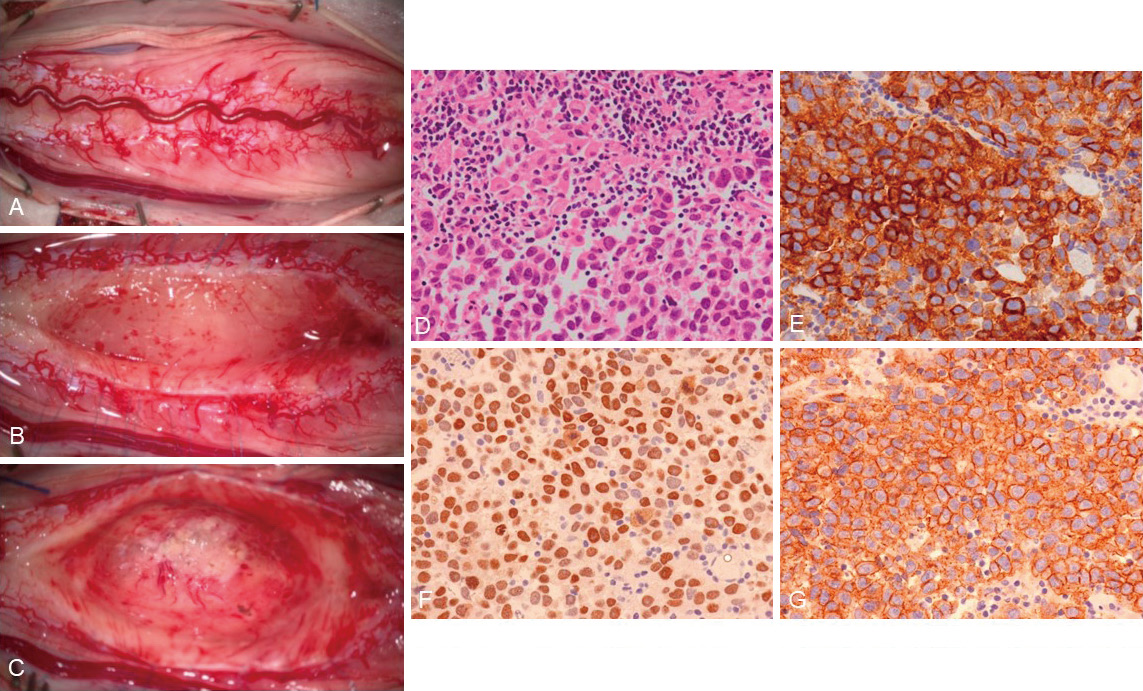
    We performed surgical treatment for cerebellar metastasis of relatively rare small-cell neuroendocrine carcinoma (SCNC) of the urinary bladder. On preoperative imaging, the lesion was solitary, and the edema around the tumor was unremarkable; thus, other differential diagnoses besides a metastatic brain tumor were also considered preoperatively. Intraoperatively, the tumor was soft, and the circumference brain and boundary were indistinct and easily hemorrhagic. The tumor was grossly totally removed, and postoperative radiotherapy was added. The clinical symptoms of the patient were relieved, and he was discharged on foot. Thus far, relatively few reports have described surgical treatment of brain metastases of SCNC of the urinary bladder. We herein report a case of metastatic brain tumor due to SCNC of the urinary bladder that required surgical treatment, along with a review of the previous literature regarding its clinical features and the characteristics of intracranial lesions related to surgery, such as imaging and intraoperative findings.

  • Tatsuya HAGIOKA, Takeshi SHIMIZU, Shingo TOYOTA, Tomoaki MURAKAMI, Tak ...
    2023 Volume 10 Pages 241-245
    Published: December 31, 2023
    Released on J-STAGE: September 29, 2023
    JOURNAL OPEN ACCESS FULL-TEXT HTML

    A 54-year-old man with no medical history presented to our hospital with vomiting, left hemiplegia, and seizures. On arrival, he was experiencing generalized tonic-clonic seizures, which required him to be intubated and deeply sedated. Contrast-enhanced computed tomography revealed extensive venous sinus obstruction from the superior sagittal sinus to the bilateral sigmoid sinus and cerebral edema with intracranial hemorrhage. An intracranial pressure (ICP) monitor was immediately placed intracranially, and mechanical thrombectomy (MT) was performed under ICP monitoring. MT was immediately terminated when the venous sinus was partially recanalized enough to decrease the ICP; then, anticoagulation therapy was initiated. Postoperative follow-up angiography revealed that venous sinus obstruction and intracranial venous perfusion improved over time. Although he had intracranial hemorrhage-induced left hemiplegia and sensory deficits, his condition improved with rehabilitation, and the patient was eventually discharged home. The indication criteria and techniques for MT for cerebral venous sinus thrombosis are yet to be established. As in this case, in patients with impaired consciousness due to intracranial hemorrhage or epilepsy, preoperative ICP monitor placement is deemed useful to evaluate venous perfusion during MT and decide the treatment goal.

  • Joji INAMASU, Masashi FUJISAWA, Mizuto SATO
    2023 Volume 10 Pages 247-252
    Published: December 31, 2023
    Released on J-STAGE: September 29, 2023
    JOURNAL OPEN ACCESS FULL-TEXT HTML

    In this study, we report on a previously healthy 44-year-old man who underwent an open biopsy under general anesthesia for a tumorous lesion found in his left frontal lobe via a small supratentorial craniotomy. While both postoperative course and brain computed tomography (CT) scans had been considered unremarkable, the patient became stuporous on postoperative day (POD) 4. A brain CT obtained on that day showed a subdural hematoma with marked brain shift which we thought might have been due to postoperative bleeding; he was immediately brought to an operating theater for hematoma removal. However, no bleeding source was found, and the brain remained depressed after hematoma evacuation. Furthermore, the brain shift remained unchanged on postoperative CT. While spontaneous intracranial hypotension (SIH) was considered, imaging studies to search for possible cerebrospinal fluid (CSF) leakage in the spinal column were not performed as the patient's condition has improved. However, he became stuporous again on POD 8, which urged us to perform CT myelogram. The CT myelogram showed a massive CSF leakage at the L1-L2 level. Subsequent autologous blood patch has successfully terminated the CSF leakage, and he became fully oriented shortly after the blood patch therapy. Thus, it should be noted that SIH may occur during postoperative period of intracranial surgery, and it may manifest radiographically as a subdural hematoma indistinguishable from postoperative bleeding. SIH should also be included in a differential diagnosis of postoperative headache, regardless of its characteristics, because headache associated with SIH may not always be orthostatic.

  • Mariya HOKAZONO, Takafumi SHIMOGAWA, Akira NAKAMIZO, Koji YOSHIMOTO
    2023 Volume 10 Pages 253-257
    Published: December 31, 2023
    Released on J-STAGE: September 29, 2023
    JOURNAL OPEN ACCESS FULL-TEXT HTML

    W report the first case of hemifacial spasm (HFS) caused by vascular compression of the anterior inferior cerebellar artery (AICA) -posterior inferior cerebellar artery (PICA) common trunk anomaly at the cisternal portion of cranial nerve VII (CN VII). A 71-year-old female with a typical right HFS was admitted to our hospital. As per her magnetic resonance (MR) imaging results, no offending arteries were noted around the CN VII root exit zone (REZ). Computed tomography angiography revealed an AICA-PICA common trunk anomaly with a dominant PICA, with the rostral branch of the AICA-PICA common trunk anomaly compressing the CN VII at the cisternal portion. The patient underwent microvascular decompression (MVD), and the HFS disappeared after surgery. The amplitude of the abnormal muscle responses (AMR) disappeared immediately after complete transposition of the offending artery. However, the patient experienced mild transient facial palsy 3 days after MVD which was eventually resolved with the administration of vitamin B12. No HFS recurrence was observed during the 1-year follow-up period. The AICA-PICA common trunk anomaly has been found to cause HFS as it compressed the CN VII at the cisternal portion, and not at the REZ. AMR monitoring might be helpful for cases where the unusual vessel particularly compresses the CN VII.

  • Yoshiyuki MATSUMOTO, Ryuta NAKAE, Fumihiro MATANO, Asami KUBOTA, Akio ...
    2023 Volume 10 Pages 259-263
    Published: December 31, 2023
    Released on J-STAGE: September 29, 2023
    JOURNAL OPEN ACCESS FULL-TEXT HTML

    Ruptured cerebral aneurysms that occur in the anterior wall of the internal carotid artery (ICA) are known as blood blister-like aneurysms (BBAs); they have been reported to account for 0.3% to 1% of all ruptured ICA aneurysms. In this report, we describe the treatment of an unusual traumatic BBA (tBBA) with high-flow bypass using a radial artery graft, which resulted in a favorable outcome. A 59-year-old female suffered from an acute epidural hematoma, traumatic subarachnoid hemorrhage, and traumatic carotid-cavernous sinus fistula (tCCF) after being involved in a motor vehicle accident. Her angiography results showed tCCF and a tBBA on the anterior wall of the right ICA. On the fourth day after injury, we found rebleeding from the tBBA and performed an emergency high-flow bypass using a radial artery graft with lesion trapping as a curative procedure for the tCCF and tBBA. Postoperatively, right abducens nerve palsy appeared, but no other neurological symptoms were noted; the patient was thereafter transferred to a rehabilitation hospital 49 days after injury. Traumatic ICA aneurysms commonly occur close to the anterior clinoid process, form within 1 to 2 weeks of injury, and often rupture around 2 weeks after trauma. This case was considered rare as the ICA was likely injured and bleeding at the time of injury, resulting in a form of tBBA; this allowed early detection and appropriate treatment that resulted in a good outcome.

  • Kentaro CHIBA, Yasuo AIHARA, Yuichi ODA, Kenta MASUI, Takashi KOMORI, ...
    2023 Volume 10 Pages 265-271
    Published: December 31, 2023
    Released on J-STAGE: October 14, 2023
    JOURNAL OPEN ACCESS FULL-TEXT HTML

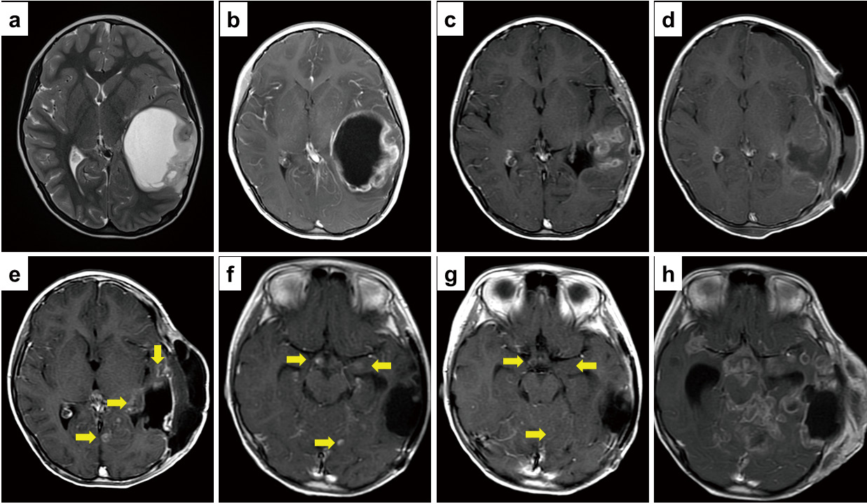
    Extracranial brain tumor metastases are extremely rare. The etiology, pathophysiology, and clinical progression of systemic metastatic brain cancer remain to be elucidated. We encountered a case of pediatric diffuse high-grade astrocytoma in a four-year-old girl with subcutaneous and lymph node metastases. Numerous metastatic lesions emerged, progressed rapidly, and were difficult to manage despite temozolomide (TMZ) administration. The patient underwent repeated surgical resection for these lesions. Conversely, the primary intracranial lesions responded well to TMZ for some time. However, the patient died 15 months after the initial diagnosis. Extracranial metastasis and highly varying effects of chemotherapy were the characteristic clinical features in this case. Our analysis did not reveal definitive histopathological and molecular factors contributing to this presentation. The lack of notable molecular pathological features illustrates the unpredictability of glioma metastasis, and the treatment for extracranial metastasis remains unknown. A gene panel analysis revealed several genetic aberrations, including PDGFRA, PIK3CA, and NBN mutations. As it is impossible to resect all frequently and rapidly progressing lesions, we stress that the prognosis of metastatic brain tumors is undoubtedly poor if these tumors are refractory to existing treatments, including chemotherapy.

  • Nobuyuki MITSUI, Manabu KINOSHITA, Junji NAKAZAWA, Hirokazu OZAKI, Ter ...
    2023 Volume 10 Pages 279-283
    Published: December 31, 2023
    Released on J-STAGE: October 14, 2023
    JOURNAL OPEN ACCESS FULL-TEXT HTML

    Endovascular treatment is a standard procedure for subclavian artery stenosis or obstruction. However, great care should be taken to avoid embolic complications to the vertebral artery, and several methods have previously been reported. Hence, as surgical procedures become increasingly complicated, unintended issues may arise during treatment. Here, the authors report a case where the filter-type protection device was caught in the stent because the patient moved during treatment, leading to open surgery to recover the filter-type protection device.

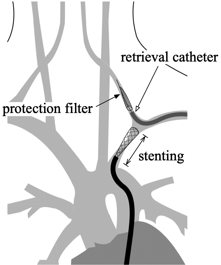
    A 78-year-old female suffering from a left subclavian steal syndrome underwent stenting due to subclavian artery stenosis. The stenotic lesion was approached via the transfemoral route, and a filter-type protection device was advanced to the vertebral artery via the transbrachial route to prevent embolic complications. As the procedure was performed under local anesthesia, the patient moved during stent deployment proximally to the left vertebral artery origin, and the stent unintentionally advanced distally, covering the vertebral artery and obstructing the retrieval catheter for the filter-type protection device to advance. Failed attempts in recovering the filter-type protection device required open surgery for retrieval. Fortunately, there was no postoperative neurological and radiographic complication, ameliorating her chief complaint.

    The retriever catheter for the protection device should be advanced beyond the vertebral artery orifice just proximal to the protection device before stenting to avoid such complications while also thoroughly considering the type of anesthesia during treatment.

  • Yuichi ODA, Kosaku AMANO, Kentaro CHIBA, Kenta MASUI, Takakazu KAWAMAT ...
    2023 Volume 10 Pages 285-289
    Published: December 31, 2023
    Released on J-STAGE: October 14, 2023
    JOURNAL OPEN ACCESS FULL-TEXT HTML

    As per the 2021 World Health Organization (WHO) Classification of Tumors of the Central Nervous System, chordoid glioma (CG) is defined as a slow-growing glial neoplasm categorized as grade II tumor. This tumor is primarily located in the anterior part of the third ventricle, often adheres to important surrounding structures, and is hemorrhagic in nature. Therefore, dissecting this tumor is extremely difficult. In this study, we present the case of a 44-year-old man who initially complained of mild headache and was diagnosed with a homogeneous gadolinium-enhanced lesion in the third ventricle via magnetic resonance imaging. The pathological diagnosis based on his biopsy at the previous hospital was CG. The patient demonstrated no neurological deficit at that time, but the tumor had gradually grown, hydrocephalus appeared 2 years after the tumor was detected, and the patient developed short memory disorder and daytime sleepiness. We resected the tumor via the anterior interhemispheric trans-lamina terminalis approach using a microscope and an endoscope. The residual tumor at the blind spot of the microscopic view was resected under an angled rigid endoscopic view using dedicated tools for transsphenoidal surgery. The tumor was grossly resected, and the histopathological diagnosis was CG. Postoperative neurological findings included slight memory disorder and hypothalamic adrenal dysfunction. No tumor recurrence was reported 3 years post resection. The endoscope-assisted anterior interhemispheric trans-lamina terminalis approach was determined useful for CG resection with minimal surgical complications and without tumor recurrence.

  • Hiroki KARITA, Kyoji TSUDA, Maya KONO, Tetsuya YAMAMOTO, Satoshi IHARA
    2023 Volume 10 Pages 291-297
    Published: December 31, 2023
    Released on J-STAGE: October 14, 2023
    JOURNAL OPEN ACCESS FULL-TEXT HTML

    Direct surgical resection remains to be the standard treatment for tuberous sclerosis complex (TSC) with subependymal giant cell astrocytoma (SEGA). Medical therapy with everolimus (mammalian target of rapamycin inhibitor or mTOR) serves as a second-line treatment for patients with SEGA who are determined to be ineligible for surgical resection. Some recent studies have reported that neoadjuvant therapy for SEGA may be a useful, novel treatment.

    In this study, we herein present a case of SEGA and demonstrate the efficacy of preoperative everolimus therapy. We have also examined the utility and safety of neoadjuvant therapy for SEGA and investigated four previously reported cases of preoperative administration of mTOR inhibitors. In these cases, everolimus was administered preoperatively to shrink the tumor although the duration of treatment varied. Afterward, gross total tumor removal was conducted in all the cases. No postoperative complications were reported during the follow-up period. These findings indicate that neoadjuvant therapy with an mTOR inhibitor can be a potential treatment for SEGA. The findings of this present study also suggested that a short administration period of about 2 months may be sufficient to achieve preoperative tumor reduction.

  • Yuhei ITO, Takao KOJIMA, Takuya MAEDA, Masazumi FUJII
    2023 Volume 10 Pages 299-302
    Published: December 31, 2023
    Released on J-STAGE: October 14, 2023
    JOURNAL OPEN ACCESS FULL-TEXT HTML

    Le Fort I osteotomy is a common surgical technique for correcting jaw deformities. Although it is generally a safe procedure, there have been cases of postoperative vascular complications, which include arteriovenous fistulas. Published reports describe the development of arteriovenous fistulas that originate from the maxillary artery after this procedure; nevertheless, no cases of arteriovenous fistulas arising from the middle meningeal artery (MMA) have been reported. We present the case of a 19-year-old woman who developed pulsatile tinnitus immediately following a Le Fort I osteotomy. Angiography with proximal flow control of the external carotid artery showed an arteriovenous fistula between the extracranial portion of the MMA and the pterygoid venous plexus. The fistula was successfully treated using coil embolization, preserving the maxillary artery, which results in tinnitus resolution. In this report, the effectiveness of endovascular treatment for arteriovenous fistulas of the MMA that occur following a Le Fort I osteotomy was demonstrated. Angiography with proximal flow interruption was effective in shunt location detection.

  • Sae YAMANAKA, Hiroshi TOKIMURA, Nayuta HIGA, Hirofumi IWAMOTO, Yosuke ...
    2023 Volume 10 Pages 303-308
    Published: December 31, 2023
    Released on J-STAGE: October 14, 2023
    JOURNAL OPEN ACCESS FULL-TEXT HTML

    Hemorrhagic pilocytic astrocytomas (PAs) are rare, accounting for 1.1%-8.0% of all PA cases. They are reported to occur more frequently in older populations, with a male predominance. In this study, we report a case of a 14-year-old boy who presented with a headache, vertigo, and diplopia. As per his brain computed tomography scan, a small hematoma was observed in the left inferior cerebellar peduncle. Follow-up magnetic resonance imaging (MRI) revealed repeated minor bleeding from the lesion and mild expansion, with no neurological deficits. Four years later, the patient developed nausea, vomiting, and left abducens palsy. MRI revealed a mulberry-shaped mass surrounded by a hypointense rim, suggesting a cavernous angioma. The lesion was surgically resected via midline occipital craniotomy with the opening of the cerebellomedullary fissure. Histopathological examination of the lesion revealed PA. Next-generation sequencing analyses revealed that PAs harbored mutations in the ARID1A, ATM, and POLE genes but not in the BRAF gene. To the best of our knowledge, there are yet no reported studies on these mutations in PAs to date. Thus, PA should be considered in the differential diagnosis of cerebellar hemorrhage, especially in young adults and children.

  • Kyosuke MATSUNAGA, Shinjiro FUKAMI, Nobuyuki NAKAJIMA, Norio ICHIMASU, ...
    2023 Volume 10 Pages 309-314
    Published: December 31, 2023
    Released on J-STAGE: November 11, 2023
    JOURNAL OPEN ACCESS FULL-TEXT HTML

    Diffuse midline glioma (DMG), H3 K27M-altered, is a tumor with a poor prognosis mainly found in children. An adolescent patient presented with thalamic hemorrhage, which initially could not be diagnosed as DMG by pathological analysis. A neoplasm in the lateral ventricle close to the previous thalamic hemorrhagic lesion was detected 12 months after the hemorrhage. Thus, endoscopic resection was performed, and a diagnosis was made. Gene expression profiling demonstrated mutation in genes, such as H3F3A and FGFR1. FGFR1 mutation was associated with intratumoral hemorrhage in low-grade gliomas and contributed to longer survival than wild-type FGFR1 in DMG H3K27M. Our findings suggest that patients with DMG, H3 K27-altered, with FGFR1 mutation may be predisposed to intratumoral hemorrhaging and/or have a longer survival time than patients without FGFR1 mutation.

  • Kenta NAKASE, Ryosuke MATSUDA, Tomoya OKAMOTO, Ichiro NAKAGAWA, Maiko ...
    2023 Volume 10 Pages 315-320
    Published: December 31, 2023
    Released on J-STAGE: November 11, 2023
    JOURNAL OPEN ACCESS FULL-TEXT HTML

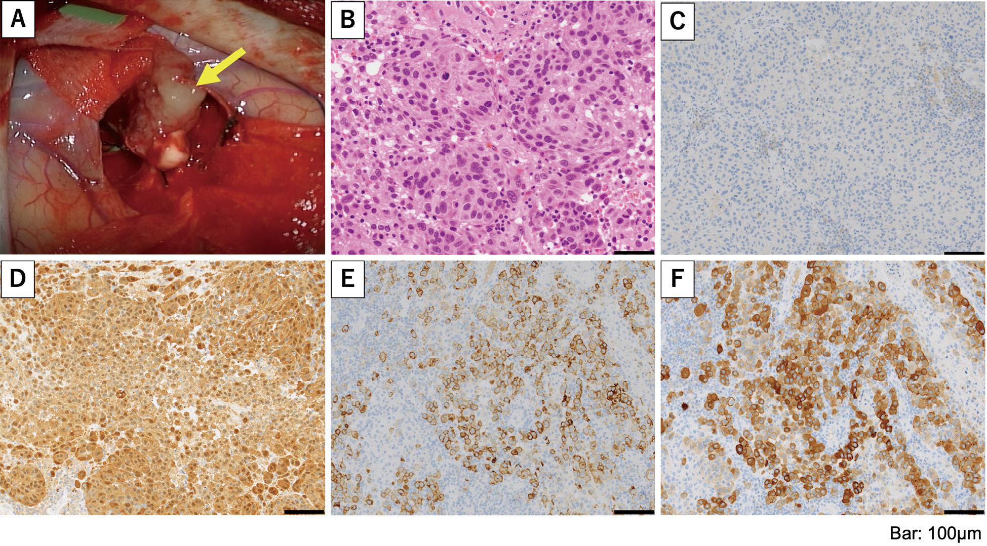
    Meningeal melanocytomas of the central nervous system, although typically benign, rarely undergo malignant transformations. A 46-year-old man presented with headache and nausea 4 years after gross total resection of a craniovertebral junction meningeal melanocytoma at another hospital. The initial clinical course was previously reported.1) Computed tomography revealed the presence of multiple intracranial mass lesions. Furthermore, magnetic resonance imaging showed multiple intracranial lesions and meningeal dissemination. A biopsy was performed for a circumflex lesion located in the right frontal lobe. Pathological examination showed anaplastic changes and a Ki-67 index of 33%. Based on the pleomorphic changes and high mitotic activity, the patient was diagnosed with primary cerebral malignant melanoma. The patient received four cycles of nivolumab (80 mg) and ipilimumab (165 mg), followed by whole-brain radiotherapy (37.5 Gy). However, the disease progressed after the third cycle. Genome analysis revealed GNAQ Q209P and SF3B1 R625C mutations, but no treatments related to these gene mutations were available. Despite the seven cycles of nivolumab therapy, the patient eventually passed away 9 months after surgery. This case was a rare example of malignant transformation and leptomeningeal melanomatosis in a meningeal melanocytoma. It highlights the importance of careful follow up after gross total resection. Identification of molecular alterations can lead to better detection of melanocytic melanomas with poor prognosis and high risk of recurrence and metastasis. It can also facilitate the development of novel therapeutic options for these patients.

feedback
Top