消化器内視鏡の進歩:Progress of Digestive Endoscopy
Online ISSN : 2189-0021
Print ISSN : 0389-9403
46 巻
選択された号の論文の65件中51~65を表示しています
症例
  • 中村 穣, 日高 道生, 小林 智子, 木幡 義彰, 渡辺 浩一, 井川 守仁, 須藤 一郎, 田口 夕美子, 窪田 良彦, 勝亦 重弘, ...
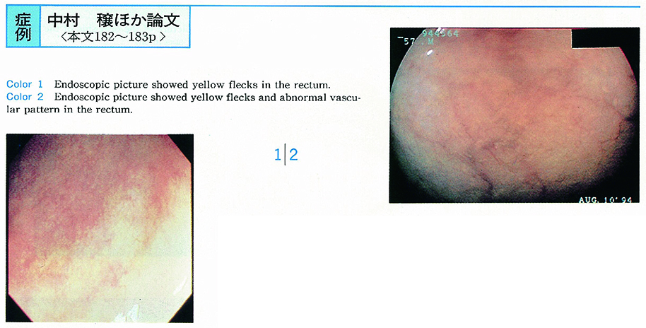
    1995 年 46 巻 p. 182-183
    発行日: 1995/06/16
    公開日: 2015/05/01
    ジャーナル フリー
    We experienced two case of Schistosomiasis Japonica showing interesting colonoscopic findings.
    Case 1 was a 63-year-old female. Her chief complaint was right hypochondralgia and she was suspected Schistosomiasis Japonica infection by characteristic liver echogram. Colonoscopic examination showed yellow flecks in the ileocecal region, sigmoid colon and mainly in the rectum. Biopsy from yellow fleck of the rectum proved Schistosomal ova in submucosal layer.
    Case 2 was a 57-year-old male. He was examined barium enema because of positive occult blood test and suspected the polyp in the sigmoid colon. Colonoscopy revealed faded mucosa with yellow flecks in the rectum with loss of vascular pattern. Schistosomal ova were seen from yellow fleck mucosal biopsy.
    We stress these yellow fleck is important finding on colonoscopy suggesting the ova of Schistosomiasis Japonica.
  • 中川 義也, 柴田 喜明, 辰口 篤志, 佐藤 順, 伊月 葉子, 藤森 俊二, 丸山 正明, 山門 進, 田口 文彦, 岸田 輝幸, 吉田 ...
    1995 年 46 巻 p. 184-185
    発行日: 1995/06/16
    公開日: 2015/05/01
    ジャーナル フリー
    A 55-year-old man was admitted to our hospital accompanied by massive bloody stool. Four years ago the patient was attacked by cerebral infarction, and has been treated with ticlopidine (100mg/day) . We could not find the souce of the hemorrhage by the upper gastrointestinal endoscopy. Colonoscopic finding was a Borrmann 1 type advanced cancer in the ileocecal region. The tumor was histologically diagnosed as carcinonid (mixed type) and the patient underwent an operation of the right half colon resection.
    The tumor was 4cm×3.5cm in size, with relatively smooth surface. Tumor cells infiltrated to the serosa. Lymph node metastasis were extended to N2. HE stain showed nodular solid nests, partially with trabecular and rosette-like structure. Grimelius arglophil reaction and Fontana-Masson argentaffin reaction were both positive in the tumor.
    Ileocecal carcinoid is very rare in Japan, compared with western countries and USA. Our review of the literature revealed only 13 cases of ileocecal carcinoid except our case in Japan. Our report was a rare case of cecal carcinoid that seemed to cause massive bleeding due to ticlopidine.
  • 尾石 樹泰, 山田 弥生, 小泉 浩一, 甲斐 俊吉, 川村 紀夫, 山口 俊和, 川上 眞寿弘, 橋本 国男, 丸山 雅一, 尾形 悦郎, ...
    1995 年 46 巻 p. 186-187
    発行日: 1995/06/16
    公開日: 2015/05/01
    ジャーナル フリー
    A 77-year-old woman visited our hospital for the post operative surveillance of the ascending colon cancer, in March 1992. She had no symptom, but multiple polypoid lesions like polyposis which were present from the anastomosis site to the splenic flexure were visualized by colonoscopy on March 1992. The polyposis-like lesions were also demonstrated by barium enema, and were considered pneumatosis cystoides intestinalis. The patient was observed for 2 months without treatment, resulting in no improvement. In June 1992 she was admitted to our institution for oxygen therapy with nasal cannula (2l/min×9hr×6days, 5l/min×12hr×15days) . This treatment was resulted in remission.
    The oxygen therapy with nasal cannula is considered to be very effective for the pneumatosis cystoides intestinalis.
  • 斉田 芳久, 長尾 二郎, 武田 明芳, 高瀬 真, 炭山 嘉伸, 剛崎 寛徳
    1995 年 46 巻 p. 188-189
    発行日: 1995/06/16
    公開日: 2015/05/01
    ジャーナル フリー
    We have experienced a post operative case with frequent attacks of abdominal pain resistant to conservative treatment and were able to treat successfully with laparoscopic adhesiolysis.
    The patient was a 71-year-old male, underwent the right hemicolectomy and sigmoidectomy for double colon cancer on May 1992. While receiving follow up care by a private physician, the patient experienced frequent attacks of abdominal pain. Because the pain was not improved by oral medication for intestinal control, the patient took habitual analgesic injections. The patient was referred to our hospital in September 1994, when his condition was evaluated as being beyond conservative medical treatment. On September 21, 1994, laparoscopic adhesiolysis was performed. The surgery lasted 55 minutes. The patient was observed passing gas immediately after the surgery and was able to eat on the first day postoperatively. The patient was discharged on the sixth day following satisfactory progress without analgesics.
    Laparoscopic adhesiolysis is a useful surgical method, as it is technically easier than laparotomy and might prevent new adhesion and postoperative pain that laparotomy may cause.
  • 佃 博, 横田 敏弘, 横山 正, 小野 裕之, 斉藤 大三, 近藤 仁, 白尾 国昭, 山口 肇, 小黒 八七郎, 石川 勉, 牛尾 恭輔 ...
    1995 年 46 巻 p. 190-191
    発行日: 1995/06/16
    公開日: 2015/05/01
    ジャーナル フリー
    A 78-year-old female was admitted to our hospital due to constipation and positive fecal occult blood test. Colonoscopy revealed four large sessile polyps with typical lobulated surface configuration at the cecum (20mm in size) , the ascending (20mm in size) , the right side transverse (20mm in size) and the left side transverse colon (30mm in size) , respectively. All tumors were removed using endoscopic mucosal resections. Histopathology of 4 tumors revealed focal adenocarcinoma in adenoma. In our present study, the incidence of case with multiple synchronous such tumors was only 4% (6/149) .
  • 高井 智子, 小林 清典, 山田 伸夫, 福田 淳, 長谷川 章雄
    1995 年 46 巻 p. 192-193
    発行日: 1995/06/16
    公開日: 2015/05/01
    ジャーナル フリー
    A very rare case of primary signet-ring cell carcinoma at an early stage of the colon is reported.
    A 77-year-old male visited to our hospital because of immunologically positive fecal occult blood test. Colonoscopic examination revealed a flat elevated lesion with central depressed area at the hepatic flexure. Biopsied specimen demonstrated signet-ring cell carcinoma, and right hemicolectomy was undergone. Resected material revealed a IIa+IIc like lesion, measuring 20 mm in diameter. Histologically, signet-ring cell carcinoma diffusely infiltrated into the deeper part of submucosal layer and invaded to the lymphatic vesseles. However, neither lymph node nor distant metastasis was observed. He is still alive in good health 2 years after the operation.
  • 横山 正, 横田 敏弘, 斉藤 大三, 白尾 國昭, 近藤 仁, 山口 肇, 杉原 健一, 姜 建宇, 赤須 孝之, 森谷 冝晧, 石川 勉 ...
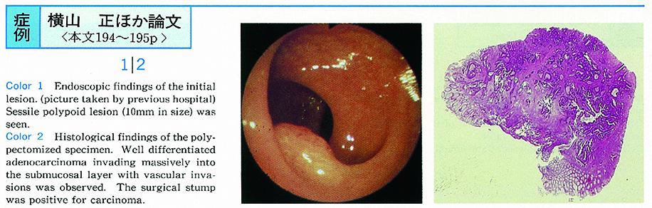
    1995 年 46 巻 p. 194-195
    発行日: 1995/06/16
    公開日: 2015/05/01
    ジャーナル フリー
    The patient was a 53-year-old woman. She received colonoscopic polypectomy 6.5 years ago for sigmoid colon polyp. The resected specimen revealed well differentiated adenocarcinoma invading massively into the submucosal layer with vascular vessel invasion. She was recommended for an additional surgical resection but she refused. She was followed up by barium enema and colonoscopy and serum CEA.
    No evidence of recurrence was detected until 6.5 years after the polypectomy, when she had bloody stool and barium enema revealed submucosal tumor-like protrusion with depression at the site of the previous lesion. Resection of the sigmoid colon was carried out successfully, and no lymph node metastasis was observed. The tumor was well differentiated adenocarcinoma invading to the serosal surface, mainly located in the submucosal layer, suggesting local recurrence of the tumor.
    This is the case having the longest period of local recurrence after polypectomy, by the review of the Japanese literature. The authors suggest that long term follow up would be necessary after polypectomy of colonic carcinoma invading into submucosal layer.
  • 山口 俊和, 出張 玲子, 小泉 浩一, 甲斐 俊吉, 川村 紀夫, 川上 眞寿弘, 橋本 国男, 丸山 雅一, 柳沢 昭夫, 加藤 洋
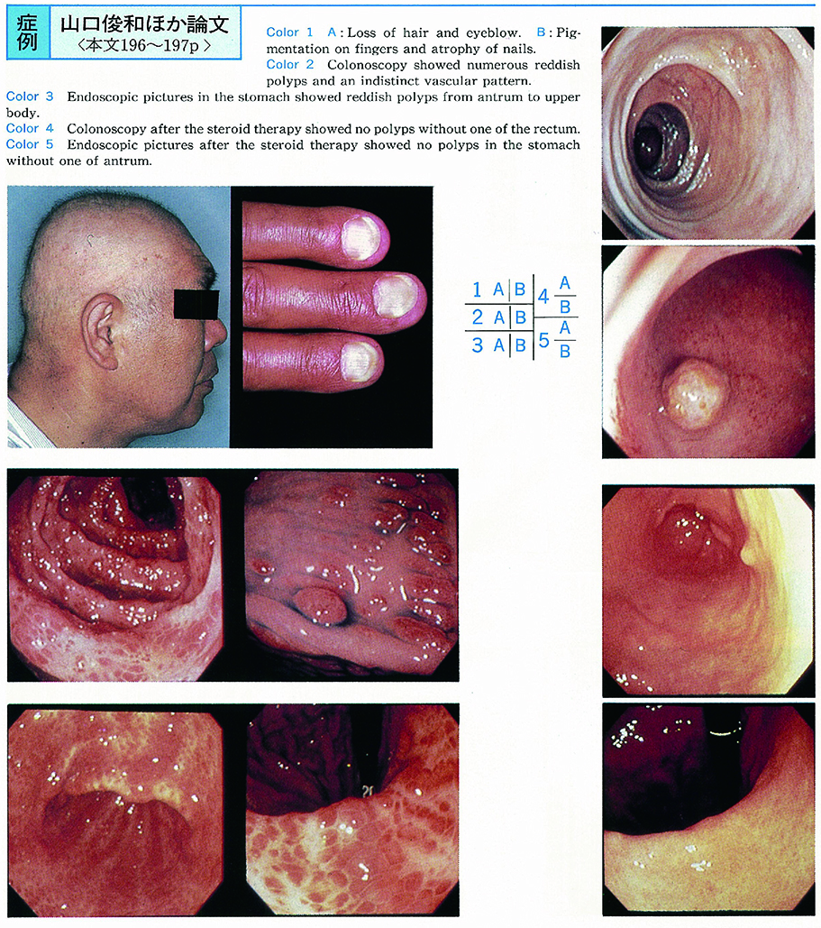
    1995 年 46 巻 p. 196-197
    発行日: 1995/06/16
    公開日: 2015/05/01
    ジャーナル フリー
    A 59-year-old male was admitted to the hospital complaining of lower abdominal pain, diarrhea of one month duration, and melena. In this case, numerous and uniform gastrointestinal polyposis, alopecia, onychotrophia, skin pigmentation, and mild hypoproteinemia were found. Histological examination of biopsy specimens from colonic polyps revealed cystic dilatation of the glans and edematous stroma. Cronkhite-Canada syndrome (CCS) was diagnosed.
    No especial changes of clinical manifestations were seen with conventional treatment (intra-venous hyperalimentation, t-AMCHA) , but diarrhea and polyposis improved remarkably with 10-days steroid therapy. Short-term steroid therapy may effective to CCS with uniform gastrointestinal polyposis, and one of its early phase.
  • 杉浦 敏昭, 渡 淳, 藤森 俊二, 田中 周, 星野 哲夫, 長尾 毅彦, 水野 杏一, 山門 進, 吉田 豊, 岸田 輝幸, 小林 正文
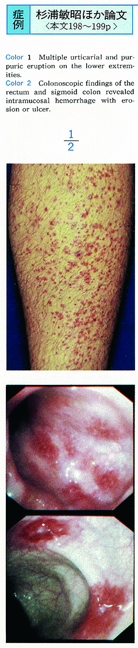
    1995 年 46 巻 p. 198-199
    発行日: 1995/06/16
    公開日: 2015/05/01
    ジャーナル フリー
    A 24-year-old man admitted to our hospital complaining abdominal pain. He suffered from melena and purpuric rash on his extremities on the second and third hospital days. Endoscopic examination of upper GI tract revealed multiple intramucosal hemorrhage in the duodenal bulb. Colonoscopic findings demonstrated intramucosal hemorrhage with erosion or ulcer through the colon. The biopsy specimens showed intramucosal hemorrhage and perivascullar cellular infiltrations. We diagnosed Schönlein-Henoch purpura from physical findings and examinations. He was treated by prednisolone 70mg/day. Abdominal pain and melena ceased in a few days.
  • 落合 保子, 今村 公美, 荻原 典和, 田中 直英, 椿 浩司, 林 洋一, 荒川 泰行
    1995 年 46 巻 p. 200-201
    発行日: 1995/06/16
    公開日: 2015/05/01
    ジャーナル フリー
    A 36-year-old male was found serum HCV antibody positive and was admitted to our university hospital for further examination. He was revealed hyperbilirubinemia with predominantly increase of the direct bilirubin on admission. An BSP excresion test revealed the increase again on 120 minutes. Laparoscopic findings showed the surface slight irregular and black patcthes. Lipofuscin granulation were found in hepatocyte.
  • 芝 祐信, 加賀谷 寿孝, 杉浦 克明, 熊 英治郎, 佐野 正明, 中濱 誠
    1995 年 46 巻 p. 202-203
    発行日: 1995/06/16
    公開日: 2015/05/01
    ジャーナル フリー
    We experienced two cases of hepatic cavernous hemangioma with whitish flat protrusion, which are confirmed on laparoscopy.
    Case 1 was a 69-year-old male. He was admitted to our hospital complaining of body weight loss. Abdominal ultrasonography showed hypoechoic mass in the liver. Enhanced CT scan of the liver showed pooling around the tumor and low density area near the surface of the liver. Laparoscopy revealed a hepatic cavernous hemangioma and whitish flat protrusion. The liver needle biopsy specimen showed myxomatous connective tissue.
    Case 2 was a 66-year-old female. She was admitted to our hospital complaining of pain in the right hypochondrium. Abdominal ultrasonography showed hyperechoic mass in the liver. Enhanced CT scan of the liver and angiography was useless for definite diagnosis. Laparoscopy revealed a hepatic cavernous hemangioma which grew extrahepatically out of the left lobe. This tumor was extirpated at surgical operation. Histological feature of the resected specimen showed a hepatic cavernous hemangioma. Histological findings of whitish flat protrusion showed fibromyxomatous tissue.
    On laparoscopy, whitish flat protrusion in two cases was similar to metastaic liver tumor, including adenocarcinoma. With great care, the difference between these would be known.
  • 佐藤 順, 山門 進, 辰口 篤志, 伊月 葉子, 藤森 俊二, 南 定, 立川 裕理, 田口 克司, 田口 文彦, 玉川 恭士, 岸田 輝 ...
    1995 年 46 巻 p. 204-205
    発行日: 1995/06/16
    公開日: 2015/05/01
    ジャーナル フリー
    A 46-year-old male with alcoholism admitted to our hospital for the rupture of esophageal varices. He was diagnosed alcoholic liver cirrhosis with esophageal varices (Cw, F2, Lm, RC (+) ) by laboratory, radiographic and endoscopic examinations. Colonoscopic findings revealed vascular ectasias, irregular vascular pattern, diffuse red spots, dilated veins and rectal varices, so we diagnosed portal hypertensive colopathy (PHC) .
    Transjugular intrahepatic portosystemic shunt (TIPS) was performed for esophageal varices with informed consent and the approval of Ethics Comittee in Nippon Medical School, portal venous pressure was decompressed from 35.3mmHg to 22.8mmHg (35.4%) . After TIPS therapy, esophageal varices was improved to its endoscopic finding with Cw, F1, RC (-) and colonoscopic findings of PHC was also improved.
    Portal hypertension is an important facter of etiology of PHC, we suggest that decompression of portal venous pressure induced the improvement of the vascular lesions in patient with PHC.
  • 山田 秀一, 宮藤 康則, 笹島 雅彦, 栗田 俊夫, 近藤 栄作, 武藤 ます江, 片山 雅彦, 蜂矢 朗彦, 瓜田 純久, 石原 学, ...
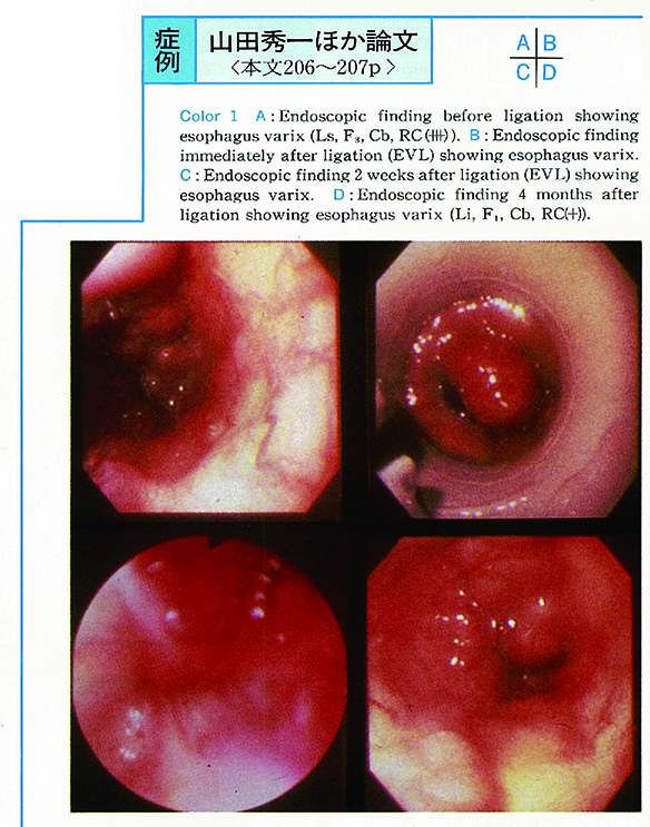
    1995 年 46 巻 p. 206-207
    発行日: 1995/06/16
    公開日: 2015/05/01
    ジャーナル フリー
    A male patient aged 3 years and 3 months had undergone surgery for biliary atresia at the age of 3 months after birth. He had hematemesis in July 1994. Under systemic anesthesia, endoscopy was performed : and a diagnosis of esophageal varices (F3, Ls, Cb, RC (⧻) ) was made. Endoscopic variceal ligation (EVL) was performed with five O-ring. Two O-rings were used for a thick varix in a direction of 0°, and one for each of varices in a direction of 3°, 6°, or 9°. Later, two more O-rings were additionally used. No marked ulcer or stenosis occurred. No hepatic dusfunction was manifested, either. EVL was believed to be very effective for esophago-gastric varices complication pediatric biliary atresia.
  • 神沢 輝実, 伊沢 友明, 江川 直人, 榊 信廣, 石渡 淳一
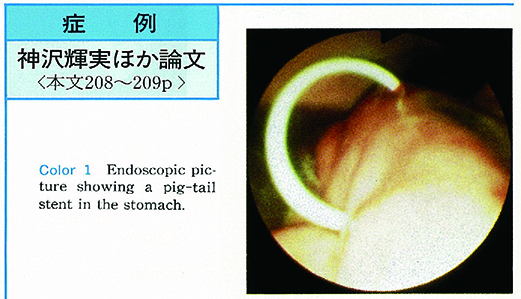
    1995 年 46 巻 p. 208-209
    発行日: 1995/06/16
    公開日: 2015/05/01
    ジャーナル フリー
    A 29-year-old female suffered from acute pancreatitis with unknown etiology. She recovered by conservative treatment, but a pseudocyst measuring 10 cm was formed in the tail of the pancreas. ERP showed no communication between the cyst and the main pancreatic duct, but the cyst increased in the course. As the patient was a young female and there was a possibility of the recurrence of acute pancreatitis, we performed endoscopic cystgastrostomy. The cyst disappeared in a few days following the procedure and the patient is well eight months after.
  • 坂本 仁, 秋田 泰, 田中 滋城, 仲又 進, 倉林 幹雄, 武田 方利, 野津 史彦, 新村 和平, 吉川 望海, 三田村 圭二, 上田 ...
    1995 年 46 巻 p. 210-211
    発行日: 1995/06/16
    公開日: 2015/05/01
    ジャーナル フリー
    A 70-year-old male who had been under observation for chronic alcoholic pancreatitis with pancreatolith and pancreatic cyst was admitted to our hospital because of epigastralgia. CT scan showed a pancreatolith in the head of the main pancreatic duct and multiple pancreatic cysts communicating with the dilated main pancreatic duct. A fistula was found in the duodenal bulb by endoscopy. Endoscopic fistulography revealed a pancreato-duodenal fistula and the dilated pancreatic duct containing mucus. Diagnosis of mucin producing adenocarcinoma was made by biopsied specimen. Pancreatectomy was performed and the resected specimen indicated that the pancreatolith was impacted in the head of the main pancreatic duct and the pancreato-duodenal fistula was formed in the body of the pancreas. The tumorous tissues were histopathologically characterized by papillary adenocarcinoma which were mainly presented in main and large pancreatic ducts and invaded into pancreatic parenchyma, duodenum and stomach.
feedback
Top