
51 巻
選択された号の論文の71件中1~50を表示しています
掲載論文カラー写真集
-
1998 年 51 巻 p. 2-20
発行日: 1998年
公開日: 2015/01/22
PDF形式でダウンロード (22778K)
内視鏡の器械と技術
-
1998 年 51 巻 p. 56-58
発行日: 1998/03/06
公開日: 2015/01/22
PDF形式でダウンロード (524K)
臨床研究
-
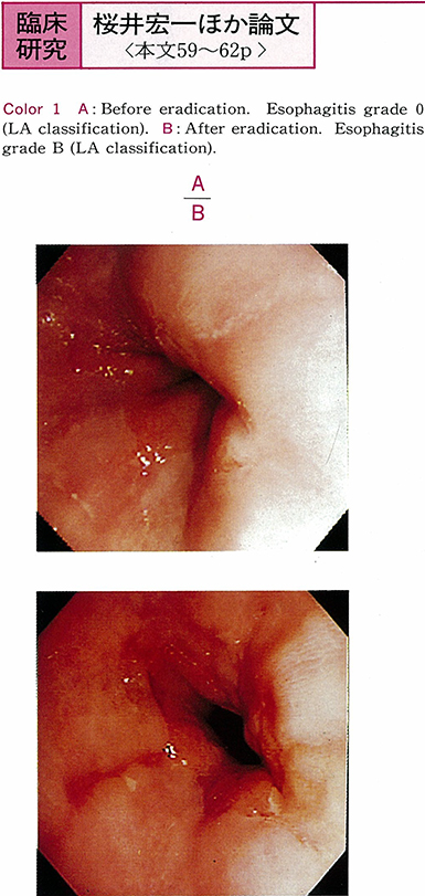
1998 年 51 巻 p. 59-62
発行日: 1998/03/06
公開日: 2015/01/22
 PDF形式でダウンロード (497K)
 -
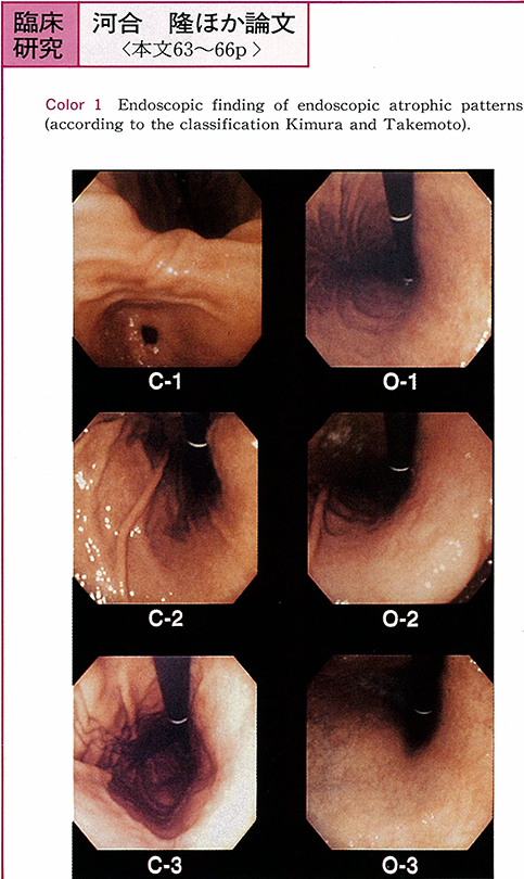
1998 年 51 巻 p. 63-66
発行日: 1998/03/06
公開日: 2015/01/22
 PDF形式でダウンロード (505K)
 -
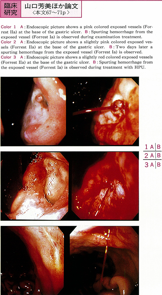
1998 年 51 巻 p. 67-71
発行日: 1998/03/06
公開日: 2015/01/22
 PDF形式でダウンロード (733K)
 -
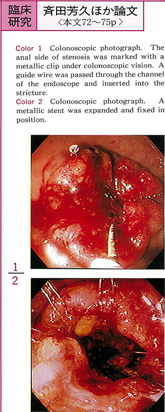
1998 年 51 巻 p. 72-75
発行日: 1998/03/06
公開日: 2015/01/22
 PDF形式でダウンロード (1120K)
 -
1998 年 51 巻 p. 76-80
発行日: 1998/03/06
公開日: 2015/01/22
PDF形式でダウンロード (1552K)
症例
-
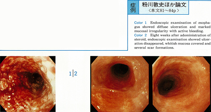
1998 年 51 巻 p. 81-84
発行日: 1998/03/06
公開日: 2015/01/22
 PDF形式でダウンロード (1803K)
 -
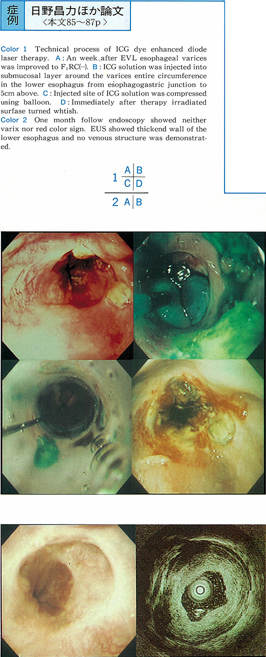
1998 年 51 巻 p. 85-87
発行日: 1998/03/06
公開日: 2015/01/22
 PDF形式でダウンロード (539K)
 -
1998 年 51 巻 p. 88-91
発行日: 1998/03/06
公開日: 2015/01/22
 PDF形式でダウンロード (793K)
 -
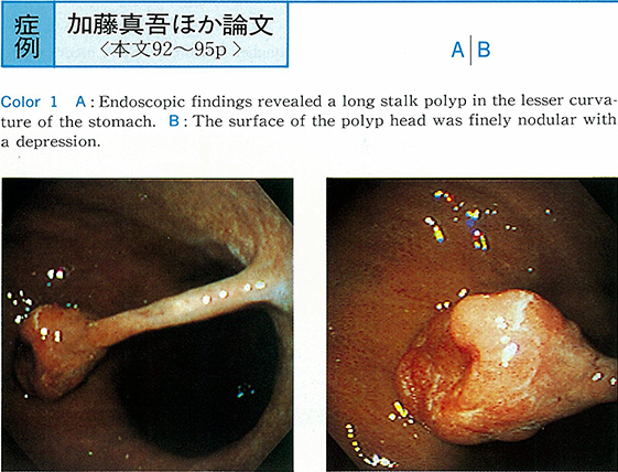
1998 年 51 巻 p. 92-95
発行日: 1998/03/06
公開日: 2015/01/22
 PDF形式でダウンロード (1404K)
 -
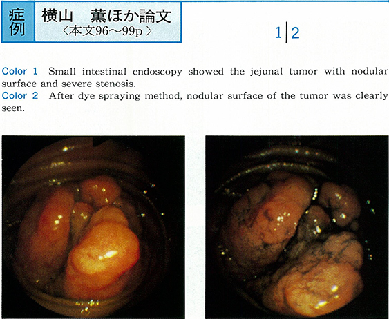
1998 年 51 巻 p. 96-99
発行日: 1998/03/06
公開日: 2015/01/22
 PDF形式でダウンロード (1636K)
 -
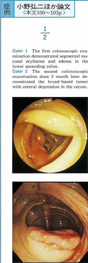
1998 年 51 巻 p. 100-103
発行日: 1998/03/06
公開日: 2015/01/22
 PDF形式でダウンロード (1758K)
 -
1998 年 51 巻 p. 104-107
発行日: 1998/03/06
公開日: 2015/01/22
 PDF形式でダウンロード (1064K)
 -
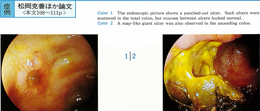
1998 年 51 巻 p. 108-111
発行日: 1998/03/06
公開日: 2015/01/22
 PDF形式でダウンロード (874K)
 -
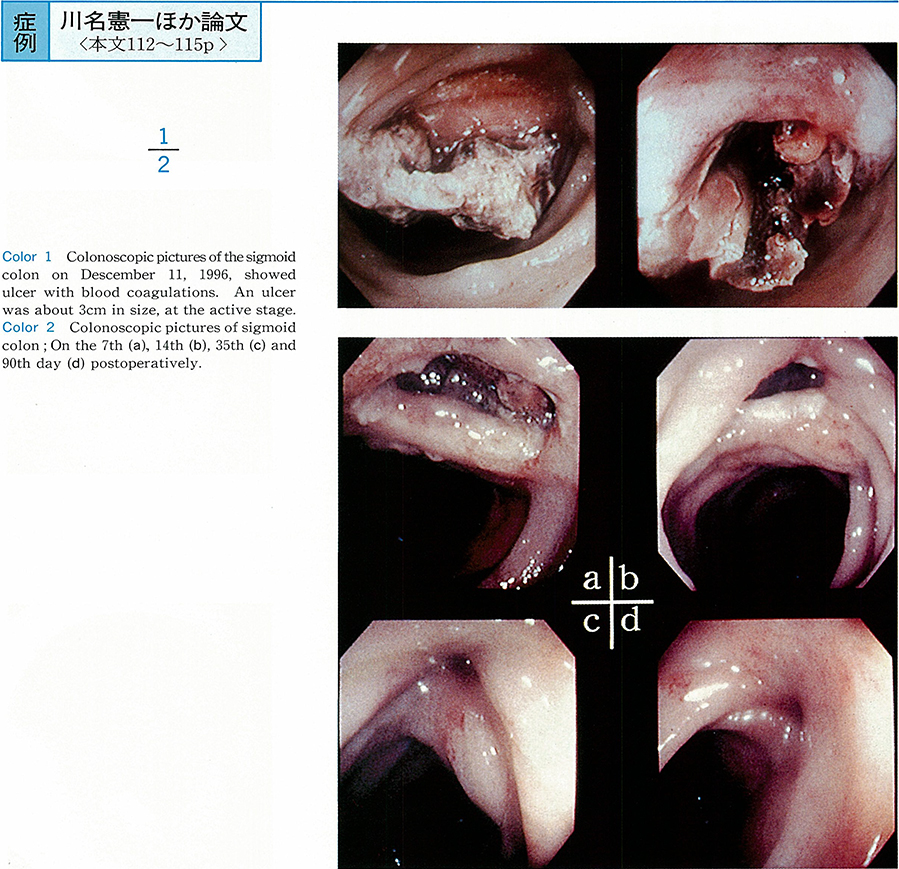
1998 年 51 巻 p. 112-115
発行日: 1998/03/06
公開日: 2015/01/22
 PDF形式でダウンロード (857K)
 -
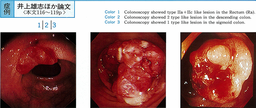
1998 年 51 巻 p. 116-119
発行日: 1998/03/06
公開日: 2015/01/22
 PDF形式でダウンロード (1202K)
 -
1998 年 51 巻 p. 120-123
発行日: 1998/03/06
公開日: 2015/01/22
 PDF形式でダウンロード (1147K)
 -
1998 年 51 巻 p. 124-127
発行日: 1998/03/06
公開日: 2015/01/22
 PDF形式でダウンロード (1857K)
 -
1998 年 51 巻 p. 128-132
発行日: 1998/03/06
公開日: 2015/01/22
PDF形式でダウンロード (2507K)
内視鏡の器械と技術
-
1998 年 51 巻 p. 134-135
発行日: 1998/03/06
公開日: 2015/01/22
 PDF形式でダウンロード (190K)
 -
1998 年 51 巻 p. 136-137
発行日: 1998/03/06
公開日: 2015/01/22
 PDF形式でダウンロード (681K)
 -
1998 年 51 巻 p. 138-139
発行日: 1998/03/06
公開日: 2015/01/22
PDF形式でダウンロード (581K)
臨床研究
-
1998 年 51 巻 p. 140-141
発行日: 1998/03/06
公開日: 2015/01/22
PDF形式でダウンロード (239K) -
1998 年 51 巻 p. 142-143
発行日: 1998/03/06
公開日: 2015/01/22
PDF形式でダウンロード (300K) -
1998 年 51 巻 p. 144-145
発行日: 1998/03/06
公開日: 2015/01/22
PDF形式でダウンロード (325K) -
1998 年 51 巻 p. 146-147
発行日: 1998/03/06
公開日: 2015/01/22
 PDF形式でダウンロード (448K)
 -
1998 年 51 巻 p. 148-149
発行日: 1998/03/06
公開日: 2015/01/22
 PDF形式でダウンロード (247K)
症例
-
1998 年 51 巻 p. 150-151
発行日: 1998/03/06
公開日: 2015/01/22
 PDF形式でダウンロード (270K)
 -
1998 年 51 巻 p. 152-153
発行日: 1998/03/06
公開日: 2015/01/22
 PDF形式でダウンロード (552K)
 -
1998 年 51 巻 p. 154-155
発行日: 1998/03/06
公開日: 2015/01/22
 PDF形式でダウンロード (1172K)
 -
1998 年 51 巻 p. 156-157
発行日: 1998/03/06
公開日: 2015/01/22
 PDF形式でダウンロード (990K)
 -
1998 年 51 巻 p. 158-159
発行日: 1998/03/06
公開日: 2015/01/22
 PDF形式でダウンロード (488K)
 -
1998 年 51 巻 p. 160-161
発行日: 1998/03/06
公開日: 2015/01/22
 PDF形式でダウンロード (184K)
 -
1998 年 51 巻 p. 162-163
発行日: 1998/03/06
公開日: 2015/01/22
 PDF形式でダウンロード (791K)
 -
1998 年 51 巻 p. 164-165
発行日: 1998/03/06
公開日: 2015/01/22
 PDF形式でダウンロード (213K)
 -
1998 年 51 巻 p. 166-167
発行日: 1998/03/06
公開日: 2015/01/22
 PDF形式でダウンロード (661K)
 -
1998 年 51 巻 p. 168-169
発行日: 1998/03/06
公開日: 2015/01/22
 PDF形式でダウンロード (522K)
 -
1998 年 51 巻 p. 170-171
発行日: 1998/03/06
公開日: 2015/01/22
 PDF形式でダウンロード (744K)
 -
1998 年 51 巻 p. 172-173
発行日: 1998/03/06
公開日: 2015/01/22
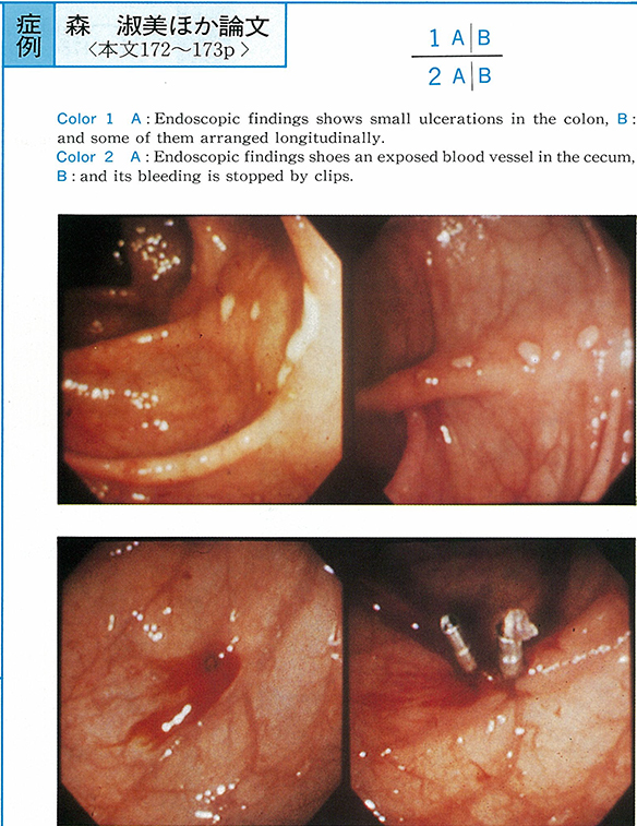 PDF形式でダウンロード (354K)
 -
1998 年 51 巻 p. 174-175
発行日: 1998/03/06
公開日: 2015/01/22
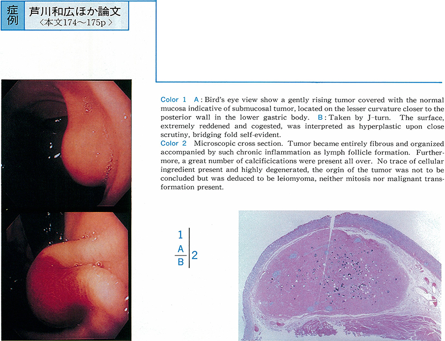 PDF形式でダウンロード (570K)
 -
1998 年 51 巻 p. 176-177
発行日: 1998/03/06
公開日: 2015/01/22
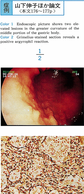 PDF形式でダウンロード (735K)
 -
1998 年 51 巻 p. 178-179
発行日: 1998/03/06
公開日: 2015/01/22
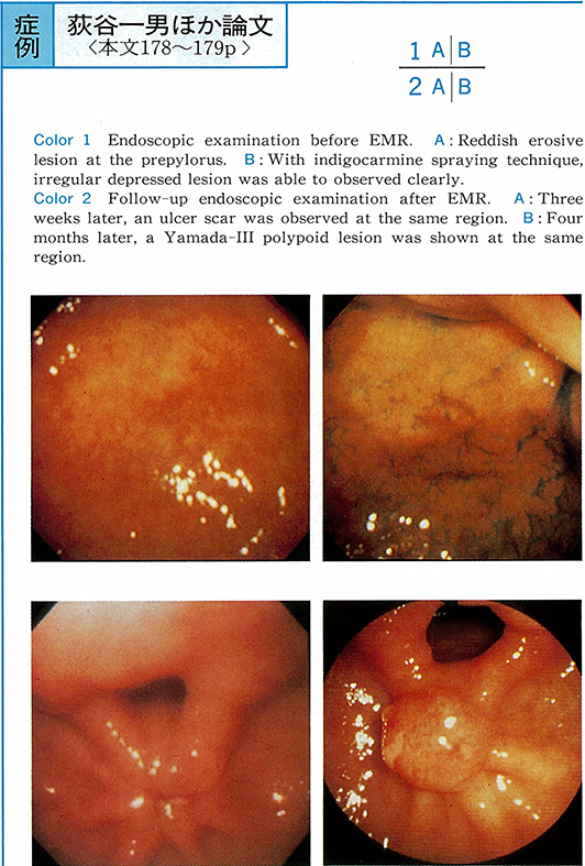 PDF形式でダウンロード (810K)
 -
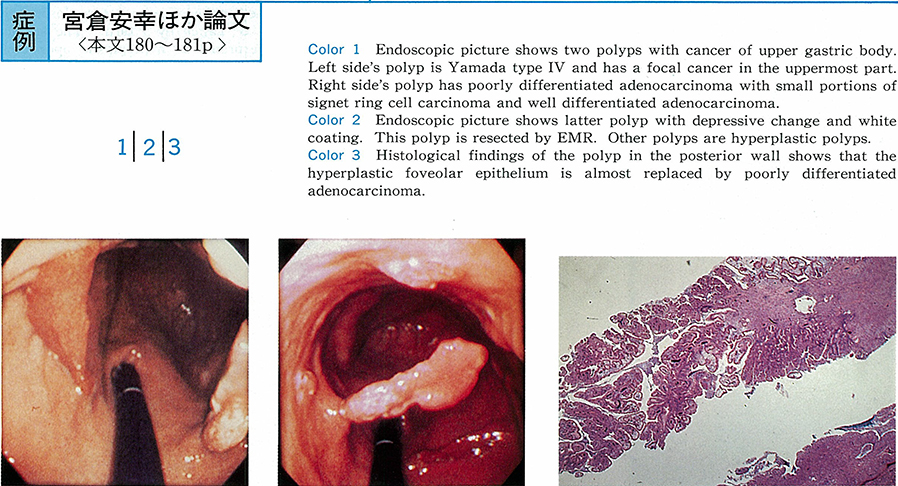
1998 年 51 巻 p. 180-181
発行日: 1998/03/06
公開日: 2015/01/22
 PDF形式でダウンロード (686K)
 -
1998 年 51 巻 p. 182-183
発行日: 1998/03/06
公開日: 2015/01/22
 PDF形式でダウンロード (850K)
 -
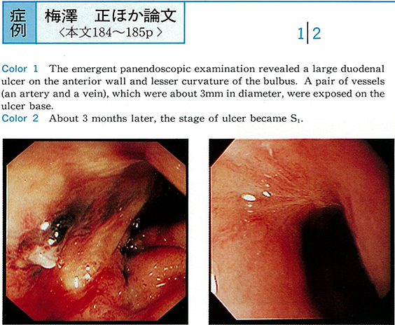
1998 年 51 巻 p. 184-185
発行日: 1998/03/06
公開日: 2015/01/22
 PDF形式でダウンロード (777K)
 -
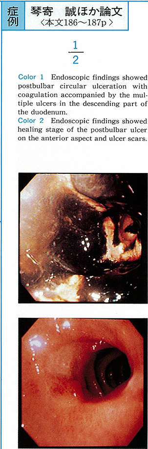
1998 年 51 巻 p. 186-187
発行日: 1998/03/06
公開日: 2015/01/22
 PDF形式でダウンロード (214K)
 -
1998 年 51 巻 p. 188-189
発行日: 1998/03/06
公開日: 2015/01/22
 PDF形式でダウンロード (382K)
 -
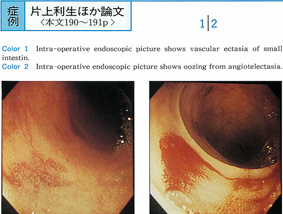
1998 年 51 巻 p. 190-191
発行日: 1998/03/06
公開日: 2015/01/22
 PDF形式でダウンロード (694K)
 -
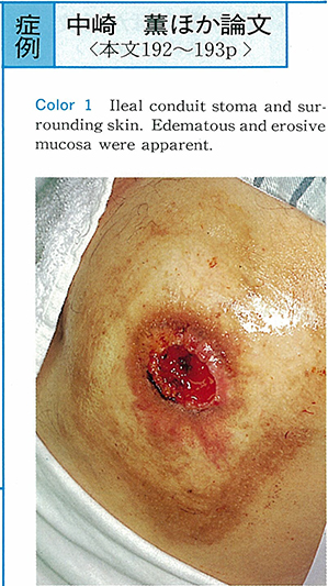
1998 年 51 巻 p. 192-193
発行日: 1998/03/06
公開日: 2015/01/22
 PDF形式でダウンロード (619K)