
45 巻
選択された号の論文の67件中1~50を表示しています
掲載論文カラー写真集
-
1994 年45 巻 p. 2-26
発行日: 1994年
公開日: 2015/05/25
PDF形式でダウンロード (49859K)
内視鏡の器械と技術
-
1994 年45 巻 p. 60-62
発行日: 1994/12/08
公開日: 2015/05/25
 PDF形式でダウンロード (720K)
 -
1994 年45 巻 p. 63-65
発行日: 1994/12/08
公開日: 2015/05/25
 PDF形式でダウンロード (745K)
 -
1994 年45 巻 p. 66-68
発行日: 1994/12/08
公開日: 2015/05/25
 PDF形式でダウンロード (1076K)
臨床研究
-
1994 年45 巻 p. 69-73
発行日: 1994/12/08
公開日: 2015/05/25
 PDF形式でダウンロード (1490K)
 -
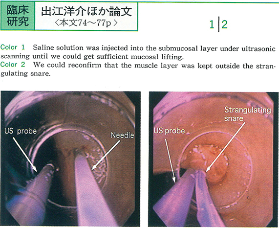
1994 年45 巻 p. 74-77
発行日: 1994/12/08
公開日: 2015/05/25
 PDF形式でダウンロード (1480K)
 -
1994 年45 巻 p. 78-82
発行日: 1994/12/08
公開日: 2015/05/25
 PDF形式でダウンロード (2127K)
 -
1994 年45 巻 p. 83-87
発行日: 1994/12/08
公開日: 2015/05/25
 PDF形式でダウンロード (1131K)
 -
1994 年45 巻 p. 88-92
発行日: 1994/12/08
公開日: 2015/05/25
 PDF形式でダウンロード (3024K)
 -
1994 年45 巻 p. 93-96
発行日: 1994/12/08
公開日: 2015/05/25
 PDF形式でダウンロード (725K)
 -
1994 年45 巻 p. 97-100
発行日: 1994/12/08
公開日: 2015/05/25
 PDF形式でダウンロード (671K)
 -
1994 年45 巻 p. 101-105
発行日: 1994/12/08
公開日: 2015/05/25
 PDF形式でダウンロード (1648K)
 -
1994 年45 巻 p. 106-109
発行日: 1994/12/08
公開日: 2015/05/25
 PDF形式でダウンロード (1144K)
症例
-
1994 年45 巻 p. 110-113
発行日: 1994/12/08
公開日: 2015/05/25
 PDF形式でダウンロード (1853K)
 -
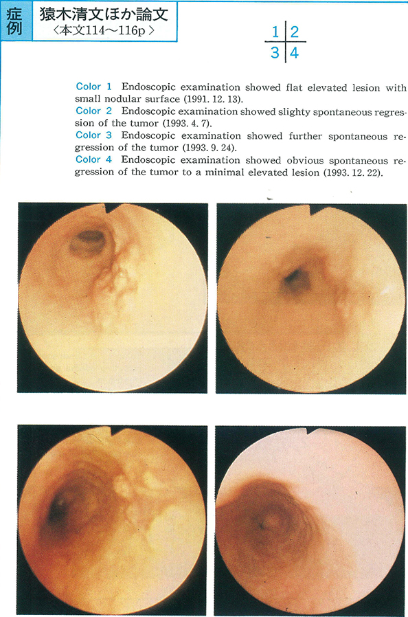
1994 年45 巻 p. 114-116
発行日: 1994/12/08
公開日: 2015/05/25
 PDF形式でダウンロード (1262K)
 -
1994 年45 巻 p. 117-120
発行日: 1994/12/08
公開日: 2015/05/25
 PDF形式でダウンロード (1934K)
 -
1994 年45 巻 p. 121-124
発行日: 1994/12/08
公開日: 2015/05/25
 PDF形式でダウンロード (2185K)
 -
1994 年45 巻 p. 125-129
発行日: 1994/12/08
公開日: 2015/05/25
 PDF形式でダウンロード (2662K)
 -
1994 年45 巻 p. 130-133
発行日: 1994/12/08
公開日: 2015/05/25
 PDF形式でダウンロード (833K)
 -
1994 年45 巻 p. 134-136
発行日: 1994/12/08
公開日: 2015/05/25
 PDF形式でダウンロード (1265K)
 -
1994 年45 巻 p. 137-140
発行日: 1994/12/08
公開日: 2015/05/25
 PDF形式でダウンロード (1886K)
 -
1994 年45 巻 p. 141-143
発行日: 1994/12/08
公開日: 2015/05/25
 PDF形式でダウンロード (2058K)
 -
1994 年45 巻 p. 145-148
発行日: 1994/12/08
公開日: 2015/05/25
 PDF形式でダウンロード (2244K)
内視鏡の器械と技術
-
1994 年45 巻 p. 150-151
発行日: 1994/12/08
公開日: 2015/05/25
 PDF形式でダウンロード (460K)
 -
1994 年45 巻 p. 152-153
発行日: 1994/12/08
公開日: 2015/05/25
 PDF形式でダウンロード (654K)
臨床研究
-
1994 年45 巻 p. 154-155
発行日: 1994/12/08
公開日: 2015/05/25
PDF形式でダウンロード (655K) -
1994 年45 巻 p. 156-157
発行日: 1994/12/08
公開日: 2015/05/25
 PDF形式でダウンロード (327K)
 -
1994 年45 巻 p. 158-159
発行日: 1994/12/08
公開日: 2015/05/25
PDF形式でダウンロード (440K) -
1994 年45 巻 p. 160-161
発行日: 1994/12/08
公開日: 2015/05/25
 PDF形式でダウンロード (1240K)
 -
1994 年45 巻 p. 162-163
発行日: 1994/12/08
公開日: 2015/05/25
 PDF形式でダウンロード (308K)
 -
1994 年45 巻 p. 164-165
発行日: 1994/12/08
公開日: 2015/05/25
 PDF形式でダウンロード (306K)
 -
1994 年45 巻 p. 166-167
発行日: 1994/12/08
公開日: 2015/05/25
 PDF形式でダウンロード (349K)
 -
1994 年45 巻 p. 168-169
発行日: 1994/12/08
公開日: 2015/05/25
 PDF形式でダウンロード (366K)
 -
1994 年45 巻 p. 170-171
発行日: 1994/12/08
公開日: 2015/05/25
 PDF形式でダウンロード (654K)
 -
1994 年45 巻 p. 172-173
発行日: 1994/12/08
公開日: 2015/05/25
 PDF形式でダウンロード (340K)
 -
1994 年45 巻 p. 174-175
発行日: 1994/12/08
公開日: 2015/05/25
 PDF形式でダウンロード (303K)
症例
-
1994 年45 巻 p. 176-177
発行日: 1994/12/08
公開日: 2015/05/25
 PDF形式でダウンロード (1041K)
 -
1994 年45 巻 p. 178-179
発行日: 1994/12/08
公開日: 2015/05/25
 PDF形式でダウンロード (740K)
 -
1994 年45 巻 p. 180-181
発行日: 1994/12/08
公開日: 2015/05/25
 PDF形式でダウンロード (292K)
 -
1994 年45 巻 p. 182-183
発行日: 1994/12/08
公開日: 2015/05/25
 PDF形式でダウンロード (1233K)
 -
1994 年45 巻 p. 184-185
発行日: 1994/12/08
公開日: 2015/05/25
 PDF形式でダウンロード (1217K)
 -
1994 年45 巻 p. 186-187
発行日: 1994/12/08
公開日: 2015/05/25
 PDF形式でダウンロード (1309K)
 -
1994 年45 巻 p. 188-189
発行日: 1994/12/08
公開日: 2015/05/25
 PDF形式でダウンロード (1127K)
 -
1994 年45 巻 p. 190-191
発行日: 1994/12/08
公開日: 2015/05/25
 PDF形式でダウンロード (1279K)
 -
1994 年45 巻 p. 192-193
発行日: 1994/12/08
公開日: 2015/05/25
 PDF形式でダウンロード (1121K)
 -
1994 年45 巻 p. 194-195
発行日: 1994/12/08
公開日: 2015/05/25
 PDF形式でダウンロード (1689K)
 -
1994 年45 巻 p. 196-197
発行日: 1994/12/08
公開日: 2015/05/25
 PDF形式でダウンロード (241K)
 -
1994 年45 巻 p. 198-199
発行日: 1994/12/08
公開日: 2015/05/25
 PDF形式でダウンロード (1407K)
 -
1994 年45 巻 p. 200-201
発行日: 1994/12/08
公開日: 2015/05/25
 PDF形式でダウンロード (939K)
 -
1994 年45 巻 p. 202-203
発行日: 1994/12/08
公開日: 2015/05/25
 PDF形式でダウンロード (1466K)