44 巻
選択された号の論文の47件中1~47を表示しています
- |<
- <
- 1
- >
- >|
掲載論文カラー写真集
-
1994 年 44 巻 p. 2-16
発行日: 1994年
公開日: 2015/05/25
PDF形式でダウンロード (17674K)
内視鏡の器械と技術
-
1994 年 44 巻 p. 39-42
発行日: 1994/06/06
公開日: 2015/05/25
PDF形式でダウンロード (650K) -
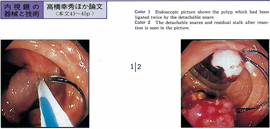
1994 年 44 巻 p. 43-45
発行日: 1994/06/06
公開日: 2015/05/25
 PDF形式でダウンロード (275K)
 -
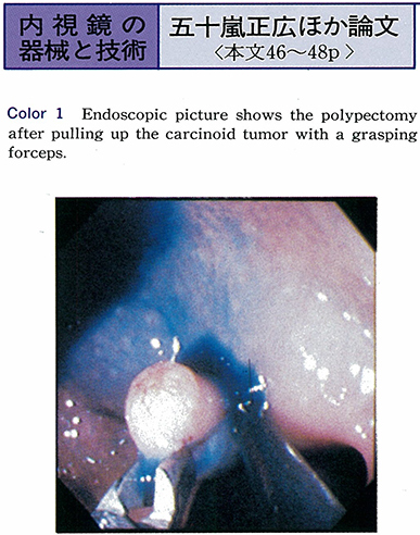
1994 年 44 巻 p. 46-48
発行日: 1994/06/06
公開日: 2015/05/25
 PDF形式でダウンロード (451K)
臨床研究
-
1994 年 44 巻 p. 49-52
発行日: 1994/06/06
公開日: 2015/05/25
 PDF形式でダウンロード (1137K)
 -
1994 年 44 巻 p. 53-56
発行日: 1994/06/06
公開日: 2015/05/25
 PDF形式でダウンロード (1093K)
 -
1994 年 44 巻 p. 57-59
発行日: 1994/06/06
公開日: 2015/05/25
 PDF形式でダウンロード (337K)
 -
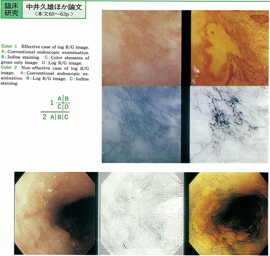
1994 年 44 巻 p. 60-63
発行日: 1994/06/06
公開日: 2015/05/25
 PDF形式でダウンロード (960K)
 -
1994 年 44 巻 p. 64-67
発行日: 1994/06/06
公開日: 2015/05/25
 PDF形式でダウンロード (929K)
 -
1994 年 44 巻 p. 68-72
発行日: 1994/06/06
公開日: 2015/05/25
 PDF形式でダウンロード (1304K)
 -
1994 年 44 巻 p. 73-76
発行日: 1994/06/06
公開日: 2015/05/25
 PDF形式でダウンロード (862K)
 -
1994 年 44 巻 p. 77-81
発行日: 1994/06/06
公開日: 2015/05/25
 PDF形式でダウンロード (995K)
 -
1994 年 44 巻 p. 82-86
発行日: 1994/06/06
公開日: 2015/05/25
PDF形式でダウンロード (599K) -
1994 年 44 巻 p. 87-91
発行日: 1994/06/06
公開日: 2015/05/25
 PDF形式でダウンロード (1453K)
 -
1994 年 44 巻 p. 92-94
発行日: 1994/06/06
公開日: 2015/05/25
 PDF形式でダウンロード (1022K)
 -
1994 年 44 巻 p. 95-97
発行日: 1994/06/06
公開日: 2015/05/25
 PDF形式でダウンロード (351K)
症例
-
1994 年 44 巻 p. 99-101
発行日: 1994/06/06
公開日: 2015/05/25
 PDF形式でダウンロード (344K)
 -
1994 年 44 巻 p. 102-105
発行日: 1994/06/06
公開日: 2015/05/25
 PDF形式でダウンロード (1036K)
 -
1994 年 44 巻 p. 106-109
発行日: 1994/06/06
公開日: 2015/05/25
 PDF形式でダウンロード (1545K)
 -
1994 年 44 巻 p. 110-113
発行日: 1994/06/06
公開日: 2015/05/25
 PDF形式でダウンロード (929K)
 -
1994 年 44 巻 p. 114-118
発行日: 1994/06/06
公開日: 2015/05/25
 PDF形式でダウンロード (1917K)
 -
1994 年 44 巻 p. 119-122
発行日: 1994/06/06
公開日: 2015/05/25
 PDF形式でダウンロード (1285K)
 -
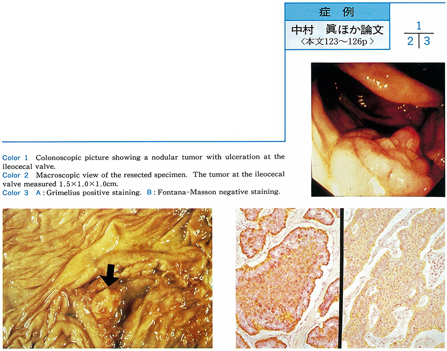
1994 年 44 巻 p. 123-126
発行日: 1994/06/06
公開日: 2015/05/25
 PDF形式でダウンロード (1083K)
 -
1994 年 44 巻 p. 127-129
発行日: 1994/06/06
公開日: 2015/05/25
 PDF形式でダウンロード (331K)
 -
1994 年 44 巻 p. 130-135
発行日: 1994/06/06
公開日: 2015/05/25
 PDF形式でダウンロード (1682K)
 -
1994 年 44 巻 p. 136-138
発行日: 1994/06/06
公開日: 2015/05/25
 PDF形式でダウンロード (663K)
 -
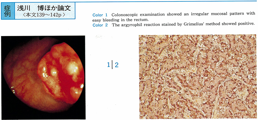
1994 年 44 巻 p. 139-142
発行日: 1994/06/06
公開日: 2015/05/25
 PDF形式でダウンロード (1649K)
 -
1994 年 44 巻 p. 143-146
発行日: 1994/06/06
公開日: 2015/05/25
 PDF形式でダウンロード (1637K)
 -
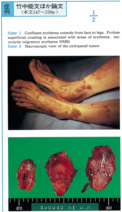
1994 年 44 巻 p. 147-150
発行日: 1994/06/06
公開日: 2015/05/25
 PDF形式でダウンロード (1450K)
 -
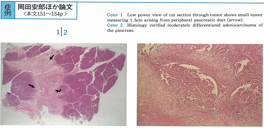
1994 年 44 巻 p. 151-154
発行日: 1994/06/06
公開日: 2015/05/25
 PDF形式でダウンロード (1720K)
内視鏡の器械と技術
-
1994 年 44 巻 p. 156-157
発行日: 1994/06/06
公開日: 2015/05/25
 PDF形式でダウンロード (182K)
 -
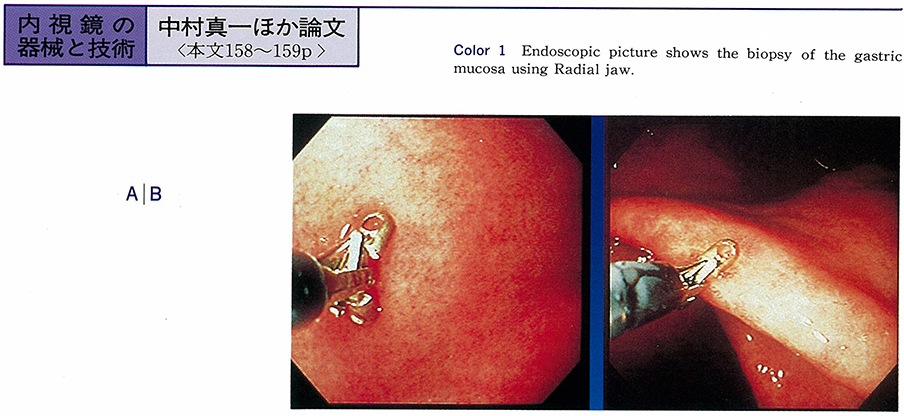
1994 年 44 巻 p. 158-159
発行日: 1994/06/06
公開日: 2015/05/25
 PDF形式でダウンロード (549K)
 -
1994 年 44 巻 p. 160-161
発行日: 1994/06/06
公開日: 2015/05/25
 PDF形式でダウンロード (495K)
臨床研究
-
1994 年 44 巻 p. 162-163
発行日: 1994/06/06
公開日: 2015/05/25
 PDF形式でダウンロード (586K)
 -
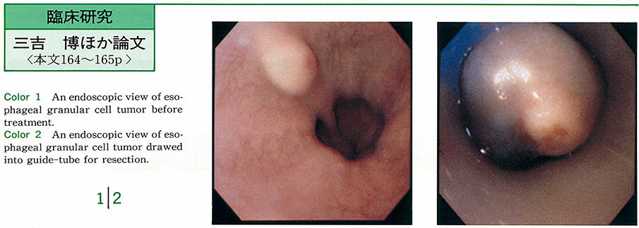
1994 年 44 巻 p. 164-165
発行日: 1994/06/06
公開日: 2015/05/25
 PDF形式でダウンロード (909K)
症例
-
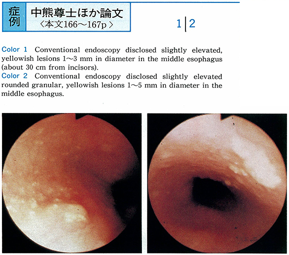
1994 年 44 巻 p. 166-167
発行日: 1994/06/06
公開日: 2015/05/25
 PDF形式でダウンロード (718K)
 -
1994 年 44 巻 p. 168-169
発行日: 1994/06/06
公開日: 2015/05/25
 PDF形式でダウンロード (750K)
 -
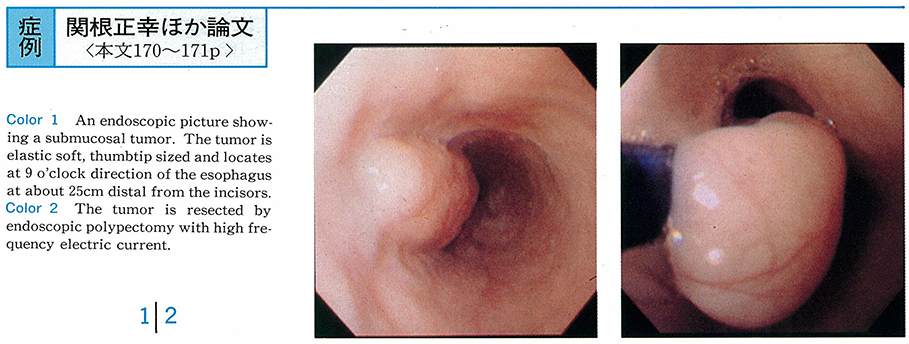
1994 年 44 巻 p. 170-171
発行日: 1994/06/06
公開日: 2015/05/25
 PDF形式でダウンロード (1194K)
 -
1994 年 44 巻 p. 172-173
発行日: 1994/06/06
公開日: 2015/05/25
 PDF形式でダウンロード (495K)
 -
1994 年 44 巻 p. 174-175
発行日: 1994/06/06
公開日: 2015/05/25
 PDF形式でダウンロード (855K)
 -
1994 年 44 巻 p. 176-177
発行日: 1994/06/06
公開日: 2015/05/25
 PDF形式でダウンロード (954K)
 -
1994 年 44 巻 p. 178-179
発行日: 1994/06/06
公開日: 2015/05/25
 PDF形式でダウンロード (1263K)
 -
1994 年 44 巻 p. 180-181
発行日: 1994/06/06
公開日: 2015/05/25
 PDF形式でダウンロード (1299K)
 -
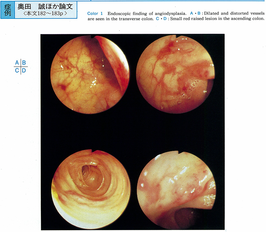
1994 年 44 巻 p. 182-183
発行日: 1994/06/06
公開日: 2015/05/25
 PDF形式でダウンロード (1055K)
 -
1994 年 44 巻 p. 184-185
発行日: 1994/06/06
公開日: 2015/05/25
 PDF形式でダウンロード (957K)
 -
1994 年 44 巻 p. 186-187
発行日: 1994/06/06
公開日: 2015/05/25
 PDF形式でダウンロード (840K)
 -
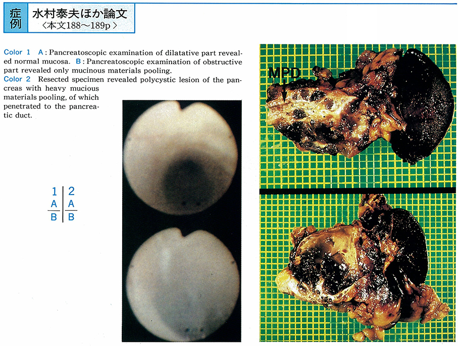
1994 年 44 巻 p. 188-189
発行日: 1994/06/06
公開日: 2015/05/25
 PDF形式でダウンロード (974K)
- |<
- <
- 1
- >
- >|