55 巻, 2 号
選択された号の論文の32件中1~32を表示しています
- |<
- <
- 1
- >
- >|
掲載論文カラー写真集
-
1999 年 55 巻 2 号 p. 1-8
発行日: 1999年
公開日: 2014/10/28
PDF形式でダウンロード (9128K)
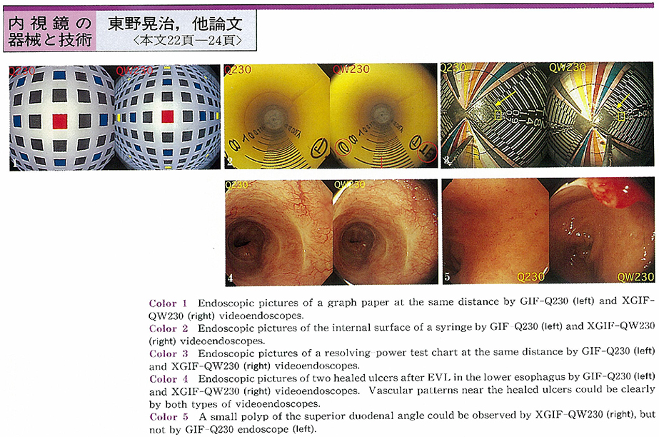
内視鏡の器械と技術
-
1999 年 55 巻 2 号 p. 22-24
発行日: 1999/11/25
公開日: 2014/10/28
 PDF形式でダウンロード (275K)
臨床研究
-
1999 年 55 巻 2 号 p. 25-29
発行日: 1999/11/25
公開日: 2014/10/28
 PDF形式でダウンロード (1296K)
 -
1999 年 55 巻 2 号 p. 30-33
発行日: 1999/11/25
公開日: 2014/10/28
 PDF形式でダウンロード (1419K)
 -
1999 年 55 巻 2 号 p. 34-37
発行日: 1999/11/25
公開日: 2014/10/28
PDF形式でダウンロード (1107K)
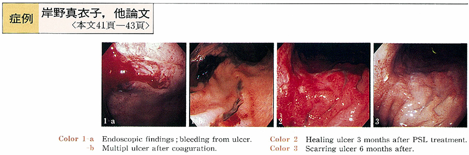
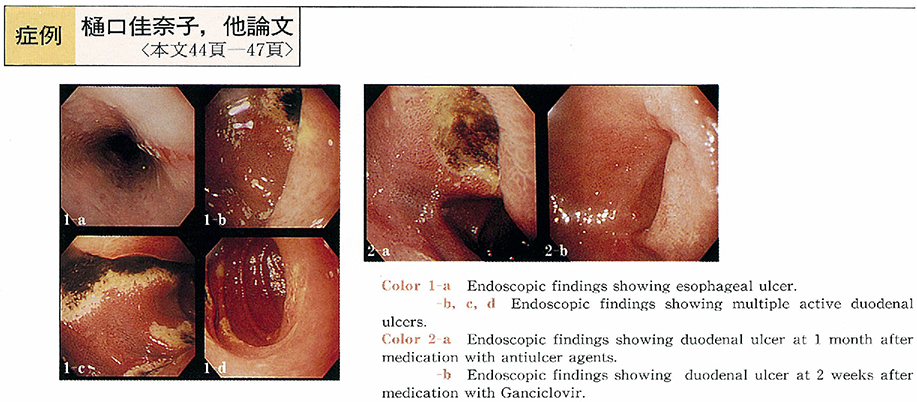
症例
-
1999 年 55 巻 2 号 p. 38-40
発行日: 1999/11/25
公開日: 2014/10/28
 PDF形式でダウンロード (821K)
 -
1999 年 55 巻 2 号 p. 41-43
発行日: 1999/11/25
公開日: 2014/10/28
 PDF形式でダウンロード (536K)
 -
1999 年 55 巻 2 号 p. 44-47
発行日: 1999/11/25
公開日: 2014/10/28
 PDF形式でダウンロード (416K)
 -
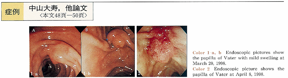
1999 年 55 巻 2 号 p. 48-50
発行日: 1999/11/25
公開日: 2014/10/28
 PDF形式でダウンロード (1046K)
 -
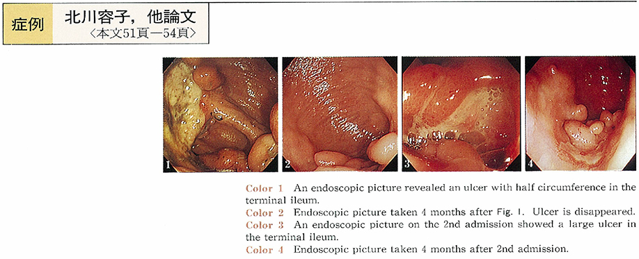
1999 年 55 巻 2 号 p. 51-54
発行日: 1999/11/25
公開日: 2014/10/28
 PDF形式でダウンロード (534K)
 -
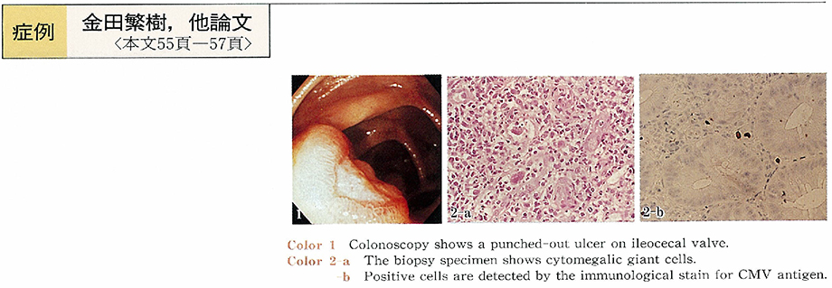
1999 年 55 巻 2 号 p. 55-57
発行日: 1999/11/25
公開日: 2014/10/28
 PDF形式でダウンロード (356K)
内視鏡の器械と技術
-
1999 年 55 巻 2 号 p. 58-59
発行日: 1999/11/25
公開日: 2014/10/28
 PDF形式でダウンロード (350K)
 -
1999 年 55 巻 2 号 p. 60-62
発行日: 1999/11/25
公開日: 2014/10/28
 PDF形式でダウンロード (1382K)
臨床研究
-
1999 年 55 巻 2 号 p. 64-66
発行日: 1999/11/25
公開日: 2014/10/28
PDF形式でダウンロード (338K) -
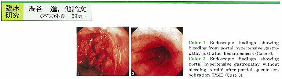
1999 年 55 巻 2 号 p. 68-69
発行日: 1999/11/25
公開日: 2014/10/28
 PDF形式でダウンロード (432K)
 -
1999 年 55 巻 2 号 p. 70-71
発行日: 1999/11/25
公開日: 2014/10/28
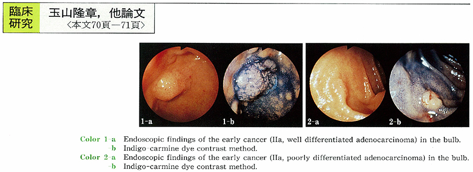 PDF形式でダウンロード (229K)
症例
-
1999 年 55 巻 2 号 p. 72-73
発行日: 1999/11/25
公開日: 2014/10/28
 PDF形式でダウンロード (258K)
 -
1999 年 55 巻 2 号 p. 74-75
発行日: 1999/11/25
公開日: 2014/10/28
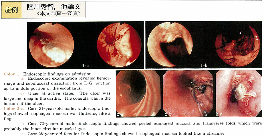 PDF形式でダウンロード (445K)
 -
1999 年 55 巻 2 号 p. 76-77
発行日: 1999/11/25
公開日: 2014/10/28
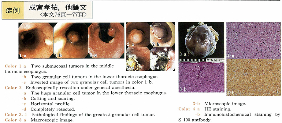 PDF形式でダウンロード (514K)
 -
1999 年 55 巻 2 号 p. 78-79
発行日: 1999/11/25
公開日: 2014/10/28
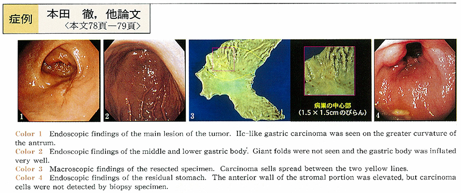 PDF形式でダウンロード (1233K)
 -
1999 年 55 巻 2 号 p. 80-81
発行日: 1999/11/25
公開日: 2014/10/28
 PDF形式でダウンロード (566K)
 -
1999 年 55 巻 2 号 p. 82-83
発行日: 1999/11/25
公開日: 2014/10/28
 PDF形式でダウンロード (764K)
 -
1999 年 55 巻 2 号 p. 84-85
発行日: 1999/11/25
公開日: 2014/10/28
 PDF形式でダウンロード (567K)
 -
1999 年 55 巻 2 号 p. 86-87
発行日: 1999/11/25
公開日: 2014/10/28
 PDF形式でダウンロード (702K)
 -
1999 年 55 巻 2 号 p. 88-89
発行日: 1999/11/25
公開日: 2014/10/28
 PDF形式でダウンロード (485K)
 -
1999 年 55 巻 2 号 p. 90-91
発行日: 1999/11/25
公開日: 2014/10/28
 PDF形式でダウンロード (376K)
 -
1999 年 55 巻 2 号 p. 92-93
発行日: 1999/11/25
公開日: 2014/10/28
 PDF形式でダウンロード (566K)
 -
1999 年 55 巻 2 号 p. 94-95
発行日: 1999/11/25
公開日: 2014/10/28
 PDF形式でダウンロード (535K)
 -
1999 年 55 巻 2 号 p. 96-97
発行日: 1999/11/25
公開日: 2014/10/28
 PDF形式でダウンロード (679K)
 -
1999 年 55 巻 2 号 p. 98-99
発行日: 1999/11/25
公開日: 2014/10/28
 PDF形式でダウンロード (664K)
 -
1999 年 55 巻 2 号 p. 100-101
発行日: 1999/11/25
公開日: 2014/10/28
 PDF形式でダウンロード (582K)
 -
1999 年 55 巻 2 号 p. 102-103
発行日: 1999/11/25
公開日: 2014/10/28
 PDF形式でダウンロード (623K)
- |<
- <
- 1
- >
- >|