
54 巻
選択された号の論文の49件中1~49を表示しています
- |<
- <
- 1
- >
- >|
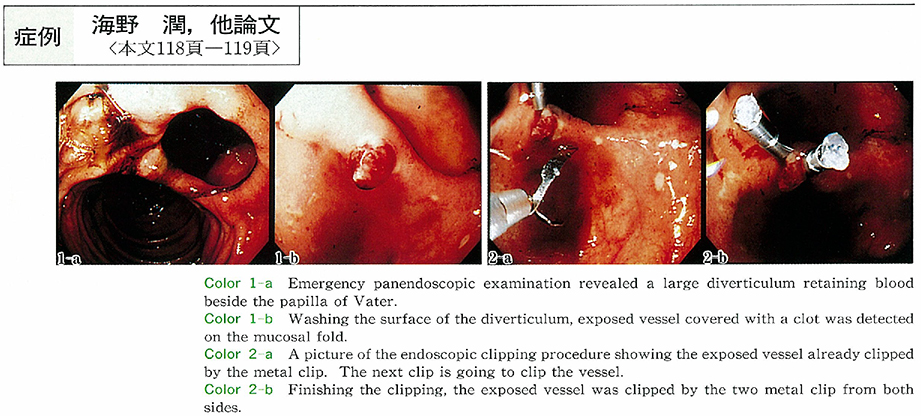
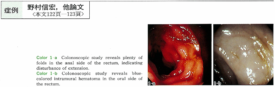
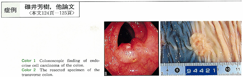
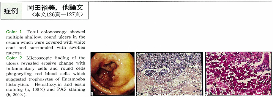
掲載論文カラー写真集
-
1999 年 54 巻 p. 1-11
発行日: 1999年
公開日: 2014/10/28
PDF形式でダウンロード (11424K)
臨床研究
-
1999 年 54 巻 p. 40-42
発行日: 1999/08/15
公開日: 2014/10/28
 PDF形式でダウンロード (361K)
 -
1999 年 54 巻 p. 43-47
発行日: 1999/08/15
公開日: 2014/10/28
PDF形式でダウンロード (765K) -
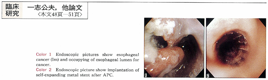
1999 年 54 巻 p. 48-51
発行日: 1999/08/15
公開日: 2014/10/28
 PDF形式でダウンロード (407K)
 -
1999 年 54 巻 p. 52-56
発行日: 1999/08/15
公開日: 2014/10/28
PDF形式でダウンロード (573K) -
1999 年 54 巻 p. 57-61
発行日: 1999/08/15
公開日: 2014/10/28
 PDF形式でダウンロード (1337K)
症例
-
1999 年 54 巻 p. 62-64
発行日: 1999/08/15
公開日: 2014/10/28
 PDF形式でダウンロード (677K)
 -
1999 年 54 巻 p. 65-68
発行日: 1999/08/15
公開日: 2014/10/28
 PDF形式でダウンロード (782K)
 -
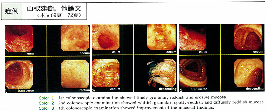
1999 年 54 巻 p. 69-72
発行日: 1999/08/15
公開日: 2014/10/28
 PDF形式でダウンロード (897K)
 -
1999 年 54 巻 p. 73-76
発行日: 1999/08/15
公開日: 2014/10/28
 PDF形式でダウンロード (956K)
 -
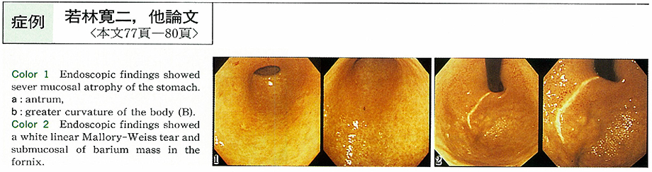
1999 年 54 巻 p. 77-80
発行日: 1999/08/15
公開日: 2014/10/28
 PDF形式でダウンロード (843K)
 -
1999 年 54 巻 p. 81-84
発行日: 1999/08/15
公開日: 2014/10/28
 PDF形式でダウンロード (757K)
内視鏡の器械と技術
-
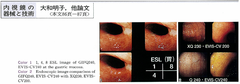
1999 年 54 巻 p. 86-87
発行日: 1999/08/15
公開日: 2014/10/28
 PDF形式でダウンロード (257K)
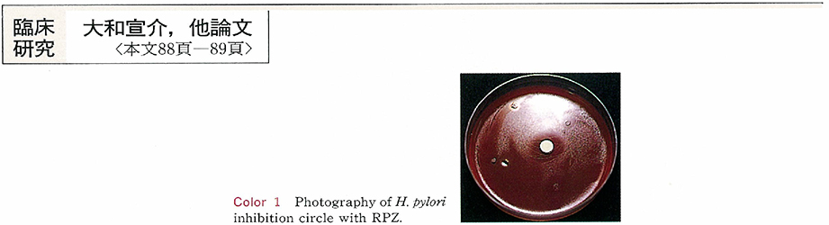
臨床研究
-
1999 年 54 巻 p. 88-89
発行日: 1999/08/15
公開日: 2014/10/28
 PDF形式でダウンロード (254K)
 -
1999 年 54 巻 p. 90-91
発行日: 1999/08/15
公開日: 2014/10/28
PDF形式でダウンロード (263K) -
1999 年 54 巻 p. 92-93
発行日: 1999/08/15
公開日: 2014/10/28
PDF形式でダウンロード (240K) -
1999 年 54 巻 p. 94-95
発行日: 1999/08/15
公開日: 2014/10/28
 PDF形式でダウンロード (409K)
 -
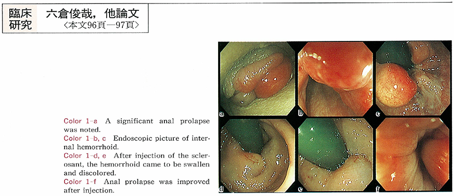
1999 年 54 巻 p. 96-97
発行日: 1999/08/15
公開日: 2014/10/28
 PDF形式でダウンロード (249K)
 -
1999 年 54 巻 p. 98-99
発行日: 1999/08/15
公開日: 2014/10/28
PDF形式でダウンロード (251K) -
1999 年 54 巻 p. 100-101
発行日: 1999/08/15
公開日: 2014/10/28
PDF形式でダウンロード (154K)
症例
-
1999 年 54 巻 p. 102-103
発行日: 1999/08/15
公開日: 2014/10/28
 PDF形式でダウンロード (1038K)
 -
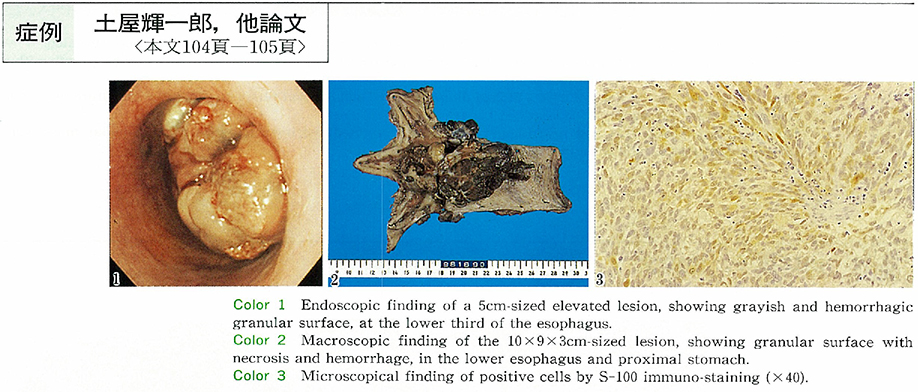
1999 年 54 巻 p. 104-105
発行日: 1999/08/15
公開日: 2014/10/28
 PDF形式でダウンロード (914K)
 -
1999 年 54 巻 p. 106-107
発行日: 1999/08/15
公開日: 2014/10/28
 PDF形式でダウンロード (714K)
 -
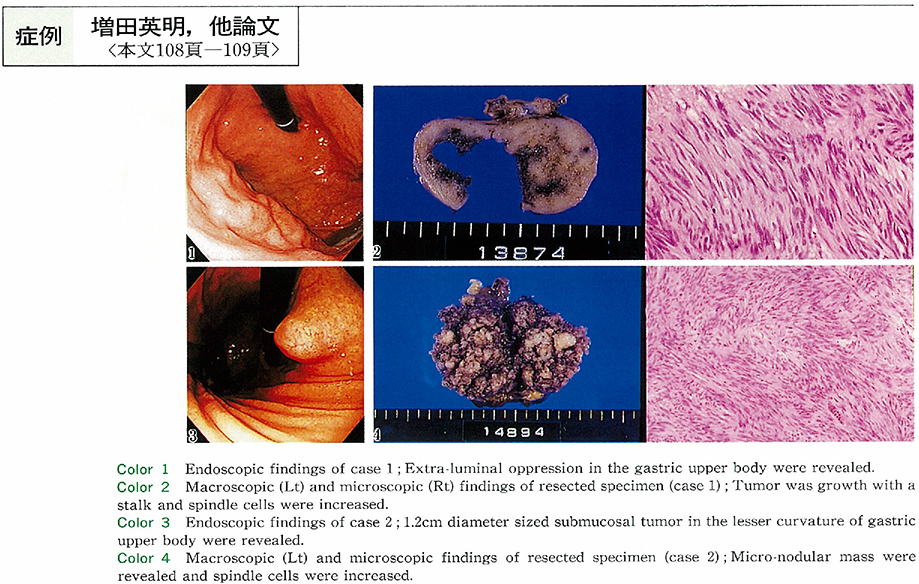
1999 年 54 巻 p. 108-109
発行日: 1999/08/15
公開日: 2014/10/28
 PDF形式でダウンロード (567K)
 -
1999 年 54 巻 p. 110-111
発行日: 1999/08/15
公開日: 2014/10/28
 PDF形式でダウンロード (402K)
 -
1999 年 54 巻 p. 112-113
発行日: 1999/08/15
公開日: 2014/10/28
 PDF形式でダウンロード (456K)
 -
1999 年 54 巻 p. 114-115
発行日: 1999/08/15
公開日: 2014/10/28
 PDF形式でダウンロード (1028K)
 -
1999 年 54 巻 p. 116-117
発行日: 1999/08/15
公開日: 2014/10/28
 PDF形式でダウンロード (470K)
 -
1999 年 54 巻 p. 118-119
発行日: 1999/08/15
公開日: 2014/10/28
 PDF形式でダウンロード (494K)
 -
1999 年 54 巻 p. 120-121
発行日: 1999/08/15
公開日: 2014/10/28
 PDF形式でダウンロード (277K)
 -
1999 年 54 巻 p. 122-123
発行日: 1999/08/15
公開日: 2014/10/28
 PDF形式でダウンロード (537K)
 -
1999 年 54 巻 p. 124-125
発行日: 1999/08/15
公開日: 2014/10/28
 PDF形式でダウンロード (652K)
 -
1999 年 54 巻 p. 126-127
発行日: 1999/08/15
公開日: 2014/10/28
 PDF形式でダウンロード (569K)
 -
1999 年 54 巻 p. 128-129
発行日: 1999/08/15
公開日: 2014/10/28
 PDF形式でダウンロード (407K)
 -
1999 年 54 巻 p. 130-131
発行日: 1999/08/15
公開日: 2014/10/28
 PDF形式でダウンロード (939K)
 -
1999 年 54 巻 p. 132-133
発行日: 1999/08/15
公開日: 2014/10/28
 PDF形式でダウンロード (801K)
 -
1999 年 54 巻 p. 134-136
発行日: 1999/08/15
公開日: 2014/10/28
 PDF形式でダウンロード (498K)
 -
1999 年 54 巻 p. 138-139
発行日: 1999/08/15
公開日: 2014/10/28
 PDF形式でダウンロード (378K)
 -
1999 年 54 巻 p. 140-141
発行日: 1999/08/15
公開日: 2014/10/28
 PDF形式でダウンロード (561K)
 -
1999 年 54 巻 p. 142-143
発行日: 1999/08/15
公開日: 2014/10/28
 PDF形式でダウンロード (719K)
第67回日本消化器内視鏡学会関東地方会 後抄録
シンポジウム
-
1999 年 54 巻 p. 144-146
発行日: 1999年
公開日: 2014/10/28
PDF形式でダウンロード (1479K)
ワークショップ
-
1999 年 54 巻 p. 146-148
発行日: 1999年
公開日: 2014/10/28
PDF形式でダウンロード (1010K) -
1999 年 54 巻 p. 149-150
発行日: 1999年
公開日: 2014/10/28
PDF形式でダウンロード (300K)
小ラウンドテーブル・ディスカッション
-
1999 年 54 巻 p. 151-152
発行日: 1999年
公開日: 2014/10/28
PDF形式でダウンロード (1225K) -
1999 年 54 巻 p. 152-153
発行日: 1999年
公開日: 2014/10/28
PDF形式でダウンロード (955K) -
1999 年 54 巻 p. 154-155
発行日: 1999年
公開日: 2014/10/28
PDF形式でダウンロード (313K) -
1999 年 54 巻 p. 156-157
発行日: 1999年
公開日: 2014/10/28
PDF形式でダウンロード (308K)
VTRセッション
-
1999 年 54 巻 p. 158-159
発行日: 1999年
公開日: 2014/10/28
PDF形式でダウンロード (315K)
一般演題
-
1999 年 54 巻 p. 160-193
発行日: 1999年
公開日: 2014/10/28
PDF形式でダウンロード (7045K)
- |<
- <
- 1
- >
- >|