Progress of Digestive Endoscopy(1972)
Online ISSN : 2189-0021
Print ISSN : 0389-9403
Volume 56, Issue 2
Displaying 1-33 of 33 articles from this issue
Technology and instrument
  • Yoko Hoshino, Atsushi Mitsunaga, Eichi Suzuki, Maiko Kishino, Hiroyuki ...
    2000 Volume 56 Issue 2 Pages 26-28
    Published: May 15, 2000
    Released on J-STAGE: October 27, 2014
    JOURNAL FREE ACCESS
    We now report our new network system for medical imaging information. Our institute has adopted the digital and imaging communication in medicine (DICOM) since October 1999 for saving endoscopic image data. DICOM is the network standard, which was developed for communication of medical information by digital image files attached with personal or other medical information on each patient, regardless of kinds or types of the imaging instruments. The adoption of this system resulted in saving image files by digital data in high quality with worselessness as well as saving a large quantity of data into small space, which are created in routine work in the hospital. Moreover the standardization of this system in the institute can : 1) unify medical information of other image information including computed tomograph, ultrasonograph etc, 2) simplify to construct network communication system within the institute, 3) simplify to exchange image files among institutes mutually. In the actual medical practice, this network system would make it possible easily for medical staffs to receive patients' information and to see a variety of needed. We can also utilize those data for presentation in the case conference in the hospital or making slides in the meeting and so on. We believe that this system will be useful for communications among hospitals and clinics if it is utilized widely, resulted in reducing the medical regional differences in future.
    Download PDF (411K)
Clinical study
  • Takeshi Matsuhisa, Norio Matsukura, Nobutaka Yamada
    2000 Volume 56 Issue 2 Pages 29-33
    Published: May 15, 2000
    Released on J-STAGE: October 27, 2014
    JOURNAL FREE ACCESS
    Helicobacter pylori (Hp) infection and gastric mucosa in Thai cases were studied comparing with Japanese cases. We used 435 cases of Thai examined by ourselves in Chiang Mai, Kingdom of Thailand, and 2,007 cases of Japanese diagnosed by the same criteria in our department.
    1) According to the comparison of Hp infection rate between Thai and Japanese after match of age, sex and endoscopic diagnosis, an infection rate of Thai (84.6%) was significantly higher than that of Japanese (70.0%) (p=0.0001) .
    2) Chronic inflammation, neutrophile activity and Hp score in specimen No.1 (greater curvature of the lower antrum) and No.3 (lesser curvature of the lower body) , taken in accordance with triple site gastric biopsy, were higher in Thai than in Japanese. On the other hand, glandular atrophy and intestinal metaplasia score at the point of No.1 and 3 specimen and endoscopic gastric mucosa score in lesser curvature of the lower body were higher in Japanese than in Thai. It was revealed that glandular atrophy and intestinal metaplasia by Hp infection were particular changes to Japanese.
    3) Pepsinogen (PG) I/II ratio in Hp positive Japanese was lower than that of Thai (each 3.3, 4.8, p<0.0001) . It was also recognized that atrophic gastritis is very popular disease for Japanese by the difference of PG I/II ratio.
    Download PDF (610K)
Case report
  • Yukiko Fukuda, Hiroyuki Imaeda, Hirotoshi Ebinuma, Shingo Miyaguchi, Y ...
    2000 Volume 56 Issue 2 Pages 34-37
    Published: May 15, 2000
    Released on J-STAGE: October 27, 2014
    JOURNAL FREE ACCESS
    We report a patient with multiple elevated gastric adenomas.
    A 67-year-old woman was admitted to our hospital due to endoscopic polypectomy for elevated polypoid lesion sized about 5 cm in diameter, at the lesser curvature of upper gastric body on April in 1995. The removed lesion histologically revealed as tubular adenoma with severe dysplasia. Secondly, she was admitted due to endoscopic mucosal resection for another flat polypoid lesion sized about 8 mm in diameter, at the lesser curvature of upper gastric body on November in 1995. The removed lesion histologically revealed as tubular adenoma with focal cancer. Thirdly, she was admitted due to endoscopic polypectomy for elevated polypoid lesion sized about 3 cm in diameter, at the fornix on May in 1998. The removed lesion histologically revealed as tubular adenoma with moderate dysplasia.
    Elevated gastric adenomas often contain cancer, so it is important that these adenomas are resected by endoscopy for diagnosis and therapy.
    Download PDF (1590K)
  • Masako Kato, Akira Mizuki, Tatsuhiko Hayashi, Nobuhiro Tsukada, Hideki ...
    2000 Volume 56 Issue 2 Pages 38-41
    Published: May 15, 2000
    Released on J-STAGE: October 27, 2014
    JOURNAL FREE ACCESS
    The patient was a 57-year-old woman, diagnosed with RA in 1996. She was admitted to the hospital in December 1997 with a 4-mo history of intractable diarrhea. At colonoscopy there was a pancolitis, resembling ulcerative colitis especially in the transverse colon. All biopsy specimens from the colon, duodenum, and stomach showed intramucosal deposits of eosinophilic material that was antiserum against amyloid A protein-positive and Congo red-positive with apple-green birefringence. Congo red staining was abolished by prior treatment with potassium permanganate, indicating that this was amyloid of AA type. These findings established the diagnosis of amyloidosis secondary to rheumatoid arthritis. The patient had a response to therapy with MTX, colchicine and PSL, which was evaluated through clinical manifestations and endoscopic findings, and she was discharged from the hospital 6 mo later. Repeat colonoscopy at 12 mo showed a marked resolution of colitis ; colonic biopsy specimens were almost negative for amyloid. It is considered that high activity of RA induces a rapid increase of SAA and promotes the deposit of amyloid, which might contribute to the onset of intractable diarrhea, and control of the underlying RA activity results in the resolution of secondary amyloidosis with clinical and biopsy remission.
    Download PDF (659K)
Technology and instrument
  • Hideki Sakurai, Hirohumi Gonda, Yuji Fujii, Eiichiro Seki, Yoh Sekine, ...
    2000 Volume 56 Issue 2 Pages 42-43
    Published: May 15, 2000
    Released on J-STAGE: October 27, 2014
    JOURNAL FREE ACCESS
    We developed a transparent hood on tip of colonoscope to put it into clinical use. A number of physician have reported usefulness of hood such as shortening of time required during colonoscopy, reducing dead-angle positions during observation as well as enabling to get the position during endscopic surgery. The disadvantage of the ordinary plastic hood is that the field of vision is obstructed by itself and irrigated fluid collected in hood. We made the hood with oblique front shape, side-holes and external flame of the hood tip. The oblique shape lowered the obstruction of visual field in relation to the position of CCD lens in colonoscope. Side-holes of hood drained irrigated fluid in hood and decreased the obstruction of visual field. The external flame of tip worked easy to hook the colon fold and decreased the blind field. We conclude that colonoscope wearing a transparent hood is very useful procedure not only a diagnostic procedure, but also a therapeutic measure.
    Download PDF (241K)
  • Hideki Sakurai, Hirohumi Gonda, Yuji Fujii, Eiichiro Seki, Yoh Sekine, ...
    2000 Volume 56 Issue 2 Pages 44-45
    Published: May 15, 2000
    Released on J-STAGE: October 27, 2014
    JOURNAL FREE ACCESS
    Hemorrhage and perforation are the most common complications associated with polypectomy of the colon. Various methods has been developed to prevent hemorrhage after polypectomy. Detachable snare is a device for preligation developed by Hachisu et al. in 1989. This procedure, however, cannot locate a snare on a pedicle of polyp properly. We contrive the procedure to ligate polyp using a stay clip to hang the detachable snare easily and accurately, and modified the ligator of detachable snare to synchronize the permanent ligating with the releasing of the temporary ligation. We applied the procedure to 20 pedunculated polyps in 17 cases. No bleeding was found after polypectomy in every case. The modified procedure is very useful and safety method for preventing hemorrhages after polypectomy for a pedunculated polyp.
    Download PDF (625K)
Clinical study
  • Masakatsu Uchihara, Namiki Izumi, Osamu Noguchi, Yasuhiro Asahina, Jun ...
    2000 Volume 56 Issue 2 Pages 46-47
    Published: May 15, 2000
    Released on J-STAGE: October 27, 2014
    JOURNAL FREE ACCESS
    Prevalence of HIV infection in Japanese has gradually increased in recent years ; therefore, physicians should give great attention to the prevention of transmission of infectious agents including HIV via endoscopy. Routine screening of serum HIV antibody as well as serum HBs antigen and HCV antibody, and serum test for syphilis has been performed prior to gastrointestinal endoscopy since 1994 in Musashino Red-Cross Hospital, and the calculated positive rate of HIV was revealed to be 0.0084 % (2 out of 23782 patients) . Ten patients have been diagnosed as HIV positive in the same period ; therefore, screening test before gastrointestinal endoscopy accounted for 2 out of 10 patients diagnosed as HIV positive in the hospital. One of these patients was diagnosed as amebic colitis thereafter. To prevent endoscopic transmission of infection including HIV, proper cleaning and disinfection procedure based on the guideline of the Japan Gastrointestinal Endoscopy Society is essential. However, routine screening including HIV antibody prior to endoscopy is recommended to prevent unexpected transmission of infectious agents including HIV.
    Download PDF (238K)
  • Yasuo Inoue, Takahiro Hayama, Takeshi Matsuhisa
    2000 Volume 56 Issue 2 Pages 48-49
    Published: May 15, 2000
    Released on J-STAGE: October 27, 2014
    JOURNAL FREE ACCESS
    Gastrointestinal endoscopy was performed in 275 cases of gastritis such as gastric erosion, erosive gastritis and superficial gastritis. In these cases, we examined psychological test, fifty-item neurotic index (FNI) and four-factor depression index (FDI) , and taste for nonessential grocery (alcohol) .
    1) According to the abdominal symptom, FNI and FDI score were higher in symptomatic group than non-symptomatic group. In many cases of superficial gastritis, there were abdominal complaint and FNI, FDI score were the highest in any other gastritis.
    2) DI score was high in the group of no Helicobacter pylori infection.
    3) FNI and FDI score were higher in no drinker group than drinker group.
    Download PDF (268K)
  • Hiroyuki Imaeda, Hirotoshi Ebinuma, Yoshikazu Tsuzuki, Yukiko Fukuda, ...
    2000 Volume 56 Issue 2 Pages 50-51
    Published: May 15, 2000
    Released on J-STAGE: October 27, 2014
    JOURNAL FREE ACCESS
    From April 1992 to July 1998, 133 patients with upper gastrointestinal bleeding were treated by endoscopic hemostasis. These 133 included 119 cases of gastroduodenal ulcer, 6 of Mallory-Weiss syndrome, 2 of gastric cancer and 6 of gastric vascular ectasia. We carried out hemostasis by injection of pure ethanol or hypertonic saline-epinephrine (HSE) , or hemoclipping for the cases of gastroduodenal ulcer, Mallory-Weiss syndrome and gastric cancer. For those of gastric vascular ectasia, we also did it by injection of pure ethanol and electrocoagulation, or endoscopic ligation using O-ring. Of the 119 cases of gastroduodenal ulcer bleeding, 3 were finally treated by TAE due to difficulty of endoscopic hemostasis and another 3 died of cardiovascular disorders after treatment. Of the 38 who underwent hemostasis by injection of pure ethanol 13 had enlargement of ulcer, but not the episode of re-bleeding. Enlargement of ulcer after injection of pure ethanol was significantly correlated with amount of ethanol. One of the 10 cases treated by injection of HSE had rupture of esophagus. One of the 3 cases treated by ligation had re-bleeding because of slipping out of O-ring. It is important to make sure of limitation for indicating endoscopic hemostasis and to make care of complication.
    Download PDF (272K)
  • Shigeyuki Hayashi, Masaki Ikeda, Yasufumi Imamura, Kennji Tominaga, Ak ...
    2000 Volume 56 Issue 2 Pages 52-53
    Published: May 15, 2000
    Released on J-STAGE: October 27, 2014
    JOURNAL FREE ACCESS
    Ultrasonographic study was examined using Endoscopic ultrasonography (EUS) in cases with extramural lesions of the colon with obvious endoscopic findings. From January 1991 to Jun 1999, 11 cases of extramural lesion of colorectum with obvious mucosal change were examined using either ultrasonic scope (CF-UM3, CF-UM20, CF-UMQ200) or ultrasonic probes (UM-2R, UM-3R, UM-3D3R) with water filling method. Subjects were composed by advanced gastric cancer (2 cases) , ovarian cancer (5 cases) , cervical cancer (2 cases) , sigmoid colon fistula of the bladder (1 case) , teratoma (1 case) . These lesions were located in the left transverse colon (2 cases) , sigmoid colon (1 case) , rectum (8 cases) .
    Most of cases were well visualized, but one case of severe colonic stenosis by recurred cervical cancer was not well visualized the extramural area with ultrasonic probe because of echo attenuation. EUS image was similar to the histological findings in each cases as follows : Irregularly mucosa in the stenotic transverse colon was noted in advanced gastric cancer with peritoneal invasion. Localized thickness of the colon wall was noted in this area. Extramural tumor growth were well visualized by EUS in gynecological tumor. Tumor invasion to the colonic wall was clearly imaged. In the case of post operative fistula (bladder- sigmoid colon) , localized thickness of the proper muscle layer was imaged clearly by EUS and following operation was successfully performed. Compare to the CT or MRI image, EUS seemed to have a advantage of localized definite diagnoses. These information were quite helpful to understand the lesion before operation. Definite EUS diagnosis was useful to diagnose extramural lesions exactly.
    Download PDF (388K)
  • Jun Unno, Sakiko Arai, Hisashi Okuyama, Yoshiyuki Tawa, Yoshiyuki Wata ...
    2000 Volume 56 Issue 2 Pages 54-55
    Published: May 15, 2000
    Released on J-STAGE: October 27, 2014
    JOURNAL FREE ACCESS
    We examined the diagnostic process of our 4 amoebic colitis cases in order to clarify the effectiveness of the colonoscopic examination and microscopic examination of the fresh biopsy sample for the diagnosis of an amoebic colitis. Without a colonoscopic examination, all findings such as symptoms, physical examination, laboratory tests and Lower GI series would not be able to strongly indicate the possibility of an amoebic colitis. It is also said that anti-amoeba antibodies cannot necessarily be detected serologically. So we feel that a colonoscopic examination, which can reveal the characteristic mucosal appearance for an amoebic colitis, is important in order to give a strong indication of the condition. For definite diagnosis of an amoebic colitis, it is necessary to find an amoeba in the colonic mucosa. But the success rate of correct diagnosis by ordinary methods, such as stool examination and histopathological examination of biopsy samples, is not high. According to our research and another reported case, microscopic examination of the fresh biopsy sample is thought to be helpful for an accurate diagnosis.
    Download PDF (600K)
Case report
  • Yujiro Uchiyama, Hiroshi Kakutani, Shouryoku Hino, Keiichi Ikeda, Kazu ...
    2000 Volume 56 Issue 2 Pages 56-57
    Published: May 15, 2000
    Released on J-STAGE: October 27, 2014
    JOURNAL FREE ACCESS
    Recently, celiac plexus nerve block using endoscopic guided puncture has been reported. We performed this technique for a patient who had the pain uncontrolled by Morphine. After this procedure, pain relief and reduction in narcotic use was obtained in this case. In this technique, it is possible to observe the entire procedure in real time. And anterior approach to celiac plexus can make less risk of neurologic complication. Additionally, close proximity of the instrument from gastric lumen allows precise needle placement and avoidance of the organs. Furthermore, this procedure can be quickly performed. EUS CPN is safer and easier technique and should be performed in earlier stage, and more often in malignant cases. And we may try to extend its application to pain control in benign disease and during endoscopic intervention.
    Download PDF (685K)
  • Tomonari Tominaga, Makoto Suga, Toshiya Ishii, Yukimitsu Mamata, Hiros ...
    2000 Volume 56 Issue 2 Pages 58-59
    Published: May 15, 2000
    Released on J-STAGE: October 27, 2014
    JOURNAL FREE ACCESS
    Four patients with hyperplastic gastric polyps were enrolled in this study. Two of them showed anemia due to multiple hemorrahagic gastric polyps, one suffered from multiple recurrent gastric polyps, and one showed a large gastric polyp with severe redness. All the cases were infected with Helicobacter pylori (H. pylori) . Although endoscopic polypectomy was performed in some of them before eradication, H. pylori eradication was started in all cases with successful elimination. By nine months after eradication, almost all of the polyps in each patient disappeared. The consecutively biopsied sample from the polyp revealed the improvement of hyperplastic change followed by disappearance of inflammatory cells infiltration after eradication of H. pylori. These results may reflect an important role of chronic active gastritis caused by H. pylori infection in hyper plastic polyps.
    Eradication of H. pylori is considered to be the first line therapy against hyperplastic polyps because of its effectiveness and non-invasiveness.
    Download PDF (554K)
  • Masakazu Kamihira, Yukio Yoshida, Hiroyuki Miyatani, Satoshi Asano, Ma ...
    2000 Volume 56 Issue 2 Pages 60-61
    Published: May 15, 2000
    Released on J-STAGE: October 27, 2014
    JOURNAL FREE ACCESS
    A 53-year-old woman presented alopecia, nail changes and nonbloody diarrhea in April 1998. When the patient visited our medical center in June, alopecia had been improved. Upper gastrointestinal endoscopy demonstrated multiple gastric polyps in the whole stomach and colonoscopy revealed polyposis from the cecum to rectum. Histologic examination of biopsy specimens from the polyps in the stomach and colon revealed hyperplastic changes of the gland, edema and inflammatory infiltration of neutrophiles, eosinocytes, etc. in the interstitium. On the basis of these findings, the patient was diagnosed as Cronkhite-Canada syndrome, but was not treated with steroids or antiplasmins because of improvement of alopecia and nail changes. Helicobacter pylori was found by culture and histological examination. The patient was treated with one week course of omeprazole, amoxicillin and clarithromycin. One year later, Helicobacter pylori was eradicated, and polyposis of the gastrointestinal tract almost disappeared. The patient has no recurrence of alopecia or nail changes.
    Download PDF (220K)
  • Takeshi Yasuda, Hirotaka Kashimura, Hiroshi Nimura, Sumio Takayama, Te ...
    2000 Volume 56 Issue 2 Pages 62-63
    Published: May 15, 2000
    Released on J-STAGE: October 27, 2014
    JOURNAL FREE ACCESS
    A 63-year-old man visited our hospital complaining of left lower abdominal pain. Endoscopic examination revealed three elevated lesions with reddish, depressed areas in the middle of the gastric body. Around this area were several small elevated lesions resembling submucosal tumor. Endoscopic ultrasonography revealed a hypoechoic area in the submucosal layers. Total gastrectomy was performed. Pathological diagnosis was multiple early gastric cancers with diffuse heterotopic submucosal cysts (DHSCs) . DHSCs are paracancerous lesions that often appear with multiple gastric cancers. If DHSCs are detected on endoscopic ultrasonography, the possibility of multiple gastric cancers should be considered.
    Download PDF (593K)
  • Satoshi Nimura, Masahiro Ikegami, Hiroshi Nimura, Hirotaka Kashimura, ...
    2000 Volume 56 Issue 2 Pages 64-65
    Published: May 15, 2000
    Released on J-STAGE: October 27, 2014
    JOURNAL FREE ACCESS
    A sixty-year-old man was admitted for a close evaluation of gastric carcinoma which was pointed out by histological examination. Endoscopy demonstrated multiple elevated lesions (10mm) in the fundic gland mucosa. The mucosectomy specimens revealed small carcinoid tumors and many endocrine cell micronests (ECMs) in the marked atrophic fundic glands. The gastric acid secretion evaluated with Congo-Red spraying technique demonstrated achlorhydria. In the laboratory tests hypergastrinemia (3620pg/ml) and marked elevation of serum anti-gastric parietal cell antibodies were noticed. Total gastrectomy was performed, with a preoperative diagnosis of multiple gastric carcinoids with type A gastritis. Postoperative histological examination of the stomach revealed marked atrophic fundic glands with numerous ECMs and submucosal (sm3) invasion of carcinoid tumors, but no metastasis to lymph nodes. The serum gastrin level returned to normal a month after operation.
    Download PDF (1150K)
  • Tatsuya Yoshida, Kyonsu Son, Takeshi Kurihara, Takuya Nojiri, Jun Asak ...
    2000 Volume 56 Issue 2 Pages 66-67
    Published: May 15, 2000
    Released on J-STAGE: October 27, 2014
    JOURNAL FREE ACCESS
    A 69-year-old male patient had anorexia since May 1999. Endoscopic examination revealed a IIa+IIc gastric carcinoma and endoscopic mucosal resection (EMR) was successfully performed. Next day, It falls to Hb 6.0g/dl and melena appeared. So, endoscopic examination was performed to search gastric, duodenal, and colorectal lesion which cause bleeding, but it was not found.
    We underwent the arteriography of SMA, and it showed the extravasation in the jejunal branch. The tip of a 3.0Fr Tracker catheter was introduced into the bleeding artery selectively, and the artery embolization was performed from this point using the emborization coil. Bleeding was stopped. It was usually reported that the bleeding and the perforation was the complication of the EMR. In this case, first, we thought the cause of melena was the bleeding from the operative scar of the EMR, but it was not found. It was the bleeding from jejunal artery. (It seemed the angioectasia of jejunum.)
    Download PDF (462K)
  • Tateki Yamane, Hiroyuki Kato, Toru Furuya, Makoto Nakamura, Takayuki I ...
    2000 Volume 56 Issue 2 Pages 68-69
    Published: May 15, 2000
    Released on J-STAGE: October 27, 2014
    JOURNAL FREE ACCESS
    A 56-year-old man was admitted to our hospital because of tarry stool. He had experienced resection of a nasal malignant melanoma at the age of 52. Radiographic and endoscopic examination revealed a submucosal tumor with deep ulcer at the 2nd portion of the duodenum. Pathological findings of the biopsy specimen from the tumor showed amelanotic malignant melanoma. Even though partial melanin pigmentation was seen in the past resected specimen from the nasal cavity pathologically, the duodenal lesion was considered as metastasis. In addition, multiple metastases to the bilateral adrenal glands and the cerebrum were noticed by CT scan examination. Gastrointestinal metastasis of malignant melanoma is sometimes found at autopsy, but it is rare to be diagnosed because it develops rarely as a digestive symptom. It is thought that this case is interesting because metastatic lesion of the duodenum was recognized radiographically and endoscopically.
    Download PDF (686K)
  • Toshiyuki Furukawa, Yasuhiro Asahina, Kosei Hamano, Nobuhiko Kanazawa, ...
    2000 Volume 56 Issue 2 Pages 70-71
    Published: May 15, 2000
    Released on J-STAGE: October 27, 2014
    JOURNAL FREE ACCESS
    An 85-year-old woman was admitted with massive upper gastrointestinal hemorrhage. She had a history of small upper gastrointestinal bleeding on 11 days before admission. On admission, physical examination revealed a pulsatile abdominal mass and her hematocrit was 16.8 per cent. An emergency endoscopic examination revealed no bleeding source in the esophagus and stomach but bleeding from pulsatile submucosal elevation at the third portion of the duodenum. A CT scan demonstrated a 6-cm abdominal aortic aneurysm. Emergency laparotomy revealed an abdominal aortic aneurysm with aortoduodenal fistula at the level of third portion. The fistula was taken down and closed, and the aneurysm was replaced by a graft. Histology of the resected aorta showed the features of atherosclerotic aneurysm with fibrin plug at the fistula. No complication including infection and rebleeding occurred, and endoscopy at 14 days after surgery revealed improvement of the duodenal fistula. Aortoduodenal fistula is rare but invariably fatal without precise diagnosis and prompt surgical treatment. The presence of antecedent nonexsanguinating hemorrhage before hypovolemic shock, socalled“herald bleed”is present in two-thirds of the patients, making it important to establish diagnosis before fatal exsanguination. Physician should consider the diagnosis of aortoduodenal fistula in patients with even mild upper gastrointestinal bleeding of uncertain etiology and abdominal pulsatile mass.
    Download PDF (574K)
  • Maiko Kishino, Atsushi Mitsunaga, Yoko Hoshino, Hiroyuki Konishi, Shin ...
    2000 Volume 56 Issue 2 Pages 72-73
    Published: May 15, 2000
    Released on J-STAGE: October 27, 2014
    JOURNAL FREE ACCESS
    A 65-year-old man had been followed up in our hospital due to liver cirrhosis, meanwhile endoscopic examination revealed a minute depressed lesion at the descending portion of duodenum. Although the histological diagnosis from the biopsy specimen was adenoma, group III, we concluded it malignant tumor because of its depressed shape and irregular margin. Endoscopic mucosal resection (EMR) was performed and from pathological examination this lesion was diagnosed as well differentiated adenocarcinoma of the duodenum. There reported only 14 cases of depressed early duodenal cancer in Japan, and 4 out of 14 cases were treated by EMR successfully.
    Download PDF (517K)
  • Kazuto Tsuboi, Jun Tsutsumi, Yasuhiro Tabata, Yoshiyuki Furukawa, Kats ...
    2000 Volume 56 Issue 2 Pages 74-75
    Published: May 15, 2000
    Released on J-STAGE: October 27, 2014
    JOURNAL FREE ACCESS
    Because of its very low incidence and lack of specific symptoms, carcinoma of the small intestine is seldom diagnosed preoperatively.
    Ten months prior to admission, a forty four years old male began passing tarry stool. Colonoscopy at the other hospital disclosed only a diverticulosis in the ascending colon. He continued to deteriorate with several more episodes of melena. At the time of admission, hemoglobin was 8.9gm/dl and tarry stool was still present. Endoscopic examinations of the upper and lower GI tracts failed to identify bleeding source. CT, angiogram of SMA, and barium swallow were noncontributory. Small bowel follow through via long intestinal tube, however visualized an annular constricting lesion at 60cm from the ligament of Treitz. Presence of this lesion was confirmed by fibroptic jejunoscopy as an annular constricting type lesion. Biopsy yielded well differentiated adenocarcinoma. Partial resection of the small intestine with regional lymphadenectomy was performed. This case suggested feasibility of fibroptic jejunoscopy for making a definite diagnosis of carcinoma of the small intestine.
    Download PDF (393K)
  • Hiroyuki Konishi, Atsushi Mitsunaga, Maiko Kishino, Yoko Fukazawa, Shi ...
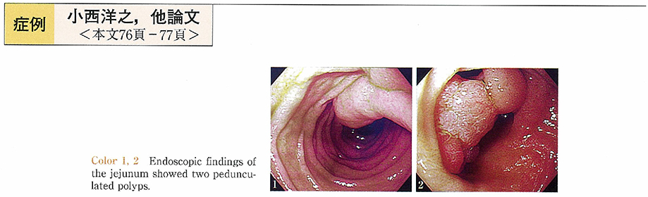
    2000 Volume 56 Issue 2 Pages 76-77
    Published: May 15, 2000
    Released on J-STAGE: October 27, 2014
    JOURNAL FREE ACCESS
    A 72-year-old male with no complaint was accidentally pointed out elevated lesion in jejunum by barium meal. Small intestinal radiography showed two pedunculated polyps in about 3cm distant from the legament of Treitz. They were sized about 3cm in diameter. We could observe the lesions by ordinary endoscopy, and histopathological diagnosis of the biopsied specimen was tubular adenoma. Because there was a possibility of bleeding and intussusception in the future, the lesions were removed by endoscopic polypectomy without any complication. Histopathological study of the resected specimen revealed tubular adenoma with mild atypia. We reported this case because the intestinal adenoma which was resected endoscopically without any complication was extremely rare.
    Download PDF (619K)
  • Kazufumi Suda, Miki Adachi, Kouichi Sakagawa, Kota Okinaga, Fumihiko T ...
    2000 Volume 56 Issue 2 Pages 78-79
    Published: May 15, 2000
    Released on J-STAGE: October 27, 2014
    JOURNAL FREE ACCESS
    A 74-year-old man underwent colonoscopic examination on November 20, 1998. A polyp of the sigmoid colon was removed by snare polypectomy. The specimen was not retrieved and pathological examination was not done. After 7 months, he complained of right abdominal pain and lumbar pain. CT scan revealed multiple hepatic and paraaortic lymph node swelling, and bone scintigram revealed hot lesions in his spines and right leg. Follow up examination of CT scan showed rapid progression of tumors and tumor marker (CEA and CA19-9) increased rapidly.
    At surgery, a lot of hepatic tumors were observed and lymph node swelling spread from the mesentery of sigmoid colon to the paraaortic region. Mesenteric wall of proximal sigmoid colon was invaded of the tumor. These findings suggested that the tumors were metastases of the sigmoid colon cancer which was polypectomized in the past. Partial colectomy and hepatic biopsy was performed. The tumors were moderate to poorly differentiated adenocarcinoma histologically. The patient died 58 days after operation.
    In case when the histological examination of colonoscopically removed polyp was not available, colonoscopic findings of the lesion should be reviewed meticulously. If there were some possibility of carcinoma, surveillance examination not only for local recurrence but also for distant metastasis should be performed.
    Download PDF (618K)
  • Sumito Mizuguchi, Takashi Ohno, Satoshi Murakami, Tadashi Kitamura, Te ...
    2000 Volume 56 Issue 2 Pages 80-81
    Published: May 15, 2000
    Released on J-STAGE: October 27, 2014
    JOURNAL FREE ACCESS
    We reported a rare case of neurinoma of the cecum. A 55-year-old female who complained of right lower abdominal distention was admitted to our hospital. Physical examination revealed solid and movable tumor, about 30 mm in size, in right lower abdomen without tenderness. Tumor markers, CEA and NSE, were within normal range. Abdominal ultrasonography, CT, MRI, barium enema and colonoscopic examination revealed a submucosal tumor of the cecum that measured 31×28×23 mm. This tumor was slightly hypervascular on angiography and negative on Ga scintigraphy. These findings suggested that this tumor was neurogenic tumor. Since patient had a symptom and the possibility of malignancy could not be excluded, the tumor was removed by a part of cecal resection assisted by laparoscopy. HE histological examination showed that the tumor was composed of fibroblast-like spindle cells and the characteristic of nuclear palisading was observed. Immunohistochemical staining showed that the tumor cells were positive for S-100 protein, NSE, but negative for desmin, smooth muscle actin, CD34. On the basis of HE histological and immunohistochemical findings, a diagnosis of neurinoma was made.
    We think that immunohistochemical examination is important for differential diagnosis of the neurogenic tumor, myogenic tumor and gastrointestinal stromal tumor (GIST) .
    Download PDF (555K)
  • Toru Tezuka, Yuji Inoue, Shigeru Suzuki, Mamoru Suzuki, Ken Takasaki
    2000 Volume 56 Issue 2 Pages 82-83
    Published: May 15, 2000
    Released on J-STAGE: October 27, 2014
    JOURNAL FREE ACCESS
    We experienced a case of nodule aggregating lesion in the appendical orifice. A 74-year-old woman was admitted to our hospital because a cecal tumor was detected colonoscopically and the biopsy of the same lesion was malignant (group V) , following a positive fecal occult blood test. When she was 28 years old, she underwent sterilization, with appendectomy.
    Colonoscopic findings showed a nodule aggregating lesion in the appendical orifice. We considered endoscopic mucosal resection (EMR) for this lesion, but it was high risk of perforation and remnant, so we selected laparotomy. We performed partial resection of the ceacum by auto-suture device only one time. She was discharge after 7 days. Histological examination revealed that the tumor was well differentiated tudular adenocarcinoma which limited in the mucosa.
    Download PDF (467K)
  • Toshiaki Suzuki, Makoto Suga, Tomonari Tominaga, Yukimitsu Mamada, Tos ...
    2000 Volume 56 Issue 2 Pages 84-85
    Published: May 15, 2000
    Released on J-STAGE: October 27, 2014
    JOURNAL FREE ACCESS
    A 63-year-old female was admitted to our hospital with complaints of lower abdominal pain, melena and purpura. Colonoscopic examination revealed severe total colitis lesion with redness, edema and erosions. We treated the patient supportive therapy, abdominal pain and melena were improved immediately. Ten days later, on the second colonoscopic examination showed markedly improved colitis lesion. On 15days after hospitalization, although abdominal pain already disappeared, proteinuria was noted. We suspected Schönlein-Henoch nephritis and started use of steroid. In a few days, proteinuria and purpura were disappeared. A month later, the 3rd colonoscopic examination revealed normal mucosal lesion. Next day, she discharged. She is free from recurrence now.
    We reported severe colitis lesion in Schönlein-Henoch purpura patient with old age who showed typical clinical course.
    Download PDF (455K)
  • Kyonsu Son, Tatsuya Yoshida, Takeshi Kurihara, Takuya Nojiri, Jun Asak ...
    2000 Volume 56 Issue 2 Pages 86-87
    Published: May 15, 2000
    Released on J-STAGE: October 27, 2014
    JOURNAL FREE ACCESS
    This report concerns our experience with a case of intractable femoral fistula caused by a residual foreign object, where the fisteloscopy was helpful.
    The patient is a male aged 35. On having fallen from a high elevation in August 1998, some pieces of bamboo stuck into the left femur. Surgical operation was carried out twice in another hospital. The patient visited our department because persistent pyorrhea and pain at the thign. Contrast imaging of fistula, computed tomography and enema examination revealed a communication between the foreign body and the large intestine. The endoscopy through the femoral fistula allowed to identify the foreign object as a piece of bamboo, which was difficult to remove because of its large size. On laparotomy, the bamboo piece was recognized in the fistula of sigmoid colon, prompting partial sigmoidectomy. The foreign object in the thigh was removed under observation with fistula-endoscope. The endoscopy was also useful for postoperational checking for residual foreign body.
    Download PDF (665K)
  • Chiyuki Shikano, Hideaki Suzuki, Tetsuo Isozaki, Yasutoshi Mitsui, Har ...
    2000 Volume 56 Issue 2 Pages 88-89
    Published: May 15, 2000
    Released on J-STAGE: October 27, 2014
    JOURNAL FREE ACCESS
    A 16-year-old female was admitted to our hospital because of lower abdominal pain, diarrhea and bloody stool. Abdominal CT examination revealed marked symmetric wall thickening in the cecum, the ascending colon and the transverse colon. Colonoscopic findings showed severe mucosal hemorrhage with marked edema and narrowing of the cecum and ascending colon. Mucosal edema, redness, erosion and hemorrhage were observed from the cecum through the sigmoid colon. The severity of these findings tend to increase on the proximal side of the colon. Subsequently Escherichia coli O-157 was cultured in the stool and hemolytic uremic syndrome (HUS) was caused. Her symptoms and laboratory data improved with antibiotics, sufficient transfusion and diuretics.
    Escherichia coli O-157 infection bring about hemorrhagic colitis and HUS. The presence of O-157 in the stool culture is necessary for diagnosis, but it seems to be possible to suspect O-157 infection by knowing colonoscopic findings.
    Download PDF (621K)
  • Hitoshi Koshikawa, Toshiaki Suzuki, Harue Shimizu, Yasuo Okada, Toshih ...
    2000 Volume 56 Issue 2 Pages 90-91
    Published: May 15, 2000
    Released on J-STAGE: October 27, 2014
    JOURNAL FREE ACCESS
    A 16-years-old male was admitted to our hospital complaining of epigastric pain and high grade fever. He had total colectomy with ileostomy done three months prior to admission for uncontrolled ulcerative colitis by conservative treatment. Upper GI X-ray examination showed poorly pliable stomach with wall stiffness. Endoscopy of upper gastrointestinal tract detected the whole gastric mucosa covered with thick white-coat and mucus. The biopsy specimens taken from the white-coated mucosa were reported as showing marked inflammatory cell infiltration and crypt abscess similar to the histological features of ulcerative colitis.
    Both symptoms and gastric lesions did not respond to H2-receptor antagonist, but were dramatically alleviated by steroid. There have been reported 4 cases of gastric lesions complicating with ulcerative colitis including our own case. Macroscopic findings are different from each other, but crypt abcess is seen in all cases. It is the fact that histological features of gastric lesions are similar to those of ulcerative colitis in all cases and dramatic response to steroid was seen in 2 cases.
    The pathogenesis, however, of gastric lesions is a big problem awaiting to be solved.
    Download PDF (672K)
  • Hiroshi Shiozaki, Masahiko Fukuda, Toshiyuki Tahara, Isao Nakata
    2000 Volume 56 Issue 2 Pages 92-93
    Published: May 15, 2000
    Released on J-STAGE: October 27, 2014
    JOURNAL FREE ACCESS
    A 59-year-old male, with RA and pulmonary fibrosis, visited to our hospital because of interstitial pneumonia. He had recieved predonisolone (PSL) 5mg/day, methotrexate (MTX) , and non-steroidal anti-inflammatory drugs (NSAIDs) . After he started to take PSL 40mg/day, his condition recovered. When the dosage of PSL was tapered to 25mg/day, he was admitted because of the respiratory infection. Antibiotic treatment and withdrawal of MTX led to improvement of his condition. Then diarrhea occured, and methicillin-resistant Staphylococcus aureus (MRSA) was isolated from his fecal sample. Oral administration of vancomycin was effective but melena was manifested. Colonoscopic examination showed multiple longitudinal ulcers in the transverse colon, and multiple round ulcers from rectum to transverse colon. Biopsy specimen from the lesion showed ulcer with inflamed granulation tissue microscopically, but neither epithelioid granuloma nor amyloid substance was seen. Adiministration of mesalazine, tapering of PSL (to 10mg/day) , and withdrawal of NSAIDs were done. His symptom was relieved, and after 12 weeks, colonoscopic finding revealed that the ulcers were cured to scars. According to the literature, these ulcers may have been mainly caused by intestinal circulatory disturbance accompanying RA. MRSA and NSAIDs might have somewhat contributed to ulceration, in this case.
    Download PDF (441K)
  • Shoryoku Hino, Hiroshi Kakutani, Keiichi Ikeda, Yujiro Uchiyama, Kazuk ...
    2000 Volume 56 Issue 2 Pages 94-95
    Published: May 15, 2000
    Released on J-STAGE: October 27, 2014
    JOURNAL FREE ACCESS
    A 60 year-old-man was refered to our hospital complaining upper abdominal pain and pyrexia. Pancreatic pseudocyst had been noted in the region of the tail since Hassab operation was performed for esophageal varices 7 years ago. CT revealed remarkable increasing of the size of the cyst. Endoscopic examination showed no bulge in the stomach. Firstly, nasocystic dorainage was performed. 19G needle was passed into the cyst under EUS guidance. After guide wire was inserted through the needle, nasocystic tube was placed. Although the 3rd day CT revealed the pseudocyst reduced in size, the patients removed nasocystic tube by himself. Then cystgastrostomy was performed. After the cyst was punctured under EUS gidance, the site was dilated with balloon under duodenoscope. Then an endoprosthesis was placed. After the procedure, CT revealed disappearance of pseudocysts and the patient became free of symptoms. Endoscopic ultrasonography guided drainage was safer and more effective procedure for the treatment of pancreatic pseudocyst.
    Download PDF (512K)
  • Yoshinari Miyakawa, Hiroshi Kakutani, Shoryoku Hino, Keiichi Ikeda, Yu ...
    2000 Volume 56 Issue 2 Pages 96-97
    Published: May 15, 2000
    Released on J-STAGE: October 27, 2014
    JOURNAL FREE ACCESS
    Recently endoscopic pancreatic stenting have been attempted for pancreatic duct strictures in chronic pancreatitis. But in technically difficult cases for stenting, the alternative methods such as surgical treatment have been selected. A 51-year-old male presented with severe epigastric pain radiating to the back since 1991. ERCP demonstrated chronic pancreatitis and pancreatic divisum. Then choledochojejunostomy and cholecystectomy was performed in 1995. In 1999 he was admitted because of the increasing frequency and intensity of abdominal pain. The extracorporeal ultrasonography and contrast-enhanced abdominal CT scan revealed dilated pancreatic duct, pancreatic calcification and large pancreatic pseudocyst without septations. Using curved linear array echoendoscope the cystic puncture was performed with the needle and the fluid was aspirated. The transgastric drainage with the stent was attempted for the cyst, but the stent could not be passed through the wall. Then the transpapillary stenting was attempted, but the minor papilla could not be identified. Therefore the percutaneous puncture for dilatated pancreatic duct was performed. Then the minorpapilla was identified endoscopically with the guidewire inserted via extracorporeal route. The percutaneous approach with extracorporeal ultrasound guidance could be considered as the alternative method with safty in case that minor papilla can not be identified in the patient with chronic pancreatitis.
    Download PDF (707K)
feedback
Top