Progress of Digestive Endoscopy(1972)
Online ISSN : 2189-0021
Print ISSN : 0389-9403
Volume 50
Displaying 1-50 of 76 articles from this issue
Clinical study
  • Hiroshi Koyama, Yukihiko Naruki, Mamoru Nishino, Masahiko Sasajima, Ei ...
    1997 Volume 50 Pages 147-151
    Published: June 06, 1997
    Released on J-STAGE: February 17, 2015
    JOURNAL FREE ACCESS
    We evaluated the endoscopic findings and histopathological data after eradication of Helicobacter pylori (HP) . One hundred and two HP-positive patients consisted of 30 gastric ulcer, 23 duodenal ulcers, 11 gastroduodenal ulcers and 38 chronic gastritis were treated. Fifty-five patients were treated with 600mg of clarithromycin (CAM) , 30mg of lansoprazole (LAZ) and 2.0g of ecabet sodium (E-Na) in a day for 14 days (A group) . Fourty-seven patients treated with 2000mg of amoxicillin (AMPC) , 30mg of LAZ and 2.0g E-Na in a day for 14 days (B group) .
    Endoscopy was performed before and after eradication. Each two gastric biopsies were collected from site of upper body, lower body and antrum in greater curvature. One piece was examined for HP-culture and other was used for the pathological study. 13C-urea breath test (UBT) was performed using 100mg of 13C-labelled urea. Expiration was collected at 0, 5, 10, 20, 30, 60 minutes and 13CO2/12CO2 was measured by laser spectroscopy. UBT was carried out before and 0, 2, 4, 12 weeks after treatment.
    HP was eradicated in 70.5% (72/102) . 54.5% in A group and 89.3% in B group were eradicated. In endoscopic study before treatment, low grade of flat erosive gastritis showed in effective cases compared with non-effective cases. Erythematous gastritis improved after eradication of HP. In histopathological study, neutrocyte and monocyte infiltration were improved after treatment.
    In conclusion, AMPC group (B group) demonstrated higher effective than CAM group (B group) . UBT can be useful in observing presence of HP in the stomach after treatment.
    Download PDF (573K)
  • Takeshi Matsuhisa, Takahiro Hayama, Shotaro Iida, Akane Arai, Izumi Ku ...
    1997 Volume 50 Pages 152-156
    Published: June 06, 1997
    Released on J-STAGE: February 17, 2015
    JOURNAL FREE ACCESS
    A rate of eradication of Helicobacter pylori (H. pylori) infection and change of serum gastrin and pepsinogen (PG) I, II levels were studied.
    1) In many regimens, we got a high eradication rate with 1 week triple eradication method. It is consisted of PPI (proton pump inhibitor) +tinidazole (TNZ) +amoxicillin (AMPC) or clarithromycin (CAM) . An eradication rate was each 76.9% and 90.0%.
    2) In 181cases (peptic ulcer, atrophic gastritis and other diseases) , an eradication rate occupied 77.3% including additive and re-eradication.
    3) Serum PGII levels dropped and PGI/II ratio rose by eradication. Serum gastrin levels rose significantly in succeeded cases, too.
    Download PDF (569K)
  • Naoki Igarashi, Koichiro Kumai, Soichiro Isshiki, Yoichiro Ishikawa, T ...
    1997 Volume 50 Pages 157-159
    Published: June 06, 1997
    Released on J-STAGE: February 17, 2015
    JOURNAL FREE ACCESS
    The clinical management of elevated lesions classified as Group III (borderline between benign and malignant on gastric biopsy by JRSGC) were analyzed. No cancer in adenoma was found among 101 resected specimens, consisting of well differentiated adenocarcinomas (51 cases) and adenomas (50 cases) obtained by gastrectomy (24 cases) , laparoscopic wedge resection of the stomach (4 cases) , or endoscopic mucosal resection (EMR) (73 cases) at our department between 1990 and 1994.
    Difficulties remain in the histological classification of endoscopic biopsy specimens of malignant lesion. EMR is recommended for Group III lesions smaller than 20mm in diameter ; larger lesions require procedures such as laparoscopic wedge resection of the stomach.
    Download PDF (367K)
  • Koji Fujita, Yoshihide Otani, Yoichirou Ishikawa, Soichirou Isshiki, Y ...
    1997 Volume 50 Pages 160-163
    Published: June 06, 1997
    Released on J-STAGE: February 17, 2015
    JOURNAL FREE ACCESS
    Nineteen cases with early gastric cancer were treated with salvage gastrectomy after endoscopic mucosal resection (EMR) in Keio University Hospital from 1990 to 1995. The residual cancer cells and lymph node metastasis were evaluated in comparing with the findings of EMR-specimens.
    Macroscopic findings included one type I case, seven type IIa cases, nine type IIc cases, one IIa+IIb case, and one type IIa+IIc case. Histologically, 9 cases were well, 9 cases were moderately, and one case was poorly differentiated adenocarcinoma. The depth of the tumor invasion were mucosal cancer for nine cases, and the other ten cases had submucosal invasion. The reasons for salvage gastrectomy were'positive surgical margin’for 7 cases,'unclear surgical margin’for 2 cases, and'submucosal invasion with positive surgical margin’for 10 cases.
    In the resected specimens, the remnant cancer cells were observed in seven of nine cases (78%) for mucosal cancer, and eight of ten cases (80%) for submucosal cancer. Lymph node metastasis was found in only one case of 78-year-old male out of ten submucosal cancer cases. Although this case had positive surgical margin with submucosal invasion in the EMR-specimen, the salvage operation was not performed and the laser irradiation was added because of poor respiratory ability and the patients choice. While no cancer cells were obtained in the follow-up gastrofiberscopy for 22 months after the initial EMR, the patient developed common biliary duct stones (CBDS) . And since the preoperative gastrofiberscopy proved Group IV cells in the EMR-scar, the distal gastrectomy was performed combined with the extirpations of CBDS. The surgical specimen showed the subserosal invasion with positive lymph nodes metastasis in the second group.
    When the surgical margin of the EMR-specimens was positive, the additional surgical resection should be performed, because the cancer cells would remain around the lesions. Our indication for EMR includes well differentiated type, mucosal invasion, no ulceration, and size less than 10 mm, which warrants complete resection the lesion by the single EMR.
    Download PDF (730K)
  • Kazutomo Togashi, Fumio Konishi, Tsuneo Ishizuka, Shingo Senba, Hirosh ...
    1997 Volume 50 Pages 164-167
    Published: June 06, 1997
    Released on J-STAGE: February 17, 2015
    JOURNAL FREE ACCESS
    We have practiced endoscopic mucosal resection (EMR) for the lesions which have a possibility of incomplete resection by snare polypectomy or hot biopsy, or the lesions which required histological complete resection. The lesions apparently invading in the deep submucosal layer were excluded from the indication for EMR.
    In this report, we studied 478 cases of EMR regarding the technique and the complications after EMR. The procedures of EMR in our practice were as follows. First, we observed the lesions with dye spray technique in detail, and we decided the details of the method including the site of saline injection, the number of sites of saline injection and the injecting speed of saline according to the shape, size and site in colorectum. In some cases we pricked the lesions directly with a needle for injecting saline. When lesions were located in the caecum or the rectum, we rapidly injected saline with a full strength. When lesions were under 15 mm in size, we usually used smaller-sized snare wire (called mini-elliptic type) . When patients had a bleeding diathesis, we basically closed mucosal defect with clips to prevent delayed bleeding.
    In 478 cases of EMR, we experienced no perforation, 16 immediate bleedings after EMR and 3 delayed bleedings. 16 immediate bleedings could be controlled endoscopically and got permanent hemostasis. Two of 3 cases with delayed bleeding showed minor bleeding on colonoscopy, and another case revealed venous bleeding which was controlled by clipping. One of 20 cases with closure of mucosal defect following EMR showed delayed bleeding. There was no local recurrence except for only one patient with multinodular lesion of 40 mm in size. Residual tumor tissue was not present in the surgically resected specimens of the 4 cases in which subsequent bowel resection was indicated because of the unfavorable histology on EMR specimens.
    In conclusion, EMR is appropriate method for the purpose of getting histological complete resection, because the frequency of complication in EMR is almost as low as in that of snare polypectomy or hot biopsy.
    Download PDF (737K)
  • Toshiya Mutsukura, Hirohisa Miwa
    1997 Volume 50 Pages 168-172
    Published: June 06, 1997
    Released on J-STAGE: February 17, 2015
    JOURNAL FREE ACCESS
    The clinical effectiveness of endoscopic mucosal resection using a transparent cap (EMR-C) for early esophageal cancer and early gastric cancer has already been reported. However its effectiveness for colorectal cancer is still contraversial because of the potential risk of perforation of the colon. In order to avoid perforation, we inject 10 to 20 ml of normal saline into the submucosal layer of the cancerous lesion to elevate it from the muscle layer. By sucking the lesion into the cap, snaring of the lesion with surrounding normal mucosa is easily performed. After one second of electrocauterization, we loosen the snare to release the muscle layer and then repeat snaring to remove the lesion.
    This method enables us to resect flat or laterally spreading lesion. Moreover, the risk of perforation is thought to be minimal. After resecting the lesion, we perform electrocauterization around the resected area to destroy the residual cancerous tissue. If there is a risk of perforation or bleeding, we perform clipping after EMR-C to close the ulcer. With this method, we resected 12 cases of early colorectal cancer, and colon resection was needed only in one case because the lesion was large to be resected endoscopically. In other 11 cases, the EMR-C was successfully carried out and there were no cases of recurence or complications. We conclude that EMR-C may be a potentially effective therapeutic modality for early colorectal cancer.
    Download PDF (630K)
  • Terumi Kamisawa, Naoto Egawa, Nobuhiro Sakaki, Jun-ichi Ishiwata
    1997 Volume 50 Pages 173-176
    Published: June 06, 1997
    Released on J-STAGE: February 17, 2015
    JOURNAL FREE ACCESS
    We examined endoscopic retrograde pancreatograms of 12 cases with pancreas divisum about inferior branches from the dorsal pancreatic duct in the head of pancreas. An inferior branch over 10mm in length was demonstrated in all cases. A long inferior branch over 20mm in length was detected in 9 cases and a short inferior branch between 10mm and 19mm was in 7 cases. Only a long inferior branch was detected in 5 cases, both long and short inferior branches were in 4 cases, and only a short inferior branch was in 3 cases. A long inferior branch arised frequently from the dorsal pancreatic duct between 20mm and 26mm from the minor duodenal papilla, and a short inferior branch between 7mm and 15mm.
    Of 70 cases, in which the length of Wirsung's duct from its orifice to its junction with Santorini's duct was long and the course of the dorsal pancreatic duct formed a straight line (long type) , a long inferior branch was demonstrated in 59 cases from Santorini's duct, and a short inferior branch was in 42 cases. A long inferior branch arised frequently from Santorini's duct between 13mm and 25mm from the minor duodenal papilla, and a short inferior branch between 7mm and 14mm. These findings were similar to those of pancreas divisum.
    It was suggested that the dorsal pancreatic ducts of both pancreas divisum and long type were persistent drainage ducts of the embryologic dorsal pancreatic bud.
    Download PDF (379K)
Case report
  • Shigeru Shirota, Motomichi Urabe, Noboru Mizobuchi, Fuyumi Isayama, Ka ...
    1997 Volume 50 Pages 177-180
    Published: June 06, 1997
    Released on J-STAGE: February 17, 2015
    JOURNAL FREE ACCESS
    We reported a case of adenocarcinoma arising from Barrett's esophagus concerning a 62-year-old man with a complaint of dysphasia. The patient had a tumorous lesion in the lower esophagus shown by radiologic and endoscopic examinations. The lesion was identified as adenocarcinoma based on a biopsy. An excision of the lower esophagus and proximal gastrectomy with lymphadenectomy was operated.
    The surgical specimen showed that the lower part of the esophagus was lined by columnar epithelium with no dysplasia, and the protruded lesion was composed of papillary adenocarcinoma. The carcinoma was 5×3.3cm in size and the cancerous invasion was limited within the submucosa in which the esophageal glands were seen microscopically. In the lymphnodes, metastatic adenocarcinoma was not present.
    Immunohistochemically staining for PCNA and p53 were performed in this case. The PCNA labelling index of adenocarcinoma and Barrett's epithelium were higher than that of esophagus and gastric epithelium. These results suggested that Barrett's epithelium has a high proliferative activity.
    Download PDF (1251K)
  • Manabu Nishimaki, Yutaka Tani, Tetsuya Sanji, Masako Midorikawa, Yutak ...
    1997 Volume 50 Pages 181-184
    Published: June 06, 1997
    Released on J-STAGE: February 17, 2015
    JOURNAL FREE ACCESS
    A 51-year-old male underwent medical examination and was found to have an abnormality in gastric antrum. The patient visited to our department for more detailed examination. Gastric X-ray and endscopic findings revealed a submucosal tumor, approximately 3cm in diameter, with smooth surface, and with bridging fold.
    Endscopic ultrasonography indicated that intratumor ununiform pattern and the tumor was included in the muscle layer. Abdominal CT showed contrast enhacement of the tumor. These findings led us to suspect the mass as malignant tumor and partial gastrectomy was performed. Histological findings showed typical glomus tumors are composed of blood vessels lined by normal endothelial cells and surrounded by sheets of round to oval tumor cells without atypia.
    Download PDF (936K)
  • Yusuke Iizuka, Atsushi Mitsunaga, Megumi Uchiyama, Kouji Uchida, Yukih ...
    1997 Volume 50 Pages 185-187
    Published: June 06, 1997
    Released on J-STAGE: February 17, 2015
    JOURNAL FREE ACCESS
    A 65-year-old woman has been periodically observed in our department since 1991 because of type A gastritis with hypergastrinemia. In April 1996, by the endoscopic examination the tiny elevated lesions (Yamada's type I) were found in the upper and middle body of the stomach. The patient has no complaint and no systemic manifestations suggesting carcinoid syndrome. By the laboratory examinations, the serum gastrin level was remarkably high (4,432pg/ml) , and the anti-parietal cell antibody was positive. Gastric secretion test showed achlorhydria.
    Endoscopic ultrasonography to the tiny elevated lesions revealed the low echoic mass with a diameter of 4×2mm mainly in the third layer of the stomach. Helicobacter pylori cultured by gastric mucosa was negative. Endoscopic biopsy specimens from the tiny elevated lesions proved carcinoid cells with a trabecular structure in the deep lamina propria of the mucosa, and they were positive for Grimelius and Chromogranin-A stains. There was no metastasis to any lymphnode and the liver.
    This case suggests that it is very important to follow type A gastritis with hypergastrinemia for early discovering gastric carcinoid tumors.
    Download PDF (424K)
  • Hiroaki Sugisaka, Norichika Narimiya, Izumi Sugimoto, Yasunori Kadowak ...
    1997 Volume 50 Pages 188-191
    Published: June 06, 1997
    Released on J-STAGE: February 17, 2015
    JOURNAL FREE ACCESS
    We report a case of gastric MALT lymphoma and demonstrate the usefulness of infrared-ray electronic endoscopy for the evaluation of submucosal invasion.
    A 60-year-old man was admitted to our hospital because of hepatic dysfunction. The liver biopsy specimen revealed a slight infiltration of inflammatory cells into the liver parenchyma. An endoscopic examination showed the granular pattern of gastric mucosa on the anterior wall of the gastric body, and histological findings demonstrated MALT lymphoma, revealed a thickening of both the second and third layers of the stomach wall. Infrared-ray electronic endoscopic examination showed round pooling findings around the tumor. In our judgement, these findings revealed that lymphoma cells had invaded into the submucosal layer. Therefore, a total gastrectomy was performed. The resected specimen confirmed the diagnosis of MALT lymphoma, which had invaded into the deeper submucosal layer of the stomach.
    We conclude that the infrared-ray electronic endoscope was useful for the evaluation of tumor invasion into the submucosal region.
    Download PDF (1051K)
  • Nobuaki Shikama, Masahito Ohida, Hiroshi Imaizumi, Yoshiki Kida, Satos ...
    1997 Volume 50 Pages 192-196
    Published: June 06, 1997
    Released on J-STAGE: February 17, 2015
    JOURNAL FREE ACCESS
    A 45-year-old woman who had a gastric abnormality revealed by an endoscopy performed by a local doctor in June 1995, visited our hospital with epigastralgiain October 1995. The initial endoscopic examination only found an ulcer scar on the lesser curvature of the lower gastric body. After an inquiry of the local doctor about information of abnormalities in the stomach, the second detailed endoscopic examination revealed a IIc type early gastric carcinoma on the anterior wall of the lower gastric body which endoscopically resembled“hino-maru”type gastric telangiectasia. Pathological examination of the target biopsy specimen showed poorly differentiated adenocarcinoma.
    Furthermore, the mucosal blood volume ratios (mucosal blood volume in the reddish or pale areas of the lesion/in peripheral normal areas) were calculated by means of an image analysis system. The reddish area of this lesion had similar blood volume ratio (1.37) compared with our previously reported findings of reddish type minute gastric carcinoma (1.40) , but lower than that of“hino-maru”type telangiectasia (1.91) . No significant differences were found in the mucosal blood volume ratios among the pale areas in these three types (this case : 0.87, pale type minute gastric carcinoma : 0.76,“hino-maru”type telangiectasia : 0.84) .
    This report suggested that detailed endoscopic examination under condition of small amount of air supply or the calculation of the mucosal blood volume by image analysis was useful in distinguishing minute gastric carcinoma when encounter“hino-maru”type gastric telangiectasia.
    Download PDF (998K)
  • Ryozo Iwazaki, Akira Sawaguchi, Toshio Yamada, Tomoko Itatsu, Taro Osa ...
    1997 Volume 50 Pages 196-199
    Published: June 06, 1997
    Released on J-STAGE: February 17, 2015
    JOURNAL FREE ACCESS
    A 72-year-old female was admitted to the hospital complaining of tachypnea, anemia and abdominal discomfort. Endoscopic examination revealed a large villous appearing tumor, about 10cm in size, at the gastric angle. The tumor was friable and a barium meal study demonstrated the“soap-bubble”appearance, which is considered to be a characteristics of the villous tumor. As a histological examination of the biopsy specimen showed group III, the partial gastrectomy was performed. Macroscopical view of the resected specimen demonstrated the 10×5×3cm sized elevated tumor with villous appearance. As highly differentiated adenocarcinoma with villous structure was indentified, the tumor was diagnosed as early gastric cancer type 1, which invaded only to the mucosal layer.
    Download PDF (1103K)
  • Akiko Ohkubo, Hiroyuki Imaeda, Yoshikazu Tsuzuki, Shingo Miyaguchi, At ...
    1997 Volume 50 Pages 200-202
    Published: June 06, 1997
    Released on J-STAGE: February 17, 2015
    JOURNAL FREE ACCESS
    A 61-year-old female was admitted to our hospital because of abdominal pain and bloody stool. She had a history of using indomethacin suppository (250mg/day) during 17 years for her pain of the left hip joint due to its congenital dislocation. Colonosopic examination and radiologic study of small intestine revealed multiple erosions and small ulcers of the terminal ileum. Her symptoms got better by stopping indomethacin. She began to use indomethacin again, so she was admitted because of abdominal pain. Partial ileal resection was performed because of perforation. The resected specimen showed multiple ulcers and 3 sites of perforation. Microscopic view revealed multiple ulcers (Ul II-IV) and perforations with infiltration of lymphocytes and granulation tissues. Cases of NSAIDs induced intestinal ulcers associated with perforation which are caused by NSAIDs rarely occur, and only 4 cases have been reported in Japan.
    Download PDF (1072K)
  • Yuichi Kumano, Kazuhiro Kaneko, Keiji Mitamura, Masahiko Murakami, Mit ...
    1997 Volume 50 Pages 203-205
    Published: June 06, 1997
    Released on J-STAGE: February 17, 2015
    JOURNAL FREE ACCESS
    A 49-year-old male, who had undergone endoscopic polypectomy two years before, visited to our hospital in order to take colorectal examinations. Barium enema findings revealed a flat elevation of which size was 10mm in diameter, combining with wall-malformation of the sigmoid colon. Endoscopic findings demonstrated the flat elevation with a central shallow depression looking like a IIa+IIc type early cancer. Because the lesion was diagnosed as infiltrating into submucosal layer, surgical resection was performed.
    However, histologic findings revealed the infiltration of cancerous lesion into subserosal layer over the margin of the flat-elevated lesion. This case showed an interesting growth pattern in the development of poorly differentiated adenocarcinoma of the colon.
    Download PDF (790K)
  • Yuko Ota, Junko Fujisaki, Makiko ichinose, Masahiro Ikegami, Ken Ashik ...
    1997 Volume 50 Pages 206-209
    Published: June 06, 1997
    Released on J-STAGE: February 17, 2015
    JOURNAL FREE ACCESS
    A 69-year-old man suddenly presented anal bleeding. On digital rectal examination an elastic soft mass was palpated in the anteriol rectal wall. He was admitted to our hospital for further evaluation and treatment of the rectal tumor.
    Endoscopic examination showed a tumor of approximately 6cm in diameter with necrotic lesion, protruding from the anterior wall of the rectum. A biopsy was performed and histological examination comfirmed leiomyosarcoma. Pervic computed tomography and magnetic resonance imaging showed a heterogenous mass with irregular limits, suggesting some invasion into the capsule of the prostate. Pelvic ultrasonography showed an interruption of the fatty layer between rectum and prostate. However, at endoscopic ultrasonography, heterogenous hyperechoic tumor existed in the fourth layer and there was no evidence of invasion into the prostate. Abdomioperineal resection was performed, and there was no invasion into the prostate.
    Rectal leiomyosarcoma is a rare disease. We reported a case of leiomyosarcoma of the rectum. In this case endoscopic ultrasonography was useful for the choice of treatment.
    Download PDF (1468K)
  • Shigeru Iwase, Toshifumi Saitoh, Hideo Tsuboi, Kazuya Sugimori, Tadash ...
    1997 Volume 50 Pages 210-213
    Published: June 06, 1997
    Released on J-STAGE: February 17, 2015
    JOURNAL FREE ACCESS
    We report a case of ischemic proctitis with broad mucosal necrosis. A 52-year-old man suddenly developed melena, lower abdominal pain and high fever, and admitted to our hospital on May 13, 1996. Colonoscopic examination revealed broad rectal mucosal necrosis. Biopsy specimen showed that the lesion seems to be caused by ischemic change of rectal mucosa. Further examinations such as abdominal angiography and abdominal CT revealed that no abnormality of the internal iliac artery (IIA) nor inferior mesenteric artery (IMA) . And pelvic tumor was not found.
    No pyogenes were detected by stool culture. After the treatment of diet therapy, intravenous hyperalimentation and steroid suppository, he recovered completely and discharged on Aug 4, 1996.
    Ischemic proctitis rarely occurs, since the rectal mucosa have abundant supply of blood though several arteries, such as IIA and IMA. The 69% of the 29 cases of ischemic proctitis reported in Japan showed the complications which were related to circulatory collapse, but this case did not show such abnormalities.
    Download PDF (1610K)
  • Junji Ueda, Toshihiro Yokota, Koichi Hosokawa, Hitoshi Kondo, Takashi ...
    1997 Volume 50 Pages 214-217
    Published: June 06, 1997
    Released on J-STAGE: February 17, 2015
    JOURNAL FREE ACCESS
    A 62-year-old female was admitted with complaints of dizziness, general fatigue and mucinous diarrhea. Laboratory examination showed serum electrolyte depletion (Na 137mEq/l, K 2.2mEq/l, Cl 82mEq/l) and dehydration.
    Barium enema study revealed a huge tumor involving the entire rectum which showed a spicular formation on its surface. Colonoscopy also revealed a huge tumor covered with a lot of mucus. Furthermore, magnifying observation showed that the surface of the tumor was looks like a velvet. Because there was no finding of invasive malignancy, sphincter saving operation was done. A villous tumor, 17×9.5cm in size, revealed well differentiated adenocarcinoma limited to the mucosal layer. The clinical course after surgery was improved.
    Thus far, only 25 cases of villous tumor showing electrolyte depletion syndrome were reported in Japan. Our current case demonstrated a good suggestion for the clinical management of such cases.
    Download PDF (1578K)
  • Naohiro Kawamura, Noriyuki Asaba, Gen-ichi Koyama, Yoshihisa Sato, Sae ...
    1997 Volume 50 Pages 218-221
    Published: June 06, 1997
    Released on J-STAGE: February 17, 2015
    JOURNAL FREE ACCESS
    A 27-year-old woman was admitted because of jaundice and severe liver damage during her 28th week of pregnancy on July 30, 1992. Acute fatty liver of pregnancy (AFLP) was suspected and Caesarian operation was performed. The liver biopsy specimen showed submassive necrosis and fatty metamorphosis and was diagnosed as AFLP. On admission HCV antibody was positive. The laparoscopic examination on 61 hospital day revealed irregular surface and reddish markings on the depressed area of the liver. Liver function tests were intermittently abnormal.
    The second iaparoscopy in September 1996 showed remarkable improvement of hepatic surface, although the reddish markings were still present. The histological findings of AFLP were improved and findings were consistent with chronic hepatitis type C (F1/A1) .
    Download PDF (961K)
Clinical study
  • Akiko Soyama, Shin-ichi Satoh, Motonobu Nishimura, Nehiro Kuriyama, Ta ...
    1997 Volume 50 Pages 222-223
    Published: June 06, 1997
    Released on J-STAGE: February 17, 2015
    JOURNAL FREE ACCESS
    From 1977 to 1996 we treated endoscopically 19 cases of esophageal foreign body at Sakura National Hospital without any complication. Nineteen were 12 males and 7 females and 2 to 79 years in age. Objects of foreign bodies were a PTP in 7 cases, foods in 5, a coin in 4 and others. All foreign bodies in childhood were a coin. For the removal of pointed objects such as a PTP or denture, use of an overtube, a transparent hood and a rigid esophagoscope is important.
    Download PDF (409K)
  • Kou Shiraishi, Soji Ozawa, Nobutoshi Ando, Shikou Seki, Eiichi Nakamur ...
    1997 Volume 50 Pages 224-225
    Published: June 06, 1997
    Released on J-STAGE: February 17, 2015
    JOURNAL FREE ACCESS
    The flexible self-expandable metallicstent (Ultra flex esophageal setent system) was implanted in 5 patients with malignant stricture of the esophagus, in whom 4 patients had advanced esophageal cancer and 1 patient anastmotic stricture after total gastrectomy for advanced gastric cancer. All patients were able to swallow liquid diet and/or gruel after stent implantation, and 3 of 5 had no complaints. Two patients had fever over 38.5℃ or pain which required analgesics, but improved within 3 days.
    We conclude that flexible self-expandable metallicstent was effective for improvement of quality of life for patients with malignant stricture of the esophagus because of better improvement of stricture and less complication.
    Download PDF (729K)
  • Tadashi Hachisu, Hideo Yamada, Takesada Gotoh, Kazuyuki Matsushita, Ya ...
    1997 Volume 50 Pages 226-227
    Published: June 06, 1997
    Released on J-STAGE: February 17, 2015
    JOURNAL FREE ACCESS
    The endoscopic clip is made from austenite of stainless steel which consist of Fe, Ni, and Cr. The austenite does not have magnetism its-self, however, slight magnetism is produced in the plastic workings during manufacturing of the clips. And there were no reports with respect to the affect of endoscopic clip on MRI. Therefore, the experimental studies on physical injury and imaging artifact of the endoscopic clip associated with MRI were carried out.
    The materials in this studies were 12 endoscopic clips (MD-850) which grasped a chunk of fresh beef, and a MRI equipment used was the Gyroscan, ACS-II (1.5T) . Scanning condition was spine-echo (T1-weighted image, T2-weighted image, proton density-weighted image) and field-echo similar to clinical usage. Total time of the scanning was one hour. All clips remained on the beef throughout the scanning, and there were no physical injuries around them. However, the imaging artifact of the endoscopic clip became bigger in such an order as, T1-weighted image, proton density-weighted image, T2-weighted image and field-echo. We conclude that MRI examination being carried out with the remaining endoscopic clips is a safe procedure.
    Download PDF (794K)
  • Kazuya Yoshimoto, Yoshihiro Sakai, Hiroaki Suda, Jiro Tsunoda, Takahir ...
    1997 Volume 50 Pages 228-229
    Published: June 06, 1997
    Released on J-STAGE: February 17, 2015
    JOURNAL FREE ACCESS
    Three dimensional endoscopic ultrasonpgraphy (3D-EUS) using ultrasonic probe is gradually performed according to the development of 3-D ultrasound system. This system is able to visualize the ultrasound image of radial, linear and 3-D image also able to calculate the mass volume of the lesion. We examined the accuracy and usefulness of this mass volume calculating system.
    In the latest about 6 month, 8 cases of colorectal tumor were examined using this system made by Olympus Co. Experimental examination of mass volume calculation using phantom so called“konnyaku”also performed. Procedure of mass volume calculation were operated in the control panel of the 3D-EUS system. Precise tracing of the shape of obtained tumor image were repeated 5 times and reconstructed tumor image were made by several tumor imaged slice of 40 slices of radial image obtained by 3D-EUS. Mass volume was immediately calculated by this system and indicated in the TV monitor. Five types of phantom (round shape, cubic shape) and eight clinical cases (leiomyoma 2, lymphangioma 2, carcinoid 1, malignant lymphoma 1, colon cancer 2) were calculated the mass volume 5 times respectively. These experimental phantoms were also measured the mass volume precisely another procedure.
    The data of phantom volume were generally calculated larger than precise measured volume (range about 9% to 27%) . Concerning to the phantom shape, round shaped phantom revealed more accurate data than cubic shaped one. Data of colorectal tumor volume were calculated ranged from 0.09cm of small carcinoid to 3.85cm of advanced cancer. Mass volume calculation using ultrasound system were tried in various organs but the obtained data was not accurate because of operating soft was not sufficient. This 3D-EUS system is easily obtained the data of mass volume and operating time is very quick. This procedure will be more useful for the following up of the tumor mass volume. Further improvement and convenience of this system is much expected.
    Download PDF (516K)
  • Masato Katagiri, Akihiko Hachiya, Hiroshi Matsuzaki, Yoshio Oda, Shige ...
    1997 Volume 50 Pages 230-231
    Published: June 06, 1997
    Released on J-STAGE: February 17, 2015
    JOURNAL FREE ACCESS
    It is thought to be important to observe as many collateral veins as possible before performing B-RTO in our experience. This study was conducted to evaluate (1) whether all inlets and outlets of gastric varices may be visualized at both percutaneous transhepatic portography (PTP) and balloon-occuluded retrograde transvenography (B-RTV) , (2) the influence of collateral veins other than left infraphrenic vein on B-RTO.
    The subjects consisted of 5 cirrhotic patients with gastric varices who underwent both PTP and B-RTV before B-RTO.
    The inlets, which were visualized at PTP, were observed in 3 cases at B-RTV, while the other outlets were observed at B-RTV after PTP in 4 of 5 cases. B-RTO was performed successfully in 3 cases who were able to block all the outlets using sklerosant and microcoils. On the other hand, B-RTO was unsuccessful in 2 cases who had multiple major collaterals which were not shown at PTP and were too extensive to be blocked.
    It seems to be so difficult to observe all the inlets and outlets of gastric varices at either PTP or B-RTV suggesting that both procedures should be performed before B-RTO, which may determine the application of B-RTO.
    Download PDF (450K)
  • Yu Kageyama, Hideya Sano, Osamu Motohashi, Seiichi Takagi, Atsushi Kiy ...
    1997 Volume 50 Pages 232-233
    Published: June 06, 1997
    Released on J-STAGE: February 17, 2015
    JOURNAL FREE ACCESS
    In order to elucidate the clinical characteristics of the MALT lymphoma of the stomach this study was performed. The subjects were 9 patients operated in a period between November 1986 and December 1995. The results were the following : (1) The patients with the superficial spreading type were 7 of 9 patients with MALT lymphoma. (2) The endoscopic findings were composed of granular or nodular mucosa, and erosions or ulcers. (3) Endoscopic ultrasonography was useful for the diagnosis of the extent of tumor infiltration under the mucosa.
    Download PDF (256K)
  • Jiro Shioda, Toshikuni Okada, Ichiro Saeki, Kazuhiro Ashikawa, Yoshika ...
    1997 Volume 50 Pages 234-235
    Published: June 06, 1997
    Released on J-STAGE: February 17, 2015
    JOURNAL FREE ACCESS
    Stomach cancers in high position are more likely to be detected at rather advanced stages. Early stomach cancers in the cardia and its vicinity, in spite of the difficulty in the early detection and relatively poor prognosis, if detected at the earlier stages, could be well indicated to EMR or surgical partial resection. Therefore, early carcinomas (14 cases, 16 lesions) were analysed in comparison with those in other gland regions in order to elucidate the following two points : (1) Probability of less interventional treatment. (2) Endoscopic findings concerned on the early detection.
    To sum up, (1) EMR is to be indicated mainly at the stage of m. Very occasionally, however, is sm to be allowed under a given condition (age, general weakness, etc) . (2) m cancer involved is featured by blurred redness surrounded by subtle elevation, while sm cancer reveals erosion easy to haemorrhage indicative of fragility of the mucous membrane and sometimes accompanied by whitish coating. Moreover, lesions revealing tumor formation have all invaded sm.
    Early detection in here is still at issue despite the recent advances of endoscopic diagnosis.
    Download PDF (303K)
  • Mayumi Endo, Takahiro Hayama, Yasuo Inoue, Takeshi Matsuhisa, Nobutaka ...
    1997 Volume 50 Pages 236-237
    Published: June 06, 1997
    Released on J-STAGE: February 17, 2015
    JOURNAL FREE ACCESS
    A relation among Helicobacter pylori (H. pylori) infection, histological chronic active gastritis with biopsy specimen and endoscopic gastric mucosa in duodenal ulcer were studied.
    1) Many cases were infected with H. pylori. An infection rate occupied 88.9%.
    2) A frequency of histological chronic active gastritis in H. pylori infected cases in the greater curvature of lower antrum showed over 90%, which was same in the greater curvature and lesser cuevature of body.
    3) We got no relation between infection rate and gastric mucosa in duodenal ulcer. It was different from gastric ulcer and normal control group. In these groups, a positive rate rose with an increase of atrophic grade, and decreased it in severe atrophic group.
    Download PDF (259K)
  • Mikio Kokutou, Mitsuhiro Kida, Maya Watanabe, Satoshi Sugano, Yoshiki ...
    1997 Volume 50 Pages 238-239
    Published: June 06, 1997
    Released on J-STAGE: February 17, 2015
    JOURNAL FREE ACCESS
    Staritz et al made the first report about endoscopic papillary balloon dilation (EPBD) for treating bile duct stones in 1983. However EPBD has not gained wide acceptance. Because following study reported that EPBD complications such as pancreatitis were higher than that of endoscopic sphyncterotomy (EST) . However EPBD is recently begining to employ again in treating small bile duct stones instead of EST.
    We have introduced EPBD in 17 patients (EPBD group) with bile duct stones from May, 1996. Duodenal papilla were dilated with 8ATM and 8mm balloon for 2 minutes with endoscopic and radiologic guidance. And we have compared efficacy and complications of EPBD with 66 patients (EST group) treated by EST. Maximum size of bile duct stone in EPBD group and EST group is nearly of the same such as 12.8±6.8mm and 14.1±6.9mm. Using EPBD, it is also practicable to remove large bile duct stone after applicating mechanical lithotripsy. There was no perforated cases in EPBD and EST group. Hemorrhage requiring hemostasis, hyperamylasemia and acute pancreatitis is 0%, 17.6%, 0% in EPBD group, and 3.0%, 37.9%, 4.5% in EST group, respectively. That is, efficacy and complications of EPBD is equal to or slightly superior to that of EST.
    We concluded that EPBD is one of the best options for treating bile duct stones and will be emploied frequently in the near future.
    Download PDF (702K)
Clinical study
  • Eiichiro Seki, Hirofumi Gonda, Yuji Fujii, Yasumitsu Katsuura, Masashi ...
    1997 Volume 50 Pages 240-241
    Published: June 06, 1997
    Released on J-STAGE: February 17, 2015
    JOURNAL FREE ACCESS
    A 51-year-old female was admitted to our hospital with dysphagia. Double contrast esophagram shows a filling defect with smooth contour and sharp margins at the lower part of the esophagus. Endoscopic examination confirmed the protruded polyp (Yamada III type) with an erosion on its top at the lower esophagus. Endoscopic ultrasonography demonstrated a low echoic solid lesion at the submucosal layer. The polyp was considered a hemangioma of the esophagus. We could resect the polyp by endoscopic polypectomy with a detachable snear without bleeding and perforation. Pathological diagnosis of the resected material was cavernous hemangioma of the esophagus.
    Some endoscopic and clinical features and therapeutic choice of esophageal hemangioma were summarized based on 73 cases (including our own case) reported in Japan.
    The detachable snear was very useful for the prevention of hemorrhage in resecting and the fixing of the protruded lesion at the lower esophagus.
    Download PDF (829K)
  • Masao Kusano, Keiichirou Nagano, Masao Shinohara, Rie Umezawa, Hiroyuk ...
    1997 Volume 50 Pages 242-243
    Published: June 06, 1997
    Released on J-STAGE: February 17, 2015
    JOURNAL FREE ACCESS
    A 53-year-old female with dysphagia was admitted. A barium meal and endoscopic examination revealed a protruding tumor that arose from the lower thoracic esophagus with a longitudinal diameter of 2cm. The tumor was covered with normal esophageal mucosa and stained by iodine. Endoscopic ultrasonography revealed high echoic tumor with acoustic shadow in the submucosal layer. Computed tomography showed fatty density (-70HU) which appeared to be an intraluminal position. Therefore, the tumor was diagnosed as lipoma of esophagus and removed via endoscopy. The tumor was a soft consistent mass enclosed with a thin capsule and measuring 2.5×1.6×1.2cm. The pathological finding showed a benign esophageal lipoma consisting of mature adipose tissue.
    Benign tumors of esophagus are relatively rare in comparison with malignant tumors. The majority of benign tumors of esophagus are leiomyoma, but lipoma is rare that is account for about 2%. Only thirty-two cases of lipoma have been reported in Japan. Endoscopic resection is the best therapy for esophageal lipoma, if the site, size and stalk are satisfied with indication.
    Download PDF (891K)
  • Tohru Goto, Kouichi Hanada, Machiko Horimoto, Masahito Tanaka, Tadashi ...
    1997 Volume 50 Pages 244-245
    Published: June 06, 1997
    Released on J-STAGE: February 17, 2015
    JOURNAL FREE ACCESS
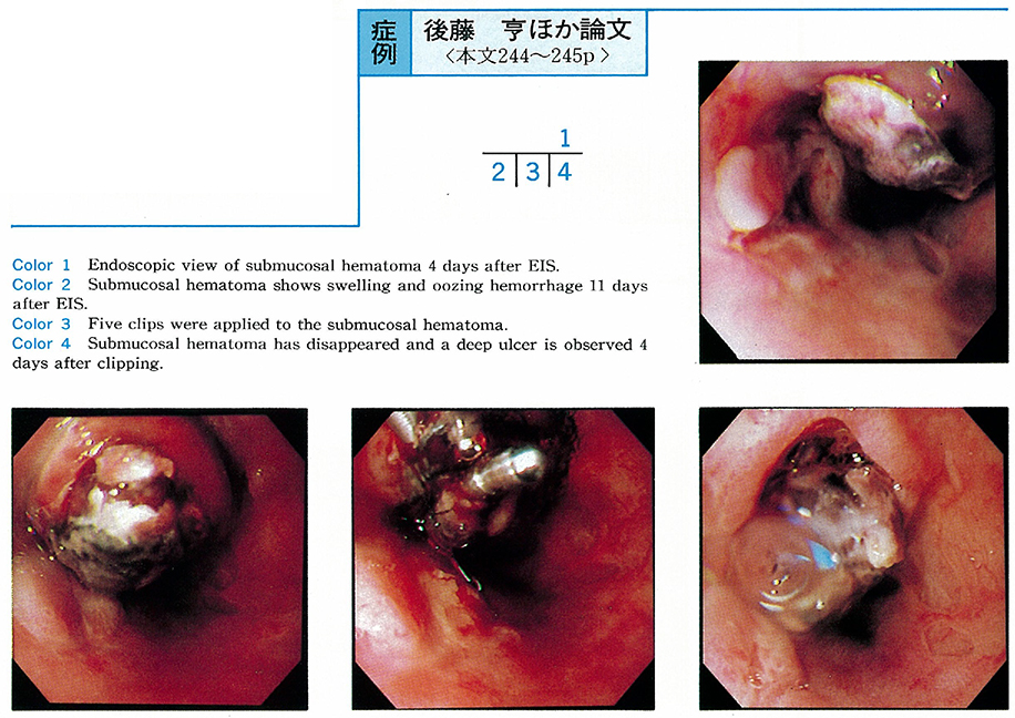
    A 64-year-old woman was diagnosed as having liver cirrhosis by biopsy 3 years previously. Esophageal varices were noted the previous year. She was treated by endoscopic variceal ligation (EVL) and EIS, but the varices recurred. Therefore, we performed endscopic injection sclerotherapy ligation (EISL) . EISL was successful and 1 week after EISL, we performed EIS by extra-vascular injection of 4ml of Aethoxysklerol.
    At 4 days after EIS, a submucosal hematoma was observed. She was treated by conservative measures, but submucosal hematoma had increased in size at 11 days. So, we tried clipping the surface of the hematoma. The hematoma had disappeared 4 days after clipping, but then a deep ulcer appeared. After one month, the ulcer was completely healed and the appearance of the varices was F0.
    We reported one successful case of clipping to treat submucosal hematoma after EIS.
    Download PDF (213K)
  • Shigeru Nakano, Akihiko Hachiya, Eisaku Kondoh, Toshio Kurita, Hiroshi ...
    1997 Volume 50 Pages 246-247
    Published: June 06, 1997
    Released on J-STAGE: February 17, 2015
    JOURNAL FREE ACCESS
    A 64-year-old female with liver cirrhosis was admitted to our hospital because of recurring esophageal varices.
    Endoscopy showed linear esophageal varices with communicating vessels to the varices on the lower esophagus as well as a slightly dilated cardiac plexus. Hepatopetal flow of paraesophageal veins via cardiac plexus was observed and esophageal variceal flow remained unclear as a result of percutaneous transhepatic portography.
    Endoscopic ligations were performed on the cardiac region to reduce blood flow toward the esophageal varices, at this time a developed esophageal varices was observed, while paraesophageal veins showed no change by EUS. Finally, EVL was performed using a spiral pattern to eradicate the varices.
    These results above suggest that esophageal varices were upward flowing rather than downward flowing, and had grester flow than that of paraesophageal veins in this case.
    Download PDF (665K)
  • Shigeru Saito, Shuichi Yamada, Mamoru Nishino, Yoshio Oda, Masato Kata ...
    1997 Volume 50 Pages 248-249
    Published: June 06, 1997
    Released on J-STAGE: February 17, 2015
    JOURNAL FREE ACCESS
    On rare occasions hemorrhagic shock develops in patients with Mallory-Weiss syndrome. We encountered a patient with Mallory-Weiss syndrome presenting with hemorrhagic shock. Hemostasis was successfully achieved with O-rings.
    The patient was a 68-year-old male who admitted our department with hematemesis after drinking a bottle of beer. When he arrived this hospital, his blood pressure was 70/40mmHg and he had a soft pulse. He had severe anemia. Emergency endoscopy following gastric lavage revealed three lacerations in the esophagogastric junction, two on the lesser curvature and one on the greater curvature. Ligation therapy with O-ring was perfomed and hemostasis was achieved completely.
    Although ulcers remained, hemorrhage did not recur to the patient. The patient was discharged without further incident. O-rings are widely used in the treatment of esophagogastric varices. We reported a case of Mallory-Weiss syndrome successfuly treated with O-ring.
    Download PDF (433K)
  • Seiji Abe, Toshitaka Takeshita, Yuji Mizokami, Takahisa Shiraishi, Kos ...
    1997 Volume 50 Pages 250-251
    Published: June 06, 1997
    Released on J-STAGE: February 17, 2015
    JOURNAL FREE ACCESS
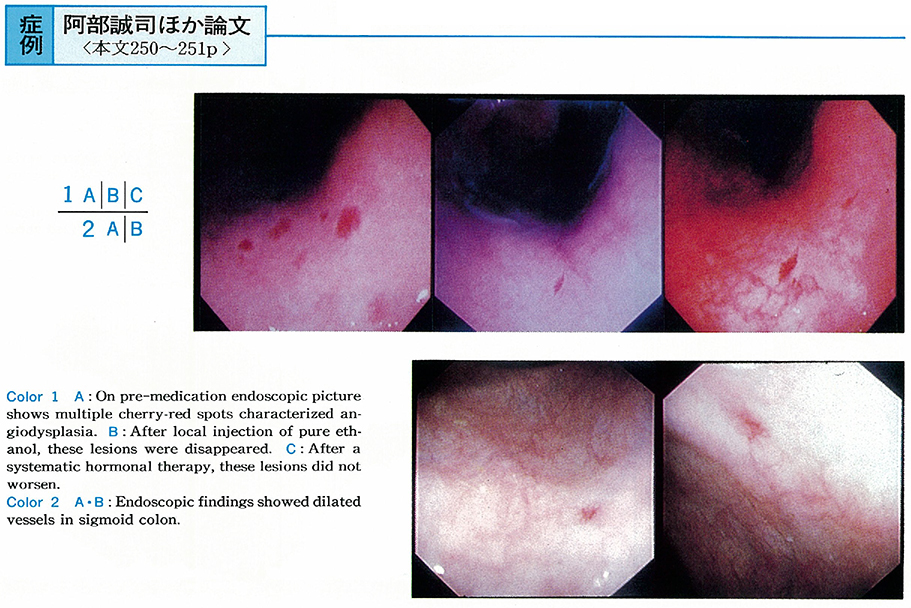
    A 71-year-old woman was hospitalized due to appetite loss and abdominal pain. Laboratory examination revealed anemia and fecal positive occult blood. Blood loss from angiodysplasia was diagnosed by endoscopically characteristic multiple gastric cherry-red spots.
    These lesions were treated endoscopically by pure ethanol injections. Treatment was successful and anemia was improved. Further more, estriol therapy was added with 2mg per day. Patient recovered completely. Local injection of pure ethanol and systemic hormonal therapy is effective treatment for bleeding from gastric angiodysplasia.
    Download PDF (452K)
  • Norio Noguchi, Tooru Honda, Ichirou Saeki, Naoya Saitou, Kimiya Takesh ...
    1997 Volume 50 Pages 252-253
    Published: June 06, 1997
    Released on J-STAGE: February 17, 2015
    JOURNAL FREE ACCESS
    A 53-year-old man complained of bloody vomiting and tarry stool on May 18th, 1996. About 5 days ago, he ate Shimesaba (raw mackerel) and so on. Endoscopic examination revealed an open and irregular ulcer with clots on the greater curvature aspect of the fornix. At that area, a codworm penetrating the gastric mucosa was found and was removed endoscopically using biopsy forceps under the direct vision.
    Histopathologically, the larva was identified as larva of Anisakis. On May 23rd, 1996, he complained of those symptoms again and was admitted to the hospital in pre-shock state. An endoscopic examination was performed immediately. There were massive blood and clots in the stomach and a ruptured vessel at the bottom of the ulcer was found. Endoscopic clippings hemostasis was performed at the vessel twice. About a month later, gastric endoscopy revealed that the ulcer was healed.
    This case was thought to be very rare that an Anisakis penetrated the gastric wall to form an ulcer with massive bleeding.
    Download PDF (264K)
  • Nehiro Kuriyama, Shin-ichi Satoh, Akiko Soyama, Motonobu Nishimura, Ke ...
    1997 Volume 50 Pages 254-255
    Published: June 06, 1997
    Released on J-STAGE: February 17, 2015
    JOURNAL FREE ACCESS
    A patient of 46-year-old male under hemodialysis owing to chronic renal failure (CRF) admitted to the hospital due to massive hematemesis and decreasing in blood pressure. An emergent endoscopy was carried out, and bleeding from a gastric elevated erosion in the antrum was found. By applying five times of clipping to the lesion, complete hemostasis was achieved.
    Erosive lesions of the upper gastrointestinal tract occurred with CRF, are known to be refractory to medical treatment. According to our data, the hemorrhage from gastric elevated erosion in CRF patient was one of the major causes of gastrointestinal hemorrhage. Usually the gastric erosions were located in the antrum and the source of active hemorrhage from them was single. Moreover, that hemorrhage tended to occur in the patients with long-term hemodialysis. We concluded that when the hemorrhage from the gastric erosion is difficult to control only by medications, it is necessary to add an endoscopic hemostasis such as clipping which does not damage the gastric mucosa so much.
    Download PDF (256K)
  • Susumu Shibuya, Takahiko Kawashima, Hiroyuki Iida, Hiroyuki Aoyagi, Na ...
    1997 Volume 50 Pages 256-257
    Published: June 06, 1997
    Released on J-STAGE: February 17, 2015
    JOURNAL FREE ACCESS
    We introduced endoscopic variceal ligation (EVL) with a giant ligator and a three-shooter for solitary gastric varix. We had 2 died cases with one autopsied case after EVL for gastric varix. Aim of this study is to determine histological changes after EVL and the indication of EVL.
    Each case was elective and emergency cases. EVL was performed for gastric varix. They had hematemesis two days after EVL and were died. It has been reported that the borderline cases (Child C terminal) for esophageal endoscopic injection sclerotherapy, had 2 out of 3 conditions ; less than albumin 2.5g/dl, more than total bilirubin 5.0mg/dl and delirium. Two reported cases was Child C terminal. First case was autospied. Histopathological findings of the autopsied specimen showed that ligated gastric varix was ulcerated without bleeding, varix between the ligated ones was thrombosed.
    Therefore, it is thought that the varix is histologically vanished after ulceration or thrombosis after EVL and the indication of EVL for gastric varix must be strictly limited than that of esophageal injection sclerotherapy in the viewpoint of liver function.
    Download PDF (268K)
  • Yujin Hoshida, Kei Shinoki, Yuichiro Yamate, Tsukasa Takeuchi, Susumu ...
    1997 Volume 50 Pages 258-259
    Published: June 06, 1997
    Released on J-STAGE: February 17, 2015
    JOURNAL FREE ACCESS
    A 70-year-old female patient with liver cirrhosis (unknown etiology) was diagnosed as hemorrhagic portal hypertensive gastropathy by upper GI endoscopy and admitted to our hospital 4 times because of iron deficiency anemia since January 1991.
    In April, 1996 (76yr) , She complained progressive dyspnea on physical exertion and fainting. Laboratory data revealed severe iron deficiency anemia. The upper GI endoscopy (June 19th) showed multiple protruding red marks with oozing type hemorrhage at cardia and antrum in the stomach. Propranolol administration was started to relieve portal hypertension at the dose of 30mgTID from July 2nd. On the 5th day, heart rate decreased 60 to 42 bpm (30%) , blood pressure 120/60 to 82/64mmHg and her symptom was getting worse. So the dose of propranolol was reduced to 20mgBID from July 15th. Heart rate decrement was 60 to 50 bpm (16.7%) , blood pressure 100/60mmHg and her symptom was improved. The upper GI endoscopic findings of PHG were improved and active hemorrhage was not observed. On hemorrhagic PHG in the elderly, adequate low dose medical treatment was suggested to be useful and safe.
    Download PDF (247K)
  • Kenji Ueda, Kaoru Nakada, Youji Harada, Shigeru Tsurui, Toshihiko Sait ...
    1997 Volume 50 Pages 260-261
    Published: June 06, 1997
    Released on J-STAGE: February 17, 2015
    JOURNAL FREE ACCESS
    The patient was a 54-year-old man who presented because of epigastric discomfort. Gastric X-ray examination and endoscopic examination of the upper gastrointestinal tract revealed a tumor of 2cm by 2cm with central depression. The patient was suspected of having a submucosal tumor. Routine biopsy yielded only normal gastric mucosal tissue so that no tumor cell was identified.
    An artificial ulcer was produceds by microwave irradiation and another biopsy was taken, with no success in the identification of tumor cells. Considering the size of the lesion and the X-ray and endoscopic findings, however, a malignant tumor could not be completely ruled out. As a result of partial gastrectomy, a definitive diagnosis of heterotopic cystic malformation of the stomach was made.
    Download PDF (695K)
  • Ichiro Saeki, Kimiya Takeshita, Masao Tani, Tooru Honda, Naoya Saito, ...
    1997 Volume 50 Pages 262-263
    Published: June 06, 1997
    Released on J-STAGE: February 17, 2015
    JOURNAL FREE ACCESS
    Case 1 : A 65-year-old male was admitted to our hospital for the operation of urinary bladder tumor. Preoperative upper gastrointestinal endoscopy revealed a protruding lesion with an ulcer in the antrum of the stomach. It was 25mm in size and the endoscopic examination using an ultrasonic probe showed homogeneous hypoechoic mass invading to the bottom of the third layer (submucosa) of the gastric wall. Partial gastrectomy was performed and the lesion was diagnosed as inflammatory fibroid polyp (IFP) histologically.
    Case 2 : A 37-year-old male was pointed out a submucosal tumor 10mm in size in the antrum of the stomach by standard endoscopy. Endoscopic ultrasonography showed homogeneous hypoechoic mass in the second and middle-third layer of the gastric wall. It was resected endoscopically and diagnosed as IFP histologically.
    It was difficult to diagnose gastric IFP endoscopically, but endoultrasonographic examination was useful in the diagnosis and treatment of these two cases.
    Download PDF (764K)
  • Tateki Yamane, Toru Furuya, Takayuki Ishii, Makoto Nakamura, Yasuo Kon ...
    1997 Volume 50 Pages 264-265
    Published: June 06, 1997
    Released on J-STAGE: February 17, 2015
    JOURNAL FREE ACCESS
    A 49-year-old male visited our hospital with a complaint of epigastric discomfort. A depressed lesion was detected in the greater curvarure of his stomach by endoscopic examination. We suspected that it was gastric early cancer, type IIc and biopsy specimen of it showed group IV (well differentiated adenocarcinoma suspected) . But specimen obtained by repeated biopsy showed group III (adenoma) .
    So we performed endoscopic mucosal resection with ligating device (EMR-L) and recognized it as adenoma. The diagnosis of depressed adenoma of the stomach is very difficult because it is similar to IIc. The method of EMR-L was useful in diagnosis of this case.
    Download PDF (778K)
  • Masao Matsumoto, Tsutomu Inomoto, Shinji Morishita, Akiko Nakamura, Ka ...
    1997 Volume 50 Pages 266-267
    Published: June 06, 1997
    Released on J-STAGE: February 17, 2015
    JOURNAL FREE ACCESS
    Recently, it has been reported that Helicobacter pylori (H. pylori) infection refered to the development of gastric mucosa-associated lymphoid tissue (MALT) lymphoma, because MALT lymphoma was improved by eradication of H. pylori.
    This patient was a 56-years-old man. Barium meal examination showed a small depressed lesion in the middle body of the stomach. Endoscopic examination revealed a scattered irregular depressed lesion in the same location. Histological study of the biopsy specimen revealed proliferation of atypical lymphoid cells and lympho-epithelial lesions in the gastric mucosa, and the diagnosis of gastric MALT lymphoma was suggested.
    H. pylori was detected by both culture and histological findings. So, we tried antibiotic therapy for H. pylori by 30mg of lansoprazole, 1500mg of amoxicillin, and 400mg of clarithromycin for 2 weeks. Six months after the therapy, eradication of H. pylori was ascertained and the MALT lymphoma lesion had disappeared endoscopically and histologically. It is suggested that H. pylori infection play a role in the pathgenesis of gastric MALT lymphoma, and that eradication of H. pylori is the first choice treatment for gastric MALT lymphoma.
    Download PDF (799K)
  • Toshiaki Okumura, Nagamu Inoue, Takeharu Hayashi, Tomoaki Ikezawa, Tet ...
    1997 Volume 50 Pages 268-269
    Published: June 06, 1997
    Released on J-STAGE: February 17, 2015
    JOURNAL FREE ACCESS
    We experienced a case with gastric leiomyosarcoma resected by laparoscopic procedure. A 37-year-old woman was referred to the hospital because of an elevated lesion of the stomach. We performed barium meal examination as well as esophagogastroduodenoscopy, indicating submucosal tumor in the posterior wall of the upper body of the stomach. The tumor was further clarified by endoscopic ultrasonography and abdominal CT, suggesting leiomyosarcoma.
    The tumor was resected by laparoscopic procedure with a sufficient surgical margin, and finally diagnosed gastric leiomyosarcoma. The patient was discharged on 9th day after the operation. The laparoscopic local resection is thought to be an useful technique for submucosal tumor.
    Download PDF (1062K)
  • Tetsuya Sanji, Yasuaki Sakai, Manabu Nishimaki, Yutaka Tani, Syouko Mi ...
    1997 Volume 50 Pages 270-271
    Published: June 06, 1997
    Released on J-STAGE: February 17, 2015
    JOURNAL FREE ACCESS
    A 51-year-old female with no complaint underwent medical examination in 1993, and gastric X-ray revealed multiple small gastric polyps on the greater curvature from upper to middle corpus. Endoscopy showed multiple appearance of small gastric polyps in that area, corresponded to Yamada II type and had same color tone as peripheral mucosa. Histopathological finding of polyps demonstrated fundic gland polyps.
    Follow-up endoscopy was performed once a year, and findings in August 1995 indicated disappearance of polyps and sessile type elevated lesion with redness, 2mm in diameter, on the greater curvature of angulus. The lesion became larger in size (10mm in diameter) and showed sub-pedunculated type in June 1996. The results of biopsy led us to diagnose the lesion as papillary adenocarcinoma.
    Endoscopic mucosal resection was performed in July 1996 and histopathological fingings revealed depth of invasion was m and no malignant cells was recognized in the cut end. From the evidence in the sets of examination and from a survey of the literature, we concluded this case as rare.
    Download PDF (857K)
  • Fuyuhiko Yamamura, Hirotaka Yamada, Tetsuya Mikami, Kazuhiro Kaneko, Y ...
    1997 Volume 50 Pages 272-273
    Published: June 06, 1997
    Released on J-STAGE: February 17, 2015
    JOURNAL FREE ACCESS
    The patient was a 62-year-old woman. In 1984, she was pointed out to have a 5mm-sized polyp in the upper body of stomach and it was diagnosed as hyperplastic polyp by a biopsied specimen. The polype enlarged to 10mm in diameter over the next 12 years and its endoscopic appearance changed to lobulated and variegated one.
    Since histological findings of biopsied specimens in 1996 showed adenocarcinoma, polypectomy was performed. Microscopic study of the resected specimen demonstrated well-defferentiated adenocarcinoma located on the surface of hyperplastic polyp. This is a very rare case of gastric hyperplastic polyp accompanied by the malignant transformation recognized by serial endoscopy during follow-up for 12 years.
    This case suggests that enlargement and lobulating tendency could be considered as sings of malignant transformation of hyperplastic polyps. Diagnostic reliability of biopsy could not be always perfect in case of malignant transfomation of gastric hyperplastic polyp, therefore, endoscopic follow-up should be performed continuously. When the signs indicated above are observed, endoscopic polypectomy should be applied regardless of negativeresults of biopsied specimens.
    Download PDF (384K)
  • Takeshi Hayashi, Yasuhito Takahashi, Yutaro Kobayashi, Yoshihiro Suzuk ...
    1997 Volume 50 Pages 274-275
    Published: June 06, 1997
    Released on J-STAGE: February 17, 2015
    JOURNAL FREE ACCESS
    Upper GI endoscopy was performed on a 75-year-old male patient complaining of abdominal discomfort on March 22. A polyp 2cm in diameter with the smooth surface, Type III (Yamada's criteria) was detected at the lesser curvature of the pyloric antrum, and was diagnosed as adenoma.
    After endoscopic polypectomy on may 30, an enlarged polyp (3cm) with lobulation was observed. Histological findings revealed adenoma including hyperplasia of the lacunar epithelium and intestinal epithelial metaplasia. Finally, the polyp was diagnosed as focal cancer in adenoma with high grade atypical cells and structure.
    This is an important case to study differentiated adenocarcinoma of the stomach.
    Download PDF (1084K)
  • Eiko Kanda, Shigeru Nakano, Toshio Kurita, Hiroshi Koyama, Eisaku Kond ...
    1997 Volume 50 Pages 276-277
    Published: June 06, 1997
    Released on J-STAGE: February 17, 2015
    JOURNAL FREE ACCESS
    A 43-year-old female patient had epigastralgia and had been pointed out SMT like elevated lesion on the anterior wall of the antrum in February 1995. Follow up study had done in April 1995, and diagnosed poorly differentiated adenocarcinoma by biopsy. On ultrasonic endoscopy, the invasive depth of the tumor showed in the muscle layer. Gastrectomy was carried out.
    There are more than 60 cases reported as SMT tumors in the stomach. We found this case as one of striking resemblance to gastric cancer and concidered about the some features of this type cancer in the stomach.
    Download PDF (1088K)
  • Jun Unno, Toshitaka Ishiko, Hisashi Okuyama, Sakiko Arai, Kie Yokoyama ...
    1997 Volume 50 Pages 278-279
    Published: June 06, 1997
    Released on J-STAGE: February 17, 2015
    JOURNAL FREE ACCESS
    An 81-year-old man visited our hospital because of loss of appitite and general fatigue. A panendoscopic examination revealed a round ulcerated lesion on the posterior wall of the gastric antrum and a pathological examination of the biopsied specimen showed a poorly differentiated adenocarcinoma. Since multiple liver tumors were detected by an abdominal CT scan and an elevated value of serum AFP, 273ng/ml, was noticed, a liver tumor biopsy was done after admission. As it was the same histology as that of the gastric carcinoma, an AFP producing gastric carcinoma with liver metastasis was highly suspected.
    Ten units of OK-432 and 250mg of 5-furuolouracil (5-FU) were injected concomitantly at the site of the gastric carcinoma through a fine needle via a panendoscopy twice with a week's interval. Ten days after the local injection, 750mg of 5-FU and 120mg of methotrexete were given intravenously. On that day the patient suddenly exhibited a fever, a chill, jaundice and hematuria. The next day he died of disceminated intervascular coagulation and multiple organ failure.
    At the autopsy, a lot of odorous gas was retained intraperitoneum and subcutaneously. There were multiple cavities in the organs such as the liver, kidney, heart and so on. A microscopic examination showed multiple rod shaped organisms on the sides of the cavities and in the vein. All these findings indicated the sepsis of gas producing organisms.
    Download PDF (796K)
  • Yukiko Yoshida, Shigeaki Nagao, Shigeaki Aono, Chizuko Okada, Kenji Ha ...
    1997 Volume 50 Pages 280-281
    Published: June 06, 1997
    Released on J-STAGE: February 17, 2015
    JOURNAL FREE ACCESS
    A 44-year-old man visited our hospital due to the epigastralgia. An endoscopic examination revealed an active gastric ulcer in the antrum of the stomoch and a healing ulcer in the duodenal bulb. Despite that either H2 receptor antagonists or proton pump inhibitors had been administered, none of them had been effective enough for ulcers to be cured. An endoscopic examination after 17 months of the conventional therapy revealed the gastroduodenal fistula, so-called the double pylorus.
    Because he was found to be positive for Helicobacter pylori by the culture of gastric biopsy specimen as well as pathologically, he received an eradication therapy of H. pylori with omeprazole 40mg plus amoxiciline 1500mg for 14 consecutive days. The therapy brought both ulcers to be cured with red scars and set the patient free from the symptom although the ulcers were recurrent after 12 weeks. Due to his request, recradication therapy was done with omeprazole 40mg, amoxicilin 1,500mg plus clarithromycin 400mg for 14 consecutive and had brought the ulcers to be cured with white scars and negative for H. pylori. The ulcers have never been recurrent and remained scars since re-eradication therapy.
    This case is considered rare and worth to report because it had been able to observe that the intractable gastroduodenal ulcers had become the gastroduodenal fistula“the double pylorus”and the eradication of H. pylori had been effective for preventing the intractable ulcers from recurring.
    Download PDF (642K)
feedback
Top