Progress of Digestive Endoscopy(1972)
Online ISSN : 2189-0021
Print ISSN : 0389-9403
Volume 48
Displaying 1-50 of 81 articles from this issue
Technology and instrument
  • Goro Kaneda, Toru Kimura, Yukihito Kokuba, Akifumi Amemiya, Fumiko Koh ...
    1996Volume 48 Pages 51-54
    Published: June 07, 1996
    Released on J-STAGE: March 20, 2015
    JOURNAL FREE ACCESS
    A problem of intragastric surgery is gas leakage from stomach to the intestine. Presently gas leakage is prevented by inserting intestinal clamp into the abdominal cavity via trocar and fixed to the duodenum or jejunum. However, due to difficulty in locating, complete blockage is not always possible and long fixation of the clamp can cause intestinal damage. For these reasons, we developed two new procedures and instruments.
    Procedure 1 : A double balloon attached to a tube is inserted into the stomach. The balloon in front is inserted into the duodenum and inflated. The rear balloon is then inflated. The two inflated balloons pinch the pyloric ring and block gas leakage.
    Procedure 2 : A pliable double disc is inserted into the oral overtube which was previously inserted. The front disc is placed in the duodenum, and then the rear disc is attached to the pyloric ring. This causes complete gas blockage.
    Download PDF (1569K)
Clinical study
  • Tadashi Hachisu, Shin-ichi Satoh, Hideo Yamada, Motonobu Nishimura, Ka ...
    1996Volume 48 Pages 55-58
    Published: June 07, 1996
    Released on J-STAGE: March 20, 2015
    JOURNAL FREE ACCESS
    We newly developed endoscopic variceal ligation using the detachable snare and the transparent cap with rim (EVLsc) . Advantages of the EVLsc are as follows ; multiple varix ligation can be achieved in a short time without over-tube, satisfactory results is available by fewer ligatures of EVLsc compared with usual EVL, better view can be obtained due to the usage of a transparent cap, and cost effective because no additional devices are needed.
    A clinical study on efficacy and compilications of EVLsc was carried out 10 cases with esophageal varices in which F-number was 2-3 and a case with cardiac varices during the period from August to December 1995 at Sakura National Hospital. Purpose of EVLsc was urgent hemostasis in 2 (including a case with cardiac varices) and prophylactic ligation of esophageal varices in 9.
    As a results, in the 2 cases who underwent hemostatic EVLsc, prompt hemostasis was available. And in the 10 cases with esophageal varices (including a case treated by hemostatic EVLsc) , improvement of F-number was obtained in all 10 cases and disappearance of the RC sign in 8 cases out of 9 (89%) at 1-2 weeks after EVLsc. There were no complications associated with EVLsc. We conclude that EVLsc is useful for treatment of esophageal or cardiac varices.
    Download PDF (1097K)
  • Shinichi Nakamura, Atsushi Mitsunaga, Youko Murata, Shigeru Suzuki
    1996Volume 48 Pages 59-63
    Published: June 07, 1996
    Released on J-STAGE: March 20, 2015
    JOURNAL FREE ACCESS
    Endoscopic variceal ligation (EVL) has been popular rapidly, because of the easy technique and useful effects. But EVL is not always safe, we have occasionally experienced some complications and bleeding cases in the patients treated by EVL. We discussed about the problems of early bleeding cases after EVL compared with the contrast no bleeding cases.
    We have been performed EVL for the prophylactic treatment of 120 patients, and recognized 5 cases (4.2%) of early bleeding and 4 cases of recurrent bleeding after EVL.
    In 5 cases of the early bleeding, there were 4 male cases and 1 female case, and the mean age of cases was 62.2 years old. There were 3 cases of liver cirrhosis associated type C hepatitis viral infection, 1 case of liver cirrhosis due to alcohol and 1 case of extrahepatic portal vein occlusion in the primary liver diseases. The form of esophageal varices before treatment were the all risky varices above F2 level with red color sign. There were 2 cases of first treatment, 2 cases of recurrence after endoscopic injection sclerotherapy (EIS) and 1 case of recurrence after operation in the therapeutic indication. There were 3 cases of class B and 2 cases of class C in the liver function by Child's classification. We didn't recognize the significant difference of liver function between the early bleeding cases and the contrast cases.
    It was 5.6 EVL sessions of the mean number for the treatment and 13.2 days of the mean period to bleeding after EVL. Bleeding points were the ulcerative lesions due to EVL. Early bleeding cases had the past history of EIS or operation for esophageal varices and the large hepatocellular carcinoma with portal thrombus. They were also the cases with general complication of diabetes mellitus, nephrotic syndrome and malignant tumor.
    In conclusion, though EVL is the less invasive and safe technique for the treatment of esophageal varices, we considered that it was necessary to be careful for the treatment and follow up of the cases with these risk factors and general complications.
    Download PDF (615K)
  • Izumi Sugimoto, Norichika Narimiya, Hiroko Hamada, Yoshihiro Itoh, Tat ...
    1996Volume 48 Pages 64-66
    Published: June 07, 1996
    Released on J-STAGE: March 20, 2015
    JOURNAL FREE ACCESS
    This study was conducted to assess the influence on esophageal varices and gastric mucosa following balloon-occluded retrograde transvenous obliteration (BRTO) in 10 patients with liver cirrhosis and gastric varices.
    Esophageal varices in 2 cases have been exacerbated by BRTO and one of them was required additional EIS. In all cases, reddish mosaic-like findings,“scaly reddening”we call, was observed in the gastric mucosa with gastric varices following BRTO. In only two cases portal hypertensive gastropathy (PHG) was deteriorated following BRTO. Newly developed? PHG have not been observed in 8 cases without PHG after the procedure. It is unlikely that obliteration of gastrorenal shunt BRTO induces deleterious effect on PHG.
    Download PDF (418K)
  • Takao Horiuchi, Hisao Tajiri, Atsushi Ohtsu, Takahiro Fujii, Narikazu ...
    1996Volume 48 Pages 67-70
    Published: June 07, 1996
    Released on J-STAGE: March 20, 2015
    JOURNAL FREE ACCESS
    To clarify the problems of endoscopic diagnosis for gastric lesions with open ulcer, we assessed the endoscopic appearance of the discrepant cases between the first endoscopic diagnosis and histopathological diagnosis. There were 550 cases which had gastric lesions with open ulcer, excluding non-epithelial tumors and received endoscopic biopsies between July 1992 and July 1995 at National Cancer Center Hospital East.
    Twenty cases of those were discrepant between the first endoscopic diagnosis and histopathological diagnosis, in which 6 cases (2.0%) were false positive and 14 (5.6%) false negative. Five of 6 false positive cases were diagnosed as advanced cancer and 8 of 14 false negative cases as IIc+III at the first endoscopic examination. In the second look diagnosis, 3 of 6 false positive cases and 8 of 14 false negative cases were diagnosed correctly. However, 3 of false positive cases could not be diagnosed even in the second look diagnosis because of showing irregularity of the ulcer margin, thinning and step-down of converging folds, or multiple ulcers with various erosions and redness. In addition, 6 of false negative cases could not be diagnosed correctly in the second look diagnosis, because one located in the prepylorus could not be observed thoroughly and the other 5 lacked malignant findings endoscopically. It was thought that the differential diagnosis was difficult endoscopically between benign ulcers and malignancies with open ulcer of the stomach in a total of 9 cases (1.6%) .
    These results suggest that careful observation combined with dye spraying method and re-examination after follow-up by anti-ulcer therapy are necessary for gastric lesions with open ulcer.
    Download PDF (537K)
  • Naoya Murase, Satoshi Okabe, Takehiro Arai, Shoji Maruyama, Masanori T ...
    1996Volume 48 Pages 71-74
    Published: June 07, 1996
    Released on J-STAGE: March 20, 2015
    JOURNAL FREE ACCESS
    We have been investigating histophathological feature under the muscularis mucosae and degree of invasion of submucosal invasive cancer for the purpose of search for the risk factors of lymph node metastasis and recurrence. We subclassified we
    ll differentiated adenocarcinoma histologically into two types i. e. simple and mixed type. We represented specific pathological features in the deepest portion of cancer as single cell infiltration (SCI) and mucinous component (MUC) . As for the degree of invasion, we classified it into three grades according to“sm depth”and“sm width”which indicated vertical and horizontal extention of carcinoma in the interstitium.
    Ninty-six cases of submucosal invasive cancer (11 cases with lymph node metastasis or recurrence) were investigated clinicopathologically. Eight of 11 cases lymph node metastasis or recurrence were located in the rectum, 9 of 11 cases showed Is-type appearance. Concerning risk factor, lymphatic invasion, venous invasion and SCI had significant relation to metastasis and recurrence. All of 11 cases showed massive submucosal invasion classed as Grade-3 (sm depth>1000µm and sm width>4 mm) .
    In order to evaluated these pathological risk factors enough, we have been performing endoscopic treatment for all lesion except one which cannot be resected curatively. According to the result of complete pathological examination, we decide the way to treat. We consider the absolute indication of subsequent intestinal resection after endoscopic resection is as follows, (1)SCI (+) , (2)lymphatic invasion (+) , (3)venous invasion (+) , (4)Graded-3, (5)lesion contains component of moderately or poorly defferentiated adenocarcinoma.
    Download PDF (547K)
  • Yoshiro Tamegai, Hidemi Ooba, Nobutaka Satoh, Noboru Komatsubara, Shig ...
    1996Volume 48 Pages 75-79
    Published: June 07, 1996
    Released on J-STAGE: March 20, 2015
    JOURNAL FREE ACCESS
    This study was carried out to find endoscopical signs of the degree of submucosal invasion with superficial type submucosal invasive cancers of the colon.
    Materials and Methods : Studies ware carried out on 47 cases of superficial type sm colonic cancer (30 males and 17 females with a mean age of 65.3 years) with 50 lesions. X-ray findings, endoscopical findings, morphological measurements, rate of lymph node metastasis, pit patterns of 23 sm cancer lesions were statistically compared between sm1 and sm2 or sm3 cancers.
    Results and Discussion : (1)Lymph nodes metastasis was found in 3 cases (6.0%) . It's metastatic rate was significantly high in these cases undergoing endoscopic resection as initial treatment, as assessed by Student's t-test. (2)When X-ray and endoscopical findings were assessed by multivariate analysis (Logistic regression) , there were significant differences in uneveness of barium fleck of the lesion surface and in marginal undulation of the depressed area difference between sm1 and sm2 or sm3 cancers. (3)Morphological measurements revealed a significant difference in the thickness of the muscularis propria and lesion height. (4)The comparison of pit patterns showed that a significant proportion of sm2 and sm3 cancers were found to have a type V pit pattern (according to Kudo's classification) .
    Download PDF (627K)
  • Kiyonori Kobayashi, Tomoe Katsumata, Hiroyuki Takahashi, Kaoru Yokoyam ...
    1996Volume 48 Pages 80-84
    Published: June 07, 1996
    Released on J-STAGE: March 20, 2015
    JOURNAL FREE ACCESS
    The purpose of this study was to evaluate the effectiveness of endoscopic ultrasonography (EUS) and submucosal saline injection method in the diagnosis of infiltration degree of submucosal carcinoma (sm-ca) . We performed EUS for 55 lesions of sm-ca, and submucosal saline injection method for 19 lesions of sm-ca. The degree of elevation by submucosal saline injection was evaluated for each lesions ; a case in which the lesion was completely elevated was defined as a positive lifting sign, a case in which the lesion was incompletely elevated was defined as a false positive lifting sign, and a case in which the lesion was not elevated was defined as a negative lifting sign.
    The results were as follows ;
    1) The depth of cancerous invasion was accurately diagnosed by EUS in 76% (42/55 lesions) of sm-ca. Accuracy rate of infiltration degree of sm-ca by EUS was 50% in sm1, 59% in sm2 and 75% in sm3 carcinomas respectively. The infiltration degree of sm-ca mostly correlated with high diagnostic accuracy of EUS. The differentiation between m-sm1 that were generally treated endoscopically and sm2-that were suitable for surgical operation was possible in 89% of sm-ca. Accordingly, EUS was useful in determining the therapeutic method of sm-ca.
    2) Lifting sign by submucosal saline injection method were all positive in sm1 carcinomas, and all negative in sm3 carcinomas. In 7 lesions of sm2 carcinomas, lifting sign were positive in 2 lesions, false positive in 2 lesions, and negative in 3 lesions.
    It was concluded that EUS and decision of lifting sign by submucosal saline injection method are useful in diagnosing the infiltration degree of sm-ca and in planning therapeutic method.
    Download PDF (1566K)
  • Shuichi Fujioka, Kimikazu Iwamoto, Ryuzo Murai, Satoshi Murata, Kazuto ...
    1996Volume 48 Pages 85-88
    Published: June 07, 1996
    Released on J-STAGE: March 20, 2015
    JOURNAL FREE ACCESS
    The goal in treatment of villous tumor of the rectum is complete excision of the tumor with preservation of the anal sphincter. Transanal endoscopic microsurgery (TEM) permits precise excision of large sessile tumors which is located 5 to 20cm from the anus. By using a operative rectoscope sealed with a gastight working insert fine visualization of a rectal tumor can be achieved. Also the manipulation of the endoscopic surgical instruments such as the high frequency knife, forceps, scissors, and suction device allows precise excision of the lesion as well as the suture of the defect of the rectal wall.
    Six patients has been underwent TEM resection for the rectal villous tumors between June 1994 and August 1995. All patients were examined by endorectal ultrasound. Villous tumors ranging from 3.2 to 8.0cm in size (mean, 4.6cm) . Mucosectomy was performed in three patients and full thickness excision in other three patients. Distal extent of the tumors ranged from 5 to 12cm from the anal verge.
    The operative duration ranged from 70 to 250 min (mean, 128min) . Postoperative histological diagnosis revealed 2 villotubular adenocarcinoma, 3 tubulovillous adenocarcinoma, and 1 tubulovillous adenoma. Five cases that diagnosed adenocarcinoma were intramucosal cancer and no invasion to the submucosal layer were seen. Complications were occurred as follows in 5 patients (83%) : 2 unknown fever, 3 dehiscence of the suture line, and 1 perforation. All of the patient were treated conservatively. The mean length of hospital stay was 10 days. No cases were converted to conventional method of resection.
    TEM is a safe and effective method for resecting favorable tumors in select cases.
    Download PDF (1244K)
Case report
  • Hisagi Yamada, Katsuhiko Iwakiri, Masaoki Yonezawa, Makoto Kotoyori, T ...
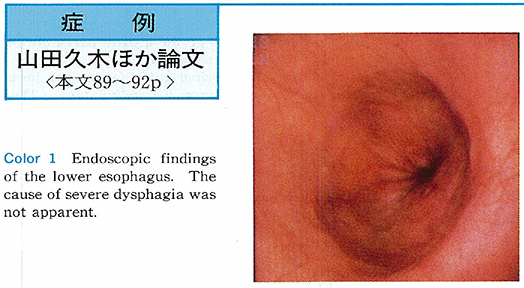
    1996Volume 48 Pages 89-92
    Published: June 07, 1996
    Released on J-STAGE: March 20, 2015
    JOURNAL FREE ACCESS
    A 72-year-old man was hospitalized with severe dysphagia and weight loss (6 kg/2 months) . Occasional dysphagia had occurred since about 20 years of age. He had been diagnosed with diffuse esophageal spasm (DES) 5 years earlier and was followed. Since July 1995, dysphagia became aggravate and prevented food intake.
    On endoscopy, the cause of severe dysphagia was unclear. Radiological findings showed segmental contraction and a pool of mucus. On conventional esophageal manometry, basal lower esophageal sphincter (LES) pressure was 28 mmHg, and LES relaxation on swallowing was incomplete. Basal esophageal body pressure was higher than intragastric pressure. Swallowing 5 ml of water showed repetitive simultaneous contraction. The mean amplitude of simultaneous contraction was 40-60 mmHg.
    These findings are characteristic of both achalasia, and DES and we diagnosed vigorous achalasia. The pathogenesis of vigorous achalasia is unclear. In this case, the possibility is that there was a transition from DES to achalasia. We consider that this case is valuable in the elucidation of the pathophysiology of vigorous achalasia.
    Regarding treatment, oral nifedipine (10 mg) was not effective so we performed pneumatic dilatation. After pneumatic dilatation, symptoms disappeared entirely.
    Download PDF (1178K)
  • Kyoya Sakimura, Hiroyuki Machino, Katsuyuki Matsumaru, Hideki Saiki, K ...
    1996Volume 48 Pages 93-96
    Published: June 07, 1996
    Released on J-STAGE: March 20, 2015
    JOURNAL FREE ACCESS
    A 60-year-old male consulted us as a follow-up after an incidence of hematemesis. The incidence had occurred following consumption of an alcoholic beverage. A diagnosis of hemorrhagic gastritis was given and the patient was treated with an antacid at an emergency hospital.
    On physical examination, no abnormal findings were detected except for reduced respiratory sounds of the lung. Laboratory data (eg, circulating blood cell counts, urine and serum chemistry, and tumor markers) were all normal. A double-contrast esophagogastric series showed an irregular esophageal mucosal border and sliding hernia of the stomach. Esophagoendoscopy showed that the esophageal mucosa assumed a gastric mucosa-like appearance at a level of 28 cm or more from the incisor teeth, which was associated with an irregular surface, ulcerous scars, and erosions. Through endoscopy and with the aid of a dye, we observed several tiny squamous islets at the anal side of the esophagus. A diagnosis of Barrett's esophagus was made. The biopsy specimen collected from Barrett's esophagus revealed well-differentiated adenocarcinoma. Subtotal esophagectomy was conducted.
    Macroscopic examination of the resected specimen showed multiple nodules and ulcer scars at the anal side of the esophagus. Under microscopy, the same specimen revealed a superficial spreading esophageal cancer measuring 6.5 by 7.5cm, which almost occupied the entire Barrett's esophagus. The cancer was generally localized within the mucosa but a small portion had invaded the submucosa region. There was no evidence of lymphatic metastasis.
    We feel that the superficial spreading type of esophageal cancer arising out of Barrett's esophagus is rare in Japan.
    Download PDF (1455K)
  • Genichi Koyama, Satoshi Wakasugi, Tatsuhiro Syoji, Satoshi Suzuki, Tsu ...
    1996Volume 48 Pages 97-99
    Published: June 07, 1996
    Released on J-STAGE: March 20, 2015
    JOURNAL FREE ACCESS
    We reported a case of intramural hematoma of the esophagus and stomach. A 73-year-old woman was admitted to our hospital complaining of nausea, vomiting and hematemesis. An emergent endoscopic examination revealed swelled mucosa spreading from whole the esophagus to lower body of the stomach, and was dark red in color. We diagnosed as intramural hematoma of the esophagus and stomach. Fasting and intravenous hyperalimentation were indicated on admission.
    She became asymptomatic until the eighth day of hospitalization. The 4th esophagoscopy performed twenty-six days later, the lesion had completely disappeared and inflamed lesion had improved.
    Williams in 1957 first reported esophageal hematoma. To our knowledge, there are a total of 70 cases reported in literature including 17 Japanese cases. Major symptoms of these cases were chest pain and slight hematemesis. In almost cases, hematomas were found in the middle and distal part of the esophagus. The diagnosis is made by endoscopic or X-ray examination. On endoscopic examination, an elevated submucosal lesion of dark red in color is charactristic. Barium meal often reveals smooth filling defects of variable length. A favorable prognosis was reported on reviewed literatures. Our particular case recovered smoothly only by conservative therapy.
    Download PDF (437K)
  • Naoyuki Yahata, Nobuyuki Ohkubo, Tadakazu Hisamatsu, Tetsu Takeuchi, A ...
    1996Volume 48 Pages 100-103
    Published: June 07, 1996
    Released on J-STAGE: March 20, 2015
    JOURNAL FREE ACCESS
    A 65-year-old female was referred to our hospital because of multiple gastric polyps and anemia for further evaluation. No obvious symptoms related carcinoid tumors were complained (such as flushing, wheezing, diarrhea and so on) .
    Upper GI series and endoscopic examination demonstrated multiple gastric polyps on the body of the stomach. A pedunculated polyp (Yamada's classification Type IV, 2.7×2.0×1.5 cm in diameter) was observed at the greater curvature of the upper gastric body. Carcinoid tumors were found histopathologically in all of the specimens biopsied from the polyps. Extensive examination had failed to demonstrate metastasis of carcinoid tumors, then the total gastrectomy was followed.
    After surgical resection, carcinoid tumors were found in the pedunculated polyp and all of the gastric polyps pathologically, and they were found in the gastric mucosa, muscularis mucosae and submucosal layer of the body of the stomach.
    We could have referred only nine reported cases of gastric carcinoid tumors with pedunculated or semi-pedunculated lesions in Japanese literature including our case. Only two cases were reported which carcinoid tumors had grown to a pedunculated polyp.
    In summary, we reported here a very rare case of multiple gastric carcinoids developed into a pedunculated polyp.
    Download PDF (1574K)
  • Youko Kitamura, Hitoshi Hashimoto, Masumi Akimoto, Seiko Sakakida, Yum ...
    1996Volume 48 Pages 104-106
    Published: June 07, 1996
    Released on J-STAGE: March 20, 2015
    JOURNAL FREE ACCESS
    The patient, a 51-year-old male, had been examined endoscopically twice a year since 1984. Early gastric cancer (type IIc) was finally detected in the posterior wall of the antrum through endoscopy for a regular check-up in July 1993, and gastrectomy was performed. It was pathologically confirmed as early gastric cancer type IIc ie, moderately-differentiated adenocarcinoma with 6×5 mm in size. Immunohistochemical staining showed p53-positive for the greater part of the carcinomatous gland. Endoscopy had previously revealed a small depressed lesion with redness in the metaplastic mucosa as the original lesion of cancer. Three time of biopsy had been carried out during the follow up period, resulting in the histological diagnosis of group II (regenerative atypism) in all cases. The p53-positive glands were detected from a biopsy specimen taken in July, 1990 (second biopsy performed three years before final) .
    It is well known that differentiated type of gastric cancer frequently shows overexpression of p53, regardless of the stage. And the case presented may indicate that immunohistochemical stainig of p53 is useful for the early detection of cancer, even in a case with benign histological diagnosis.
    Download PDF (1232K)
  • Nobuhisa Kurihara, Naoya Saito, Masao Tani, Fumio Kando, Kimiya Takesh ...
    1996Volume 48 Pages 107-110
    Published: June 07, 1996
    Released on J-STAGE: March 20, 2015
    JOURNAL FREE ACCESS
    The case was a 63-year-old male who was noted to have a submucosal tumor at the anterior wall of the duodenal bulb. Histological diagnosis of the biopsied specimen from the tumor showed the presence of carcinoid tumor, but there was no signs and symptoms of carcinoid syndrome.
    Endoscopic ultrasonography using a miniture probe demonstrated the tumor was 5×5 mm in size and located within the submucosal layer of the duodenum. The tumor was resected successfully by endoscopic mucosal resection using a cap-fitted panendoscope (EMRC) . Histologically, our case was so called classical carcinoid tumor. There was no carcinoid invasion to the resected margins, but vascular invasion was present (v1 and ly1) .
    Follow-up examinasions by endoscopy revealed no evidence of recurrence or metastatic spread of carcinoid tumor up to now, but severe follow-up examination should be performed in the future in this case.
    Download PDF (1192K)
  • Yoko Itsuki, Atsushi Tatsuguchi, Jun Sato, Shunji Fujimori, Hironori T ...
    1996Volume 48 Pages 111-114
    Published: June 07, 1996
    Released on J-STAGE: March 20, 2015
    JOURNAL FREE ACCESS
    A 73-year-old male patient came to our hospital complaining of appetite loss and body weight loss. X-ray and endoscopic examinations revealed from the stomach to the rectum multiple polypoid lesions. Physical examination and CT showed lymph nodes swelling in the whole body.
    The biopsy specimens taken from the axillary lymph nodes and gastrointestinal lesions lead to the histological diagnosis of malignant lymphoma, diffuse, small cleaved, B-cell type. Thus, as primary focus was considered GI tract, this case was diagnosed multiple lymphomatous polyposis. After chemotherapy, the X-ray and endoscopic findings were markedly improved from ascending colon to sigmoid colon.
    Download PDF (1490K)
  • Takayuki Yajima, Tokuhisa Sezaki, Gaku Hayashi
    1996Volume 48 Pages 115-118
    Published: June 07, 1996
    Released on J-STAGE: March 20, 2015
    JOURNAL FREE ACCESS
    A 48-year-old female patient visited to Chiba Cancer Center to receive a thorough examination of the transverse colon polyp. Barium enema examination revealed a smooth surfaced submucosal tumor sized 10mm in the transverse colon closed to the lienal flexure. Colonoscopic examination showed a sessile, slightly yellowish submucosal tumor. A biopsy of the tumor was performed and the tumor was histologically confirmed as granular cell tumor, and endoscopic polypectomy was applied. The resected specimen was solid and sized 10×8×6mm.
    The tumor was mainly situated in the submucosal layer and demarcated well. Histologically, the tumor cell was relatively large, and each cell had eosinophilic granules in the cytoplasm. An immunohistochemical analysis revealed positivity for S-100 protein and neuron specific enolase (NSE) . So the diagnosis of granular cell tumor of the colon was made.
    Granular cell tumor of the large bowel in Japan were reported 21 cases inclusive of our case. We discussed the characteristics of our case comparing them.
    Download PDF (1336K)
  • Yasushi Koike, Kazushige Arai, Masatoshi Kawamura, Hiroshi Ishii, Kazu ...
    1996Volume 48 Pages 119-122
    Published: June 07, 1996
    Released on J-STAGE: March 20, 2015
    JOURNAL FREE ACCESS
    A 67-year-old male was admitted to the internal medicine department of the hospital for malaise and hepatopathy in December 1994. CT and ERCP showed dilatation of the intrahepatic bile ducts and stenosis of the hilar hepatic bile ducts. The patient was transferred to the department of surgery in February 1995 after bile duct cancer of the hepatic hilum was diagnosed.
    Radiotherapy was started for extensive infiltration. Percutaneous transhepatic cholangio-drainage (PTCD) was simultaneously performed through each of the right and left intrahepatic ducts. Two Accuflex selfexpandable stents, 10 mm in diameter and 40 mm in length, were inserted into the right and left intrahepatic bile ducts. The patient was temporarily discharged from the hospital as jaundice was continuing to improve. However, he died of cancerous peritonitis on day 43. The stents remained sufficiently expanded and satisfactorily relieved jaundice until the patient died.
    One case of unresectable hilar bile duct cancer in which right and left lobes were successfully fistulized is reported here.
    Download PDF (1641K)
Technology and instrument
  • Hiroshi Miyoshi, Tamami Hanashi, Yuji Hanatani, Tatsuo Asagoe, Nobuhik ...
    1996Volume 48 Pages 124-125
    Published: June 07, 1996
    Released on J-STAGE: March 20, 2015
    JOURNAL FREE ACCESS
    We have developed endoscopic clipping method of esophageal varices using HX-3L (Olympus Optical Co Ltd, Tokyo) since 1990. Though this method have been recognized effective and minimally invasive, small bleeding during treatment or early recurrence of varices sometimes have occurred.
    Therefore, we completed a newly designed and special shaped clip for endoscopic treatment of esophageal varices. Eight patients successfully received endoscopic clipping of esophageal varices using this new type of clip.
    Download PDF (1063K)
  • Masao Tani, Kimiya Takeshita, Ichiro Saeki, Seitaku Hayashi, Haruhiro ...
    1996Volume 48 Pages 126-127
    Published: June 07, 1996
    Released on J-STAGE: March 20, 2015
    JOURNAL FREE ACCESS
    We performed endoscopic mucosal resection (EMR) using a cap-fitted panendoscope (EMRC) for 57 cases of 67 gastric lesions from August 1992 to October 1995.
    Before resection, endoscopic examination using a cap was useful for precise diagnosis of target lesions. Usual examination by panendoscope could obtain only oblique views of lesions located at the posterior wall or lesser curvature. Using a cap, front views of those lesions could be obtained, and a size of the lesion could be measured from the proportion of the cap and the lesion in diameter.
    At resection, endoscopic examination using a cap was useful for determination of resection method (an en bloc resection or a planning fractionated resection) , exact markings and submucosal injection could be made using a cap, and large specimens could be obtained using a pre-looped cap.
    When bleeding was detected from the artificial ulcer, a certain hemostasis was obtained by compression by a cap or clipping method under a good view using a cap.
    We consider that a transparent cap is useful for diagnosis, resection, and hemostasis in EMR for gastric lesions.
    Download PDF (795K)
  • Maya Matsuda, Mitsuhiro Kida, Satoshi Sugano, Ichiei Kondoh, Yukihito ...
    1996Volume 48 Pages 128-129
    Published: June 07, 1996
    Released on J-STAGE: March 20, 2015
    JOURNAL FREE ACCESS
    A new proto type EUS (XGF-UM20 20MHz) has been tested in gastroenterological diseases between November 1994 and October 1995. This EUS has switchable 20MHz and 7.5MHz transducer. The operation and appearance is of the same as GF-UM20.
    Using the 20MHz of XGF-UM20, gastroenterological diseases were revealed more fine, and clear, as compared with the image of GF-UM20 (12MHz) and stomach wall were detected as 9-layer structure under successful condition. The layer of muscularis mucosae can be shown 66.7% in gastric lesions. The diagnostic accuracy for diagnosing the depth of cancer invasion was 84.6% in early gastric cancers. Misdiagnosed cases were all depressed type of early gastric cancers. The penetration depth of ultrasound was not sufficient for advanced gastric cancers, because of extensive attenuation. The 20MHz of XGF-UM20 is considerd to be useful for diagnosing flat and small leions, especially in case with early cancer.
    Download PDF (458K)
  • Hironobu Oyanagi, Masahiko Yamada, Yoshitaka Hirahara, Yasuhiko Saitou ...
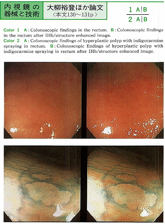
    1996Volume 48 Pages 130-131
    Published: June 07, 1996
    Released on J-STAGE: March 20, 2015
    JOURNAL FREE ACCESS
    A real time image processing system, EVIP-230, was used to improve the accuracy of colonoscopy. The assessment of crypt pattern in the normal rectal mucosa and in hyperplastic polyps was studied in 48 patients.
    Crypt pattern was demonstrated in the normal rectal mucosa in 79% by dye spraying with 5 times diluted indigocarmine and 75% by enhanced image without indigocarmine. Moreover, crypt pattern was demonstrated in hyperplastic polyps in 76% by dye spraying with 5 times diluted indigocarmine and 57% by enhanced image without indigocarmine. The demonstration of crypt pattern in the rectal mucosa and hyperplastic polyps was much higher by enhanced image using image processing system EVIP-230 than by conventional colonoscopic examination.
    The demonstration of crypt pattern by enhanced image was almost equal to dye spraying with 5 times diluted indigocarmine. Therefore, it is suggested that this method will become useful in the assessment of the lesions at routine colonoscopic examination and in observing the fine structure of the colonic mucosa.
    Download PDF (315K)
  • Nobutaka Sato, Kazuhiko Watanabe, Yoshiro Tamegai, Eita Morikoshi, Hid ...
    1996Volume 48 Pages 132-133
    Published: June 07, 1996
    Released on J-STAGE: March 20, 2015
    JOURNAL FREE ACCESS
    Small intestino-Videoendoscopy was performed in 5 patients using a small intestino-Videoendoscope (model XSIF-200) first developed by Olympus Optical Co Ltd, Tokyo. The clinical assessment of this scope are :
    (1)Scope function ; An effective length of the scope is 2,000mm and the channel for insertion of forceps used for biopsy and polypectomy is installed in its flexible part. The distaltip at the flexible part has the up/down and left/right angulation system.
    (2)Technique ; It can be carried out with the same manner as conventional intestinoscopy.
    (3)Observation ; The excellent image is provided on a TV monitor, permitting smooth cooperation with examiners for biopsy or polypectpmy.
    (4)Recordability ; Frozen images are available directly to color print photographing without monitoring and to 16mm still photographing through monitoring. Dynamic images can be used for VTR recording.
    (5)Safety and durability ; Both are excellent compared with conventional intestino-scope.
    Conclusion : This small intestino-videoendoscope was found to be superior to the conventional fiberscopes for ordinary observations and endoscopic therapy of small intestinal diseases.
    Download PDF (565K)
  • Takesada Goto, Hideo Yamada, Tadashi Hachisu, Kazuyuki Matsushita, Shi ...
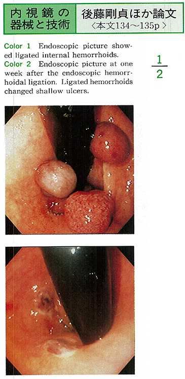
    1996Volume 48 Pages 134-135
    Published: June 07, 1996
    Released on J-STAGE: March 20, 2015
    JOURNAL FREE ACCESS
    The usefulness of endoscopic hemorrhoidal ligation using Stiegmann's ligator was discussed. The advantages of this treatment are less pain and short hospitalization due to minimal invasive operation.
    Thirty six cases were treated in this method and 33 cases (92%) showed excellent results. However, 3 cases needed following surgical operation because of post-ligated prolapsus in two patients and aggravation of external hemorrhoid in one patient. Thses 3 cases were patients with high grade hemorrhoids (Goligher's III, IV) .
    We concluded that indication of endoscopic hemorrhoidal ligation should be limited to the low and middle grade hemorrhoids (Goligher's II, III) .
    Download PDF (397K)
Clinical study
  • Katsuhiko Iwakiri, Yoshiya Nakagawa, Makoto Kotoyori, Hisagi Yamada, T ...
    1996Volume 48 Pages 136-137
    Published: June 07, 1996
    Released on J-STAGE: March 20, 2015
    JOURNAL FREE ACCESS
    We investigated whether pH and total bile acids concentration in refluxed esophageal fluid reflect those in gastric juice in patients with reflux esophagitis.
    Thirteen patients with reflux esophagitis (8 men and 5 women ; average age : 71.3 years) were examined. Reflux esophagitis was classified according to the Savary-Miller classification. Refluxed esophageal fluid and gastric juice were collected using a novel device. The total bile acids concentration and pH in the collected samples were measured using the enzymic method and a pH meter.
    Refluxed esophageal fluid could be collected in 1 of 4 patients with stage I reflux esophagitis and all patients with stage II and IV reflux esophagitis. More than 200µM of bile acids, which may be sufficient cause esophageal injury in acidic conditions could be obtained in the refluxed esophageal fluid. The relationship between total bile acids concentration and pH in refluxed esophageal fluid and gastric juice showed a positive correlation.
    We concluded that bile acids are one of the major factors in the onset of reflux esophagitis. For evaluation of aggressive factors in reflux esophagitis, gastric juice can be substituted for refluxed esophageal fluid.
    Download PDF (263K)
  • Toshiyuki Nakajima, Atsushi Nishiya, Touru Kakinuma, Taiki Iwahori, Hi ...
    1996Volume 48 Pages 138-139
    Published: June 07, 1996
    Released on J-STAGE: March 20, 2015
    JOURNAL FREE ACCESS
    We evaluated the association between flow of the splenic vein and endoscopic findings of the upper digestive tract. The subjects were 42 patients with liver cirrhosis complicated by esophageal varices in whom the clinical course were observed for 5 years. They were classified into the group who developed hepatofugal flow of the splenic vein during course (hepatofugal group) and that who did not (hepatopetal group) . Child classification, male-female ratio and average of the age before observation was similar between the two groups.
    Changes in the endoscopic findings of gastroesophageal varices during the course were slighter in the hepatofugal group than the hepatopetal group. Significant differance was observed in changes of the gastric mucosal lesions in the two groups, i. e. the hepato-fugal group was not increased but the hepatopetal group was significantly increased (p<0.05) . The hepatofugal group was significantly higher incidence of the spleno-renal shunt than the other.
    Download PDF (329K)
  • Hiroki Kawahara, Nobuhiro Masaki, Hiroshi Matsuura, Kimihiko Akimoto, ...
    1996Volume 48 Pages 140-141
    Published: June 07, 1996
    Released on J-STAGE: March 20, 2015
    JOURNAL FREE ACCESS
    Diagnosis of Helicobacter pylori (H. pylori) infection is based on the culture and isolation of this bacteria, we have developed a new endoscopic technique of targetting biopsy for isolation of H. pylori by cochineal extract spray.
    The cochineal extract is a non-toxic food-dye, its use to the human and safety having been approved in a law. In the conditions at pH>4, it indicates the distinctive color change from red to purple. H. pylori has an enzyme, urease which converts urea into ammonia. This ammonia elevates the local pH levels on the surface of gastric mucosa. Therefore, the presence of H. pylori would be expected when the color of cochineal extract sprayed on the gastric mucosa turned to purple within a few seconds. In fact, we isolated H. pylori at a high rate (90%) from the gastric mucosa where cochineal extract showed this color change.
    We conclude that this technique would be very useful in the definit diagnosis of H. pylori infection.
    Download PDF (266K)
  • Takeshi Matsuhisa, Izumi Kusama, Nobutaka Yamada
    1996Volume 48 Pages 142-143
    Published: June 07, 1996
    Released on J-STAGE: March 20, 2015
    JOURNAL FREE ACCESS
    Helicobacter pylori (H. pylori) infection in cases of after distal gastric resection was studied. We also examined relation among H. pylori and residual superficial gastritis, bile acid refluxed into the stomach.
    1) A rate of H. pylori infection in postoperative stomach shows 51.1%, which was lower than that of digestive ulcer, gastric carcinoma and control group. In cases of residual stomach, a concentration of chenodeoxycholic acid (CDCA) and ursodeoxycholic acid (UDCA) refluxed into the stomach were remarkably higher than thoes of digestive ulcer and control group. I considered the rate of H. pylori infection in postoperative stomach is low because of highly concentrated CDCA and UDCA is not adequate for habitation of H. pylori.
    2) There were no correlation among pathological inflammation, activity and superficial gastritis in postoperative stomach. Inflammation and activity were frequent in H. pylori positive cases than negative cases.
    3) A frequency of H. pylori infection in cases of after distal gastric resection by gastric carcinoma within 5 years was lower than 50.0%. A rate of infection increased 72.7% in patients who were operated over 5 years ago.
    Download PDF (272K)
  • Takeshi Matsuhisa, Toyoaki Idemitsu
    1996Volume 48 Pages 144-145
    Published: June 07, 1996
    Released on J-STAGE: March 20, 2015
    JOURNAL FREE ACCESS
    An hemoglobin index (Hb index) of gastric mucosa in digestive ulcers was studied by image processing unit SP-1000. We classified gastric ulcer cases into 2 types. One type is upper placed ulcer, which has ulcer high level than middle body. Another type is lower placed ulcer, which has ulcer under lower body.
    1) An average of Hb index of the stomach measured in cases of digestive ulcers was higher on the anterior wall of upper body.
    2) Hb index of lower placed ulcer was higher in anterior wall of upper body than in anterior wall of lower body, greater curvature of antrum.
    3) Hb index of lower placed ulcer and duodenal ulcer were higher than upper placed ulcer.
    Download PDF (271K)
  • Tadashi Yokoyama, Hajime Yamaguchi, Kuniaki Shirao, Hitoshi Kondo, Dai ...
    1996Volume 48 Pages 146-147
    Published: June 07, 1996
    Released on J-STAGE: March 20, 2015
    JOURNAL FREE ACCESS
    Eleven cases of inflammatory fibroid polyp (IFP) of the stomach were studied with respect to endoscopic diagnosis. Four cases were resected by endoscopic polypectomy and the others were surgically resected. There were seven men and four women. The mean age was 59, ranging from 44 to 71.
    Nine cases were in the antrum, and the other two were in the gastric body. Five cases were less than 10 mm, and two were over 20 mm. Five cases were classified into Yamada-I type, two into II type, four into III type. Initial endoscopic diagnosis was hyperplastic polyp in four, submucosal tumor in three, early gastric cancer in one, and gastric adenoma in one. Routine biopsy was done to eight cases, but the pathological diagnosis of IFP was not confirmed except one case. This may be because the biopsy specimens contained hyperplastic foveolar epithelium only, which covered the surface of the IFP in most of the cases.
    These findings were compatible with previously reported characteristics of IFP. Correct diagnosis of IFP on routine endoscopy were rarely obtained and diagnosis could be made on endoscopic ultrasonography and polypectomy.
    Download PDF (1124K)
  • Yumi Tsuruta, Norichika Narimiya, Hiroko Hamada, Tatsushi Maruyama, Hi ...
    1996Volume 48 Pages 148-149
    Published: June 07, 1996
    Released on J-STAGE: March 20, 2015
    JOURNAL FREE ACCESS
    Of 129 cases with malignant lymphoma experienced in our department during the past 13 years, involvement of the stomach was noted in 17 cases (13%) , of which 10 were treated with chemotherapy without gastric resection and the other 7 received surgical resection of the stomach.
    The former non-operative cases (1 with Hodgkin's disease and 9 with non-Hodgkin's lymphoma) consisted of 5 cases with primary gastric lymphoma and 5 with gastric lesions manifested as a part of generalized malignant lymphoma. Macroscopic type of these gastric lesions was endoscopically protruding type in 1, collapsing type in 1, ulcerating type in 1, giant fold type in 3 and superficially enlarged type in 4 cases.
    In 7 of 10 cases, disappearance of gastric tumors was recognized macroscopically after chemotherapy. Gastric hemorrhage occurred after chemotherapy in 3 cases invariably of primary malignancy of the stomach and bleeding from gastric lesions ocurred in none of cases with generalized malignant lymphoma. Post-chemotherapy hemorrhage was noted to occur in cases with lesions of collapsing or giant fold type, never with those of superficially enlarged type.
    From these results, it is suggested that gastric hemorrhage after chemotherapy for malignant lymphoma may be caused by damage of vessels accompanied with a loss shedding of massive tumor cells invaded into the deeper layer.
    Download PDF (280K)
  • Yasunori Hoshiya, Koichiro Kumai, Naoki Igarashi, Hideki Ishikawa, Koj ...
    1996Volume 48 Pages 150-151
    Published: June 07, 1996
    Released on J-STAGE: March 20, 2015
    JOURNAL FREE ACCESS
    The patients with gastroduodenal ulcer or AGML frequently show massive bleeding, resulting in critical status such as shock, DIC, and MOF, moreover they have often surgical risk factors. In these surgical poor risk patients, the decision of surgical treatment is difficult and important. Five hundred and 76 patients underwent emergency gastrofiberscopy for the bleeding from gastroduodenal ulcer or AGML in the period from May 1976 to Nov 1995. Effectiveness of conservative therapies and surgical treatment were analyzed.
    Effective rate of endoscopic hemostasis were over 90% for bleeding from gastroduodenal ulcer in most cases, however it was relatively low as 68.8% for bleeding with massive clot. We have been using flushing tube to remove clot, resulting in high effective rate as 80.0% in the latter period.
    Recent advances in medication and endoscopic hemostatic procedure for the bleeding from gastroduodenal ulcer has remarkably decreased the patients who need surgical treatment. The number of bleeding episode during conservative therapy firmly correlates with bad postoperative course. Our criteria for converting to surgical therapy is 3 times of massive re-bleeding during conservative therapy. Operative death has decreased after introduction of this criteria.
    Download PDF (298K)
  • Kenichi Saito, Akira Ogawa, Toshikazu Akiya
    1996Volume 48 Pages 152-153
    Published: June 07, 1996
    Released on J-STAGE: March 20, 2015
    JOURNAL FREE ACCESS
    The early stage of colorectal cancer is divided into two morphological types ; polypoid and superficial. What percentage of advanced cancer is derived from the polypoid or superficial type is a matter of concern. We designated superficial type tumors from the both morphological and histological viewpoints. Histological designation of superficial type tumor was that there was horizontal growth of cancer in the residual mucosa.
    The existence of horizontal growing mucosal cancer at the marginal area of cancer is a biomarker showing the lesion being of superficial origin. The rates of arise from superficial tumor of submucosal cancer in the residual mucosa was 29/98 (29.6%) . The cancer with invasion deeper than the muscularis propria layer usually loose cancer in the mucosa, therefore, the rates may not reflect actual incidence. The rate of submucosal cancer (29.6%) may be close to the actual rate at present.
    It may be useful to focus on the horizontal growing tumor of the colorectum for understanding the advance and development of colorectal tumors.
    Download PDF (860K)
Case report
  • Hideki Ishikawa, Yoshihide Otani, Masashi Yoshida, Naoki Igarashi, Koj ...
    1996Volume 48 Pages 154-155
    Published: June 07, 1996
    Released on J-STAGE: March 20, 2015
    JOURNAL FREE ACCESS
    The patient was a 71-year-old male. He underwent distal gastrectomy (Billroth-I) for early gastric cancer. After the operation, he was troubled with severe reflex esophagitis. Eighteen months later, for common bile duct stones and gallstones, he had second operation. In order to relieve reflex esophagitis, we changed the route of reconstruction from Billroth-I to Roux-en-Y.
    After the second operation, he has recovered from reflex esophagitis almost completely. The simultaneous pH monitoring in lower esophagus and remnant stomach was useful for understanding the pathophysiology and grade of reflex esophagitis.
    Download PDF (676K)
  • Hirohisa Katoh, Masahiko Murakami, Kazushige Arai, Mitsuo Kusano
    1996Volume 48 Pages 156-157
    Published: June 07, 1996
    Released on J-STAGE: March 20, 2015
    JOURNAL FREE ACCESS
    It has been reported that one of the effects of proton pump inhibitor (PPI) is inhibition from exocrine pancreas. We treated two cases of reflex esophagitis due to total gastrectomy with PPI and they were cured soon.
    Case 1 : A 71-year-old man was operated upon total gastrectomy (6-interposition) for early gastric cancer. He had felt aphagia and heart burn from ten days after operation. We had been following up him for three years with camostat mesilate, but his symptoms had been unchanged and the endoscopy had revealed esophagitis which was Savary-Miller (SM) stage III.
    Case 2 : Total gastrectomy (Roux-Y) for early gastric cancer was carried out to a woman who was 53 years old. Her symptoms were nausea and heart burn from eight days after operation. She had never been treating by any medicines. Four months after operation, her symptoms had been stronger and endoscopic findings was esophagitis of SM stage III.
    In both cases, their symptoms were got under control soon and esophagitises were changed to SM stage I-II with single medication of PPI for several weeks.
    Download PDF (262K)
  • Masakatsu Uchihara, Yuka Miyasaka, Kazuyoshi Nagayama, Takeshi Murakam ...
    1996Volume 48 Pages 158-159
    Published: June 07, 1996
    Released on J-STAGE: March 20, 2015
    JOURNAL FREE ACCESS
    A 67-year-old man was admitted to our hospital because of dysphagia. About 6 months ago, he admitted to another hospital due to heart failure and cerebral infarction. During the admission dysphagia had developed, and he was diagnosed as gastric cancer invading the lower esophagus.
    The operative therapy was considered to be difficult because of his heart and neurological complication. Therefore he was refered to our hospital for palliation of dysphagia. Marked stenosis was observed at the esphago-gastric junction by radiographical examination.
    Self-expanding metalic stent, the Ultraflex esophageal prosthesis was implanted endoscopically. Dysphagia improved dramatically a week after the procedure. Thereafter he was able to stay at home without dysphagia. The Ultraflex esophageal prosthesis is suggested to be useful for the palliation of cardiac cancer as well as esophageal cancer.
    Download PDF (910K)
  • Hiroshi Ishida, Tsunehisa Hirakawa, Masafumi Kobayashi
    1996Volume 48 Pages 160-161
    Published: June 07, 1996
    Released on J-STAGE: March 20, 2015
    JOURNAL FREE ACCESS
    A 15-year-old child was refferred to our hospital for recurrent abdominal pain (RAP) and nausea. Laboratory data revealed a high level of IgG antibydy to Hp in serum. Upper GI endoscopy showed antral nodularity like a goose-flesh. Phenol red stain endoscopy showed a diffuse red color change in the antrum, which emphasized antral nodularity. Antral biopsy specimen showed active chronic gastritis with a well-formed lymphoid follicle in the mucosa. Hp was found by histologic examination with Warthin-Starry staining and bacterial culture.
    The patient became symptomless after a period of one week of treatment that consisted of lansoprazole 30mg od, amoxicillin 500mg bid and metronidazole 250mg bid. A second endoscopy revealed no antral nodularity at all and biopsy specimens showed almost complete disappearance of the inflammatory process. Hp could not be identified by Warthin-Starry staining and culture. In children with RAP and/or a high level of serum Hp antibody endoscopic and histological examination should be performed for Hp infection.
    Download PDF (1062K)
  • Yoshiki Saegusa, Ryuichi Hirakawa, Takashi Oishi, Hiroyuki Ohsawa, Mit ...
    1996Volume 48 Pages 162-163
    Published: June 07, 1996
    Released on J-STAGE: March 20, 2015
    JOURNAL FREE ACCESS
    A 68-year-old man was admitted to our hospital with the chief complaint of hematemesis. Laboratory studies showed anemia and positive occult blood in the feces. Endoscopic examination revealed a bright red lesion, approximately 10mm in diameter, on the lesser curvature near the incisura angularis. Color Doppler endoscopic ultrasonography showed multiple capillary vessels and pulsatile flow in the lesion.
    Since the gastrointestinal bleeding continued and anemia progressed, the lesion was endoscopically resected using an endoscopic variceal ligation device. Such procedure is useful for the treatment the bleeding in case of angiodysplasia of the stomach.
    Download PDF (729K)
  • Hisashi Ariki, Mamoru Nishino, Hiroshi Houjou, Eisaku Kondou, Masahiko ...
    1996Volume 48 Pages 164-165
    Published: June 07, 1996
    Released on J-STAGE: March 20, 2015
    JOURNAL FREE ACCESS
    An 87-year-old male received regular outpatient treatment for hypertension, chronic renal insufficiency and chronic hepatitis. He had tarry stool and marked anemia (Hb 5.0g/dl) in December 1994, for which he received blood transfusion and administration of an iron preparation.
    Upper gastrointestinal endoscopy revealed ectatic antral capillaries extending radially, and the disease was diagnosed as GAVE. The patient was admitted in May 1995. A total of 12ml of 1% Aethoxysklerol was injected locally into 12 sites adjacent to the ectatic capillaries (1ml per site) . After two courses of this medication, the capillary ectasis showed a tendency to improve. The effectiveness of local injection of Aethoxysklerol for GAVE was reported in this paper.
    Download PDF (841K)
  • Kazuyuki Nakao, Ichiro Imoto, Teruo Shima, Hiroyuki Sakano
    1996Volume 48 Pages 166-167
    Published: June 07, 1996
    Released on J-STAGE: March 20, 2015
    JOURNAL FREE ACCESS
    A 69-year-old woman underwent surgery for primary malignant lymphoma of the stomach approximately 10 years ago at our hospital. The tumor was located on the lesser curvature side of the angulus and measured approximately 5cm×2cm. Histologically, lymphoma cells of the large cell type (Non-Hodgkin) had invaded the serosa.
    Approximately 10 years after surgery, she sometimes complained of vague abdominal fullness. A barium-meal examination revealed a bezoar in the stomach. Most gastrointestinal bezoars develop after surgery, and the most common procedure is bilateral truncal vagotomy with pyloroplasty. However, the relationship between bezoar and surgery for malignant lymphoma of the stomach is not clear, and the 5-year survival ratio in malignant lymphoma of large cell type is approximately 33%. Since there has been no report of gastric bezoars developing after lymphoma surgery, we reported this case.
    Download PDF (1347K)
  • Izumi Kusama, Syoutaro Iida, Takeshi Matsuhisa
    1996Volume 48 Pages 168-169
    Published: June 07, 1996
    Released on J-STAGE: March 20, 2015
    JOURNAL FREE ACCESS
    Hyperplastic polyp in gastric polyps is the most frequent type by pathological diagnosis. Most of them are smaller than 20 mm in diameter. We experienced three cases, which had 5 huge gastric polyps type Yamada IV more than 40 mm in size, removed in safety by endoscopic polypectomy. All of their cases had multiple polyps. It is known that huge polyps rarely combine carcinoma cells and have a risk of bleeding and stenosis. We also experienced a focal carcinoma in case 3. It is necessary to remove a huge gastric polyp type Yamada's IV as soon as possible by endoscopic polypectomy.
    Download PDF (517K)
  • Takashi Sakakibara, Natsumi Tomita, Hajime Noguchi, Takumi Ochiai, Kaz ...
    1996Volume 48 Pages 170-171
    Published: June 07, 1996
    Released on J-STAGE: March 20, 2015
    JOURNAL FREE ACCESS
    A 53-year-old male visited our hospital because of gastric carcinoid, which was 4mm sized Yamada-Itype polyp. The biopsy specimen showed carcinoid infiltrating to submucosa. Subtotal gastrectomy with cholecystectomy was performed as the diagnosis of gastric carcinoid with cholelithiasis. Pathologically no remained carcinoid of gastric surgical specimen and no lymphonoid metastasis was reported.
    It considered repeated biopsy took the all of gastric carcinoid. It revealed that endoscopic mucosal resection could be performed up to at least 4mm sized carcinoid of the stomach.
    Download PDF (518K)
  • Hiroyuki Imaeda, Yasushi Iwao, Yoshikazu Tsuzuki, Shingo Miyaguchi, At ...
    1996Volume 48 Pages 172-173
    Published: June 07, 1996
    Released on J-STAGE: March 20, 2015
    JOURNAL FREE ACCESS
    A 83-year-old female was admitted to our hospital because of right hypochondralgia and abdominal tumor. She was received gastrectomy for early gastric cancer at 78 years old.
    Abdominal CT scan showed a tumor in the gastric stump invading to the left lobe of the liver and the pancreas. Endoscopic findings showed an irregular protruded tumor with erosion and bleeding from the gastric stump to the duodenum. Biopsy specimen showed B-cell malignant lymphoma, diffuse medium cell type. Bone marrow aspiration revealed invasion of lymphoma cell. She received dimimished chemotherapy, but she died on 6 months.
    Ten cases of malignant lymphoma in the gastric stump after gastrectomy were reported in Japan. The mechanism is supposed to be concerned with metaplasia of gastric mucosa by chronic stimulation by reflux duodenal content and bile juice, depression of gastric acid and change of bacterial flora.
    Download PDF (1334K)
  • Naoki Igarashi, Yoshihide Otani, Koichiro Kumai, Masashi Yoshida, Yasu ...
    1996Volume 48 Pages 174-175
    Published: June 07, 1996
    Released on J-STAGE: March 20, 2015
    JOURNAL FREE ACCESS
    The patient is a 54-year-old male who had epigastric disconfort with the duration of 3 months. Upper GI series and endoscopic examination revealed submucosal tumor with 3cm in diameter on the posterior wall of antrum. EUS revealed the main location of the tumor is proper muscle layer. Distal gastrectomy was performed and he dicharged 14 POD uneventfully. The final pathological diagnosis was malignant lymphoma (follicular medium-sized cell type) without lymph node metastasis.
    Primary malignant lymphoma (ML) of GI tract comprises 1 to 2% of all malignancy is GI tract. The operative procedure commonly selected for ML of the stomach is principally the same as the procedure for cancer. On the other hand, leiomyosarcoma which is common in submucosal tumor can be treated with local resection. Submucosal tumor of the stomach should be treated considerably with proper method based on preoperative and intraoperative histological diagnosis.
    Download PDF (1072K)
  • Mamiko Matsumura, Tetsuya Yamaguchi, Akitake Uno, Ryuichi Kurihara, No ...
    1996Volume 48 Pages 176-177
    Published: June 07, 1996
    Released on J-STAGE: March 20, 2015
    JOURNAL FREE ACCESS
    A 34 year-old female had a tumor diagnosed as early gastric cancer, type IIc. At our outpatient clinic, the tumor was diagnosed as early gastric cancer, type IIc with multiple, irregular faded areas in anterial wall of gastric body. We recognized the tumor to be malignant lymphoma by endoscopy, but histologically it did not show any malignancy. Following another endoscopy check and subsequent histological diagnosis, which showed possible malignant lymphoma so she received total gastorectomy.
    By histological diagnosis, the tumor was not a MALT-lymphoma, the depth of the tumor was submucosal invasion, and there was no lymphnode metastasis. Helicobacter pylori was not detected before her operation. We reported a case of premalignant lymphoma, which was similar to early gastric cancer, type IIc.
    Download PDF (1076K)
  • Toshinori Kurahashi, Kazuhiro Kaneko, Nozomi Yoshikawa, Makoto Ishii, ...
    1996Volume 48 Pages 178-179
    Published: June 07, 1996
    Released on J-STAGE: March 20, 2015
    JOURNAL FREE ACCESS
    Two cases of liver cirrhosis (LC) with hepatocellular carcinoma (HCC) developed early gastric cancer. Follow up of esophagogastric varices by endoscopic examination detected IIc early gastric cancer.
    One case was 62-year-old man and the other was 65-year-old man. Their HCCs had been already treated with transarterial embolization. Their early gastric cancers were completely resected by endoscopic mucosal resection (EMR) . Histological findings revealed moderately differentiated adenocarcinoma and that partly accompanied with poorly differentiated adenocarcinoma, respectively.
    The complication of HCC with gastric cancer is reported to be relatively freguent and twenty-one cases including our two cases has been reported. These 21 cases were compared with 62 cases with early gastric cancer alone in our hospital. Seventy percent of early gastric cancer in patients with LC and HCC were located in antrum compared with 35 percent in early gastric cancer alone. Depressed type in macroscopic findings and differentiated type in histologic findings tended to be frequent. Patients with LC are usually associated with portal hypertensive gastropathy (PHG) . A causative relation of ischemic change of gastric mucosa due to PHG with gastric cancer in patients with LC could be considered.
    Download PDF (1065K)
  • Yoko Oishi, Masao Tani, Seitaku Hayashi, Fumio Kando, Naoya Saito, Kim ...
    1996Volume 48 Pages 180-181
    Published: June 07, 1996
    Released on J-STAGE: March 20, 2015
    JOURNAL FREE ACCESS
    A 72-year-old male, who complained of hematemesis, was referred a near hospital in August 1991. He was endoscopically diagnosed as kissing-type bleeding ulcer at the gastric angulus, and he recieved conservative treatment.
    The ulcer changed to the scar for a month. The histological diagnosis of the biopsied specimen was compatible with Group II. After three months, the ulcer changed to a small reddish elevated granulomatous lesion with fold concentrations around the ulcer scar.
    He was followed up by endoscope and biopsy at once in three months for two years. The lesion enlarged slowly, and in March 1994, the histological diagnosis of the biopsied specimen was compatible with Group IV. In April, the lesion changed to a multiple elevated lesion with fold concentrations and a central depression. The histological diagnosis of the biopsied specimen was compatible with Group V. The lesion was diagnosed as 0'IIa+IIc, type of gastric cancer. Distal gastrectomy, Billroth-I reconstruction and D2 lymph node dissection was performed. Histopathological diagnosis of the lesion was 0IIa+IIc, tub1, sm, Ul-IVs, n0, ly0, v0.
    Although ulcer was recovered, cancer was undeniable and we confirm again the importance of periodical endoscopic examination for gastric ulcer scar.
    Download PDF (942K)
  • Tsuyoshi Suzuki, Shin Sakurabayashi, Katsumasa Yoshino, Shuji Nishimur ...
    1996Volume 48 Pages 182-183
    Published: June 07, 1996
    Released on J-STAGE: March 20, 2015
    JOURNAL FREE ACCESS
    The patient was a male of 68 years old. The chest X-ray showed infiltrative shadow in the right upper lobe. The cytology by bronchoscopic brushing revealed small cell lung carcinoma. Although the radiation therapy showed good response, abdominal pain was appeared. Endoscopy showed an elevated lesion with a central ulceration in the upper part of body of stomach. The large mass in the upper abdomen was also detected by the abdominal CT.
    In addition the lung and abdominal tumor, the gastric elevated lesion was remarkably responsed to chemotherapy. The biopsy specimens of intraabdominal tumor showed small cell carcinoma.
    Download PDF (742K)
  • Katsuyuki Ietomi, Kazuo Miyoshi, Kyoya Sakimura, Yuichi Handa, Hideki ...
    1996Volume 48 Pages 184-185
    Published: June 07, 1996
    Released on J-STAGE: March 20, 2015
    JOURNAL FREE ACCESS
    A 16-year-old man admitted to our hospital because of severe anemia due to gastric cancer. Endoscopic findings showed that the gastric cancer was classified as Borrmann's type II. Further examinations of the colon revealed multiple polypoid lesions. Colonic polypectomy and total gastrectomy were performed.
    One year later, he was suffered from epileptic seizure due to a brain tumor. The tumor was resected, and the histological findings showed a glioblastoma multiforme. The diagnosis of Turcot's syndrome was made by the complication of adenomatous polyposis coli and glioblastoma.
    Turcot's syndrome is a rare congenital disease classified as adenomatous polyposis coli. Some reports show the complication of gastric and duodenal polyps with this syndrome. However, to our knowledge, this is the first case report of complication of gastric cancer with Turcot's syndrome.
    Download PDF (1337K)
feedback
Top