
Volume 42
Displaying 1-50 of 68 articles from this issue
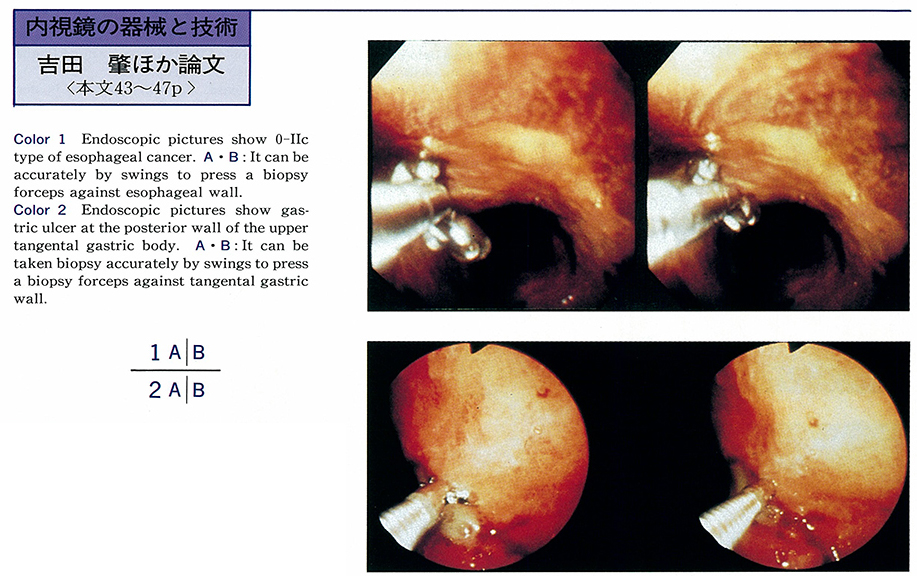
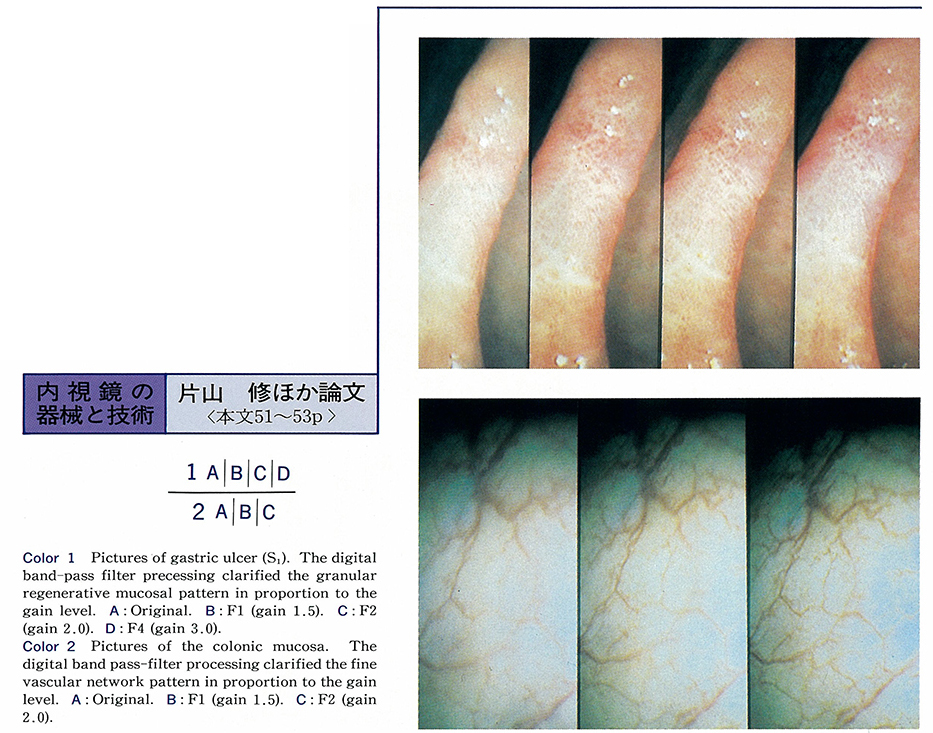
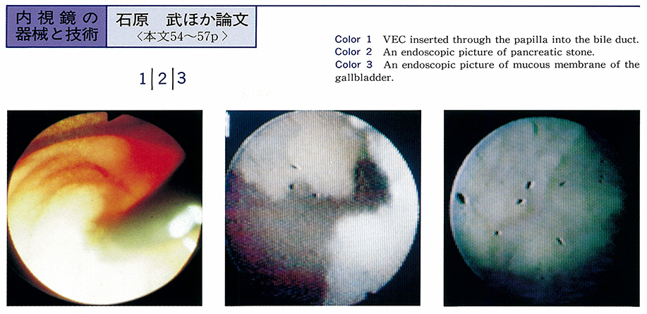
Technology and instrument
-
1993 Volume 42 Pages 43-47
Published: June 18, 1993
Released on J-STAGE: July 15, 2015
 Download PDF (1318K)
 -
1993 Volume 42 Pages 48-50
Published: June 18, 1993
Released on J-STAGE: July 15, 2015
 Download PDF (658K)
 -
1993 Volume 42 Pages 51-53
Published: June 18, 1993
Released on J-STAGE: July 15, 2015
 Download PDF (607K)
 -
1993 Volume 42 Pages 54-57
Published: June 18, 1993
Released on J-STAGE: July 15, 2015
 Download PDF (1456K)
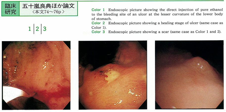
Clinical study
-
1993 Volume 42 Pages 58-62
Published: June 18, 1993
Released on J-STAGE: July 15, 2015
 Download PDF (1274K)
 -
1993 Volume 42 Pages 63-66
Published: June 18, 1993
Released on J-STAGE: July 15, 2015
 Download PDF (403K)
 -
1993 Volume 42 Pages 67-68
Published: June 18, 1993
Released on J-STAGE: July 15, 2015
 Download PDF (227K)
 -
1993 Volume 42 Pages 69-73
Published: June 18, 1993
Released on J-STAGE: July 15, 2015
Download PDF (553K) -
1993 Volume 42 Pages 74-76
Published: June 18, 1993
Released on J-STAGE: July 15, 2015
 Download PDF (382K)
 -
1993 Volume 42 Pages 77-80
Published: June 18, 1993
Released on J-STAGE: July 15, 2015
 Download PDF (413K)
 -
1993 Volume 42 Pages 81-85
Published: June 18, 1993
Released on J-STAGE: July 15, 2015
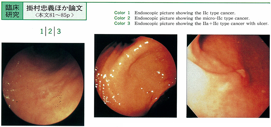 Download PDF (1653K)
 -
1993 Volume 42 Pages 86-90
Published: June 18, 1993
Released on J-STAGE: July 15, 2015
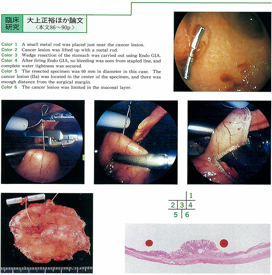 Download PDF (1377K)
 -
1993 Volume 42 Pages 91-95
Published: June 18, 1993
Released on J-STAGE: July 15, 2015
 Download PDF (1019K)
 -
1993 Volume 42 Pages 96-99
Published: June 18, 1993
Released on J-STAGE: July 15, 2015
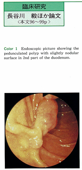 Download PDF (609K)
 -
1993 Volume 42 Pages 100-103
Published: June 18, 1993
Released on J-STAGE: July 15, 2015
Download PDF (451K) -
1993 Volume 42 Pages 104-107
Published: June 18, 1993
Released on J-STAGE: July 15, 2015
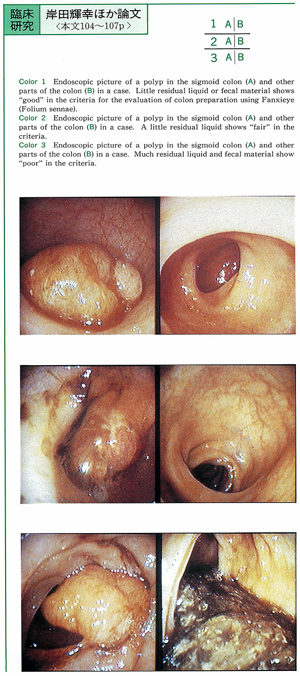 Download PDF (496K)
 -
1993 Volume 42 Pages 108-111
Published: June 18, 1993
Released on J-STAGE: July 15, 2015
Download PDF (410K) -
1993 Volume 42 Pages 112-117
Published: June 18, 1993
Released on J-STAGE: July 15, 2015
 Download PDF (1665K)
 -
1993 Volume 42 Pages 118-123
Published: June 18, 1993
Released on J-STAGE: July 15, 2015
Download PDF (770K) -
1993 Volume 42 Pages 124-128
Published: June 18, 1993
Released on J-STAGE: July 15, 2015
 Download PDF (700K)
 -
1993 Volume 42 Pages 129-132
Published: June 18, 1993
Released on J-STAGE: July 15, 2015
Download PDF (817K) -
1993 Volume 42 Pages 133-137
Published: June 18, 1993
Released on J-STAGE: July 15, 2015
Download PDF (1108K) -
1993 Volume 42 Pages 138-142
Published: June 18, 1993
Released on J-STAGE: July 15, 2015
Download PDF (606K) -
1993 Volume 42 Pages 143-147
Published: June 18, 1993
Released on J-STAGE: July 15, 2015
Download PDF (651K)
Case report
-
1993 Volume 42 Pages 148-150
Published: June 18, 1993
Released on J-STAGE: July 15, 2015
 Download PDF (677K)
 -
1993 Volume 42 Pages 151-153
Published: June 18, 1993
Released on J-STAGE: July 15, 2015
 Download PDF (835K)
 -
1993 Volume 42 Pages 154-157
Published: June 18, 1993
Released on J-STAGE: July 15, 2015
 Download PDF (1243K)
 -
1993 Volume 42 Pages 158-161
Published: June 18, 1993
Released on J-STAGE: July 15, 2015
 Download PDF (1284K)
 -
1993 Volume 42 Pages 162-164
Published: June 18, 1993
Released on J-STAGE: July 15, 2015
 Download PDF (800K)
 -
1993 Volume 42 Pages 165-168
Published: June 18, 1993
Released on J-STAGE: July 15, 2015
 Download PDF (1706K)
 -
1993 Volume 42 Pages 169-172
Published: June 18, 1993
Released on J-STAGE: July 15, 2015
 Download PDF (2315K)
 -
1993 Volume 42 Pages 173-176
Published: June 18, 1993
Released on J-STAGE: July 15, 2015
 Download PDF (1526K)
 -
1993 Volume 42 Pages 177-180
Published: June 18, 1993
Released on J-STAGE: July 15, 2015
 Download PDF (1748K)
 -
1993 Volume 42 Pages 181-184
Published: June 18, 1993
Released on J-STAGE: July 15, 2015
 Download PDF (1962K)
 -
1993 Volume 42 Pages 185-188
Published: June 18, 1993
Released on J-STAGE: July 15, 2015
 Download PDF (1503K)
 -
1993 Volume 42 Pages 189-191
Published: June 18, 1993
Released on J-STAGE: July 15, 2015
 Download PDF (1006K)
 -
1993 Volume 42 Pages 192-196
Published: June 18, 1993
Released on J-STAGE: July 15, 2015
 Download PDF (1685K)
 -
1993 Volume 42 Pages 197-201
Published: June 18, 1993
Released on J-STAGE: July 15, 2015
Download PDF (2434K) -
1993 Volume 42 Pages 202-205
Published: June 18, 1993
Released on J-STAGE: July 15, 2015
Download PDF (1600K) -
1993 Volume 42 Pages 206-208
Published: June 18, 1993
Released on J-STAGE: July 15, 2015
Download PDF (966K) -
1993 Volume 42 Pages 209-212
Published: June 18, 1993
Released on J-STAGE: July 15, 2015
Download PDF (1054K) -
1993 Volume 42 Pages 213-216
Published: June 18, 1993
Released on J-STAGE: July 15, 2015
Download PDF (1089K) -
1993 Volume 42 Pages 217-219
Published: June 18, 1993
Released on J-STAGE: July 15, 2015
 Download PDF (297K)
 -
1993 Volume 42 Pages 220-223
Published: June 18, 1993
Released on J-STAGE: July 15, 2015
Download PDF (1045K) -
1993 Volume 42 Pages 224-227
Published: June 18, 1993
Released on J-STAGE: July 15, 2015
 Download PDF (2051K)
 -
1993 Volume 42 Pages 228-231
Published: June 18, 1993
Released on J-STAGE: July 15, 2015
 Download PDF (1630K)
 -
1993 Volume 42 Pages 232-235
Published: June 18, 1993
Released on J-STAGE: July 15, 2015
 Download PDF (1120K)
 -
1993 Volume 42 Pages 236-239
Published: June 18, 1993
Released on J-STAGE: July 15, 2015
 Download PDF (1114K)
 -
1993 Volume 42 Pages 240-243
Published: June 18, 1993
Released on J-STAGE: July 15, 2015
 Download PDF (1784K)
 -
1993 Volume 42 Pages 244-247
Published: June 18, 1993
Released on J-STAGE: July 15, 2015
 Download PDF (1931K)