Vacuum and Surface Science
Online ISSN : 2433-5843
Print ISSN : 2433-5835
Volume 66, Issue 12
Special Feature : State-of-the-art Technology in Transmission Electron Microscopy
Displaying 1-12 of 12 articles from this issue
Preface
Special Feature : State-of-the-art Technology in Transmission Electron Microscopy
  • Masahide SHIMA, Masaki TANEMURA
    Article type: Introduction
    2023 Volume 66 Issue 12 Pages 682
    Published: December 10, 2023
    Released on J-STAGE: December 10, 2023
    JOURNAL RESTRICTED ACCESS

    In this special issue, we focus on transmission electron microscopy (TEM). TEM has revolutionized our understanding of materials and has found applications in various fields, including physics, materials science, medicine, and industry. This special issue presents a comprehensive overview of the latest developments in TEM.

    Download PDF (231K)
  • Takehito SEKI, Yuji KOHONO, Yuichi IKUHARA, Naoya SHIBATA
    Article type: Current Topics
    2023 Volume 66 Issue 12 Pages 683-688
    Published: December 10, 2023
    Released on J-STAGE: December 10, 2023
    JOURNAL RESTRICTED ACCESS

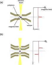
    With conventional atomic-resolution electron microscopes, atomic-scale observation of magnetic materials has been extremely difficult since the samples are inevitably placed in a strong magnetic field of 2 T or higher. To overcome this limitation, we have developed a new objective lens system that enables atomic-resolution observation while maintaining a sample in the magnetic field-free environment. The electron microscope with this new lens system facilitates atomic structure analysis of magnetic materials and is expected to enable ultra-high resolution magnetic field imaging inside materials and devices. In this review, we describe the development of elemental technologies for the Magnetic field-free Atomic-Resolution Scanning transmission electron microscope (MARS) and show the results of real-space observation of the atomic-scale magnetic field distribution inside antiferromagnetic hematite.

    Download PDF (8949K)
  • Jun-ichi KISHIKAWA, Moe ISHIKAWA, Hideto MIYOSHI, Takayuki KATO
    Article type: Current Topics
    2023 Volume 66 Issue 12 Pages 689-694
    Published: December 10, 2023
    Released on J-STAGE: December 10, 2023
    JOURNAL RESTRICTED ACCESS

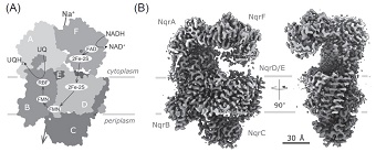
    Cryo-electron microscopy (cryo-EM) has become a powerful tool for structural analysis, owing to the recent “resolution revolution” and currently it is the mainstream technique for analyzing the structures of large molecules. The three-dimensional structural information of proteins is indispensable for structure-based drug design, particularly in the field of drug discovery.

    Hear we presents what information can be retrieved from single-particle analysis by cryo-EM, using the recently identified Na+-pumping NADH-Quinone oxidoreductase as an example. The structural analysis of the protein determined the overall structure at 2.9 Å resolution and identified the cofactors within it, which revealed the mechanism of electron transfer. Furthermore, flexible conformational changes were successfully visualized, suggesting the possibility of conformational changes associated with electron transfer. These findings demonstrate the utility of cryo-EM for structural analysis, highlighting its transformative potential in the field of drug development, particularly in understanding the mechanisms of large molecular complexes.

    Download PDF (3045K)
  • Hiroki HASHIGUCHI, Akiho NAKAMURA
    Article type: Current Topics
    2023 Volume 66 Issue 12 Pages 695-699
    Published: December 10, 2023
    Released on J-STAGE: December 10, 2023
    JOURNAL RESTRICTED ACCESS

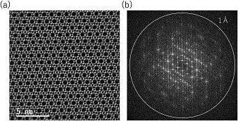
    In recent years, scanning transmission electron microscopy (STEM) with aberration corrector has become widely popular due to its ability to achieve atomic resolution and analysis. On the other hand, observation of electron-beam-sensitive materials such as catalysts and lithium-ion batteries at atomic resolution is requested. The newly developed STEM-optimum Bright Field (OBF) imaging is effective for this purpose. This method enables high-sensitivity and high-contrast observation even under low-dose conditions. In this paper, we explain the principles and practical conditions of the STEM-OBF and introduce its application results to catalyst materials such as zeolites and Metal Organic Frameworks (MOF), lithium-ion battery materials, and semiconductor devices.

    Download PDF (6835K)
  • Yuki KIMURA, Hiroyasu KATSUNO, Shizuka HIRAKAWA, Tomoya YAMAZAKI
    Article type: Current Topics
    2023 Volume 66 Issue 12 Pages 700-705
    Published: December 10, 2023
    Released on J-STAGE: December 10, 2023
    JOURNAL RESTRICTED ACCESS

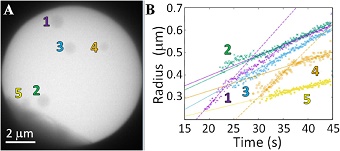
    Non-equilibrium processes such as nucleation are difficult to observe in situ using transmission electron microscopy (TEM) because of spatiotemporal stochastic process. Therefore, we have been developing a method to predict/detect nucleation events and observe under low electron doses conditions in real time with the support of machine learning. Low electron dose observation is important to avoid radiolysis of water in the observation of aqueous solutions using liquid-cell TEM. Our data-driven TEM that can suggest observation points to the operator by processing in situ observation data in real time. In other words, it is data-driven TEM in which the TEM helps the operator, rather than the TEM being driven by the data. By incorporating the two codes into the TEM's software, nucleation can now be observed efficiently.

    Editor's pick

    Download PDF (4772K)
  • Takashi MATSUMOTO, Taiji YAMAMOTO
    Article type: Current Topics
    2023 Volume 66 Issue 12 Pages 706-710
    Published: December 10, 2023
    Released on J-STAGE: December 10, 2023
    JOURNAL RESTRICTED ACCESS

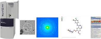
    Because of the difficulty of growing crystals large enough for single-crystal X-ray analysis, there has been considerable interest in determining structure from nanocrystals in recent years. The structure of nanocrystals can be analyzed using electron diffraction (3D ED/MicroED). This is because electron interacts more strongly with materials than X-ray. However, some of you may have given up on 3D ED/MicroED due to the need for expertise in electron diffraction or lack of experience in diffraction image processing and structural analysis.

    XtaLAB Synergy-ED, an integrated 3D ED/MicroED platform jointly developed by JEOL and Rigaku, consists of a TEM-based electron source optimized for electron diffraction, a hybrid photon counting detector, and a combination of these core technologies to maximize synergy effects, CrysAlisPro for ED.

    The Synergy-ED workflow and measurement examples are presented in this article.

    Download PDF (2018K)
  • Makoto KUWAHARA
    Article type: Current Topics
    2023 Volume 66 Issue 12 Pages 711-718
    Published: December 10, 2023
    Released on J-STAGE: December 10, 2023
    JOURNAL RESTRICTED ACCESS

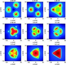
    Time-resolved measurements in transmission electron microscopy (TEM) offer the possibility of revealing ultrafast phenomenon with nanometer spatial resolution, allowing the analysis of nonuniform materials, functional materials, and advanced nano-devices. A high-quality pulsed electron beam, with high brightness, narrow energy width, and high spin-polarization, was realized by installing a semiconductor-type photocathode having a negative electron affinity surface to an electron gun in the TEM system.

    We conducted time-resolved measurements of electron energy-loss spectrum with picosecond pulsed electron beam in the TEM, and elucidated electron-phonon and phonon-phonon relaxation processes in photo-excited gold nanoparticles. Furthermore, the intensity interference of charged fermions was realized by temporally modulated spin-polarized electrons. The coherent spin-polarized electron beam facilitates the extraction of intrinsic quantum interference. These application results pave the way for a new analytical method for the ultrafast dynamics of electrons and lattices in materials, in addition to quantum measurements in electron microscopes.

    Download PDF (3659K)
  • Manabu ISHIMARU
    Article type: Review
    2023 Volume 66 Issue 12 Pages 719-724
    Published: December 10, 2023
    Released on J-STAGE: December 10, 2023
    JOURNAL RESTRICTED ACCESS

    Short-range and medium-range ordered structures of amorphous materials can be evaluated by radial distribution functions and atomic pair-distribution functions extracted by quantitative analyses of diffraction intensities. Because of the strong interaction of electrons with materials and the short wavelength of high-energy electrons, electron diffraction can produce intensity profiles up to high scattering angles comparable to those of neutron diffraction and synchrotron x-ray diffraction in a short time. Here, we present an example of electron diffraction radial distribution function analysis of amorphous boron carbide by electron diffraction techniques.

    Download PDF (6243K)
Report
Conference Report
News & Trends
feedback
Top