消化器内視鏡の進歩:Progress of Digestive Endoscopy
Online ISSN : 2189-0021
Print ISSN : 0389-9403
49 巻
選択された号の論文の73件中51~73を表示しています
症例
  • 菅原 和彦, 星原 芳雄, 山本 敬, 奥田 近夫, 木村 孝, 井口 大助, 田中 達朗, 山本 信彦, 橋本 光代
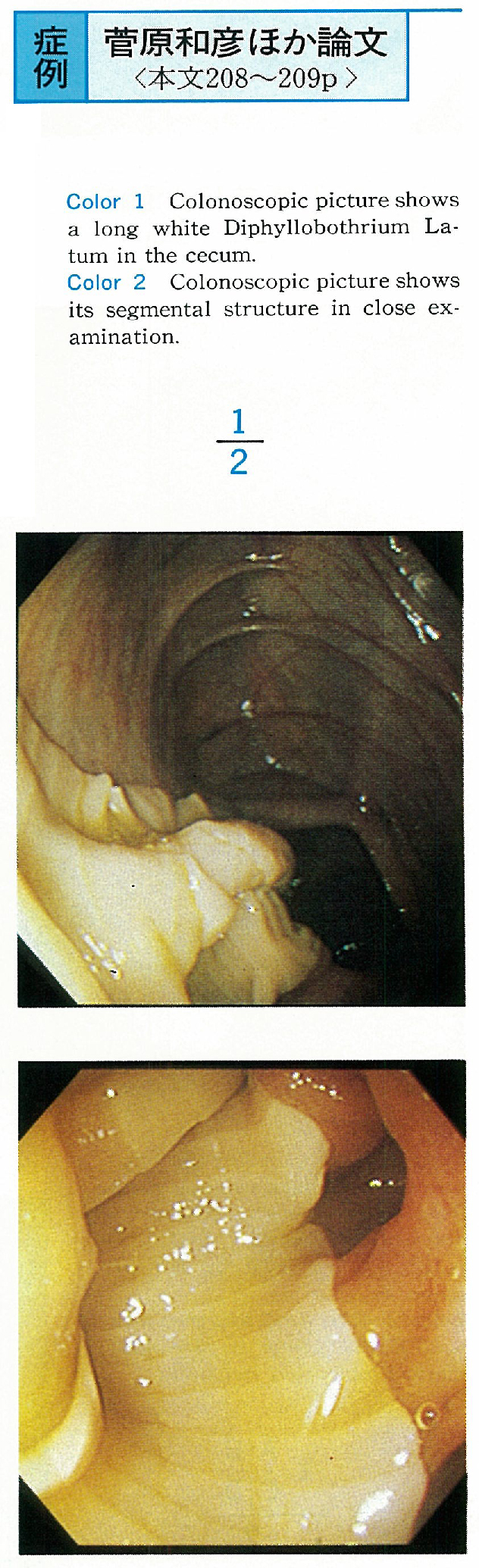
    1996 年 49 巻 p. 208-209
    発行日: 1996/12/02
    公開日: 2015/03/20
    ジャーナル フリー
    A 59-year-old man, who had lived in Sapporo and eaten raw salmon and trout until 1988, admitted our hospital because of the positive fecal occult blood test. There were no abnormal signs of his physical examinations nor the other results of laboratory datum.
    Colonoscopic examination revealed a long white Diphyllobothrium Latum in the cecum. But we could not find its head in the terminal ileum. Within one hour after administration of Gastrografin, a living Diphyllobothrium Latum (2.4 m in length) was expelled. Because we could not find its head, we administered Aminosaidine. After these therapy, the test of eggs of Diphyllobothrium Latum in the stool became negative.
    We considered this is a rare case of a Diphyllobothrium Latum unexpectedly found during colonoscopy.
  • 川嶋 一成, 有沢 淑人, 小花 光夫, 納賀 克彦, 福田 純也
    1996 年 49 巻 p. 210-211
    発行日: 1996/12/02
    公開日: 2015/03/20
    ジャーナル フリー
    A 63-year-old female who had been diagnosed as rheumatoid arthritis (RA) for 17 years, admitted to our hospital because of low grade fever and severe bloody diarrhea. Barium enema and colonoscopic examination revealed colonic pseudopolyposis and disappearance of Haustra coli. The pathological finding showed inflammatory changes which were compatible with ulcerative colitis (UC) .
    While RA was well controlled by oral administration of prednisolone, remission of UC could be obtained by increasing dosage of oral administration of prednisolone and sulfasalazopyridine.
    UC is a rare complication of RA. But on the course of RA, when anemia or severe diarrhea was detected, colon should be examined to rule out UC.
  • 代田 喜典, 柿本 應貴, 井ノ口 幹人, 上川 次郎, 永合 正浩, 小畑 満, 石田 孝雄
    1996 年 49 巻 p. 212-213
    発行日: 1996/12/02
    公開日: 2015/03/20
    ジャーナル フリー
    A 15-year-old child was admitted to our hospital due to melena. Colonoscopic examination showed a huge and irregular surfaced polypoid lesion that is called type Yamada IV in the sigmoid colon at 22cm from anal verge. The polyp was removed successfully by colonoscope. The resected specimen was 36×23×18mm in size with marked lobulation and stalk. Histologic examination revealed both juvenile polyp and tubular adenoma with severe atypia. The resection margin was free from the adenoma.
    Although the majority of colonic polyps in children are juvenile polyps, the presence of huge colonic adenoma with juvenile component in pediatric patients has also been reported by some authors. It is necessary to remove a huge colonic polyp in a child as soon as possible by colonoscope, especially that over 10 years of age.
    We conclude that colonoscopic polypectomy is necessary in this case because the resected polyp was so huge (36mm in size) and histological findings revealed pre-cancerous potential.
  • 鴨志田 敏郎, 堀田 総一, 菊池 健太郎, 茂木 秀人, 平井 信二, 岡 裕爾, 高橋 敦
    1996 年 49 巻 p. 214-215
    発行日: 1996/12/02
    公開日: 2015/03/20
    ジャーナル フリー
    A 57-year-old man was received total colonoscopy and a biopsy was performed from the head of a 5 mm polyp of the descending colon without apparent bleeding. That day he was admitted to our hospital because of melena. Next day after admission he fell into hypovolemic shock due to massive colonic hemorrhage. The second colonoscopy revealed there was not the head of the polyp, but only the stalk remained. An exposured vessel was observed at the top of the stalk.
    Polypectomy was performed for the remained stalk. After polypectomy the patient did well with supportive care. There were no vessels seemed to be a cause of massive hemorrhage in the biopsy specimen. The excised specimen by polypectomy showed a hemorrhagic vessel and thrombus. The reason why the head of a polyp dissapeared was unclear. We cannot deny the antiaggregative drug play an important role for the cause of massive colonic hemorrhage.
    Although massive hemorrhage after colonic standard biopsy is very rare, but our case indicates that massive hemorrhage is one of important complications.
  • 小野里 康博, 小林 出, 石原 弘, 吉田 誠, 茂木 健太, 坂本 輝彦, 村松 誠司, 猿谷 真也, 家崎 桂吾, 鷲田 雄二, 小林 ...
    1996 年 49 巻 p. 216-217
    発行日: 1996/12/02
    公開日: 2015/03/20
    ジャーナル フリー
    The patient was an 81-year-old woman. Colorectal endoscopy was performed due to lasting sense of abdominal fullness and, as a result well differentiated adenocarcinoma of approximately 40 mm in size was found in the ascending colon. She wished to avoid surgical resection due to her high age and so an endoscopic treatment was adopted.
    After local injection of physiological salt solution to prevent perforation, piecemeal endoscopic mucosal resection was performed followed by the endoscopic microwave coagulation therapy. The techniques were repeated 3 times in total. She has been observed for 30 months but neither residue nor recurrence has been found on endoscopy.
    Endoscopic microwave coaguration therapy is effective as an additional treatment after endoscopic mucousal resection of early colorectal cancer and local radical cure seems possible even when cancer has grown to a size unresectable as a lump.
  • 森田 尚樹, 森 潔
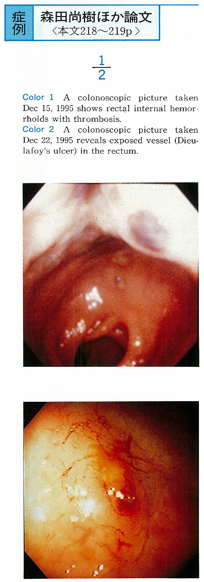
    1996 年 49 巻 p. 218-219
    発行日: 1996/12/02
    公開日: 2015/03/20
    ジャーナル フリー
    A 78-year-old woman was admitted to our hospital on Dec 14, 1995, due to anemia for the massive lower gastrointestinal bleeding. Physical examination on that day revealed no remarkable abnormality, except for internal hemorrhoids. Colonscopy performed on the next day (Dec 15) showed no abnormality of the colon and the rectal internal hemorrhoids with thrombosis. On Dec 22, however, due to her sudden and massive arterial bleeding from the anus, urgent colonoscopy was carried out and it revealed an exposed vessel with adherent clots at the rectum about 3 cm from the anal verge. Hemostasis was successfully done with endoscopic clipping to the bleeding artery and the patient was discharged from the hospital on Jan 4.
    The case presented might have Dieulafoy's ulcer in the rectum, and as far as we know, only 13 cases of this rectal disease have been reported in Japan during the past 10 years. Previous papers described that endoscopic treatment such as ethanol injection, clipping, or hypertonic saline-epinephrine (HSE) injection should be indicated for the hemostasis of this ulcer as a first choice as in the gastric cases, and transarterial embolization (TAE) for the refractory disease.
  • 森 三樹二, 岩男 泰, 厚川 和宏, 大木 英二, 大石 温子, 石井 裕正, 寺本 龍生, 渡辺 守, 日比 紀文
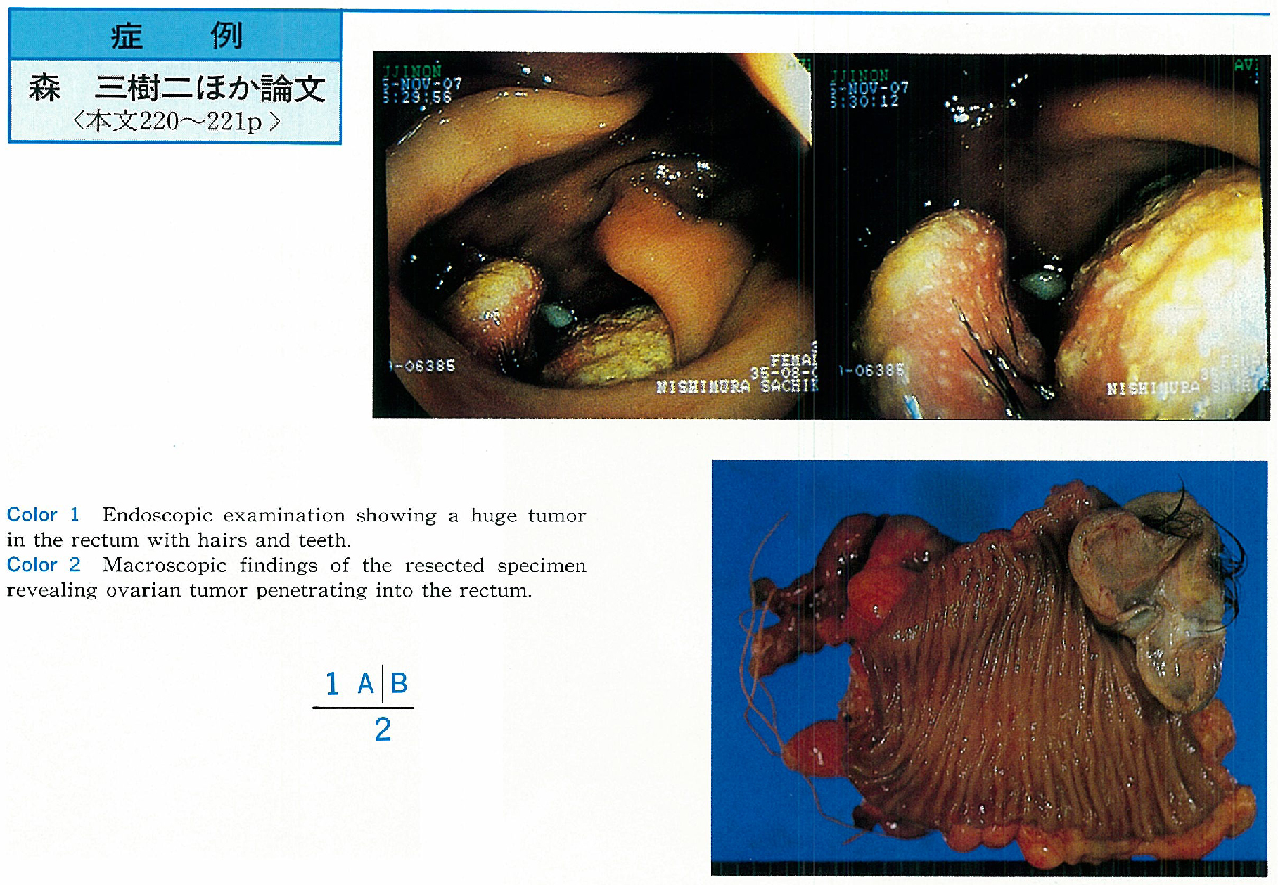
    1996 年 49 巻 p. 220-221
    発行日: 1996/12/02
    公開日: 2015/03/20
    ジャーナル フリー
    A 35-year-old woman admitted to our hospital with a chief complaint of bloody stool and lower abdominal pain. After that, she noticed that hairs were discharged from the anus. A mass lesion in diameter of 6 cm was detected in the rectum by opaque barium enema method.
    Endoscopic examination showed a white elastic tumor with several hairs and teeth in the anterior wall of the rectum. Squamous epithelium was observed by biopsy and it was diagnosed as ovarian teratoma. A 6 cm tumor of not uniform touched with wall of the rectum and penetrating into the rectum were observed by CT and MRI, we judged that this case was left ovarian teratoma penetrated into the rectum. Pathological examination proved the left ovary being mature teratoma in which various tissue produced from digerminial layers are complicatedly mixed.
    Since this patient was considered to be a rare case, this paper includes a review on similar case reported in Japan.
  • 大竹 聖子, 斉藤 民子, 塩田 淳朗, 大久保 斎, 清水 敏弘, 朝岡 昭, 田中 直英, 椿 浩司, 林 洋一, 荒川 泰行
    1996 年 49 巻 p. 222-223
    発行日: 1996/12/02
    公開日: 2015/03/20
    ジャーナル フリー
    A 58-year-old female was visited to our hospital for abdominal pain. Endoscopic findings showed gastritis appearance.
    Abdominal ultrasonography showed network pattern of the liver. Therefore she was admitted to our hospital for laparoscopic examination. Laparoscopic finding showed the yellowish white speckles and tortoise-shell pattern. Histlogical finding of liver biopsy showed Schistosomiasis Japonica ova. Therefore the patients who was living at the frequent zone of Schistosomiasis Japonica has necessity of the ultrasonography.
  • 星長 春樹, 中塚 雄久, 名知 志子, 間宮 康貴, 長田 祐二, 小泉 信人, 吉本 均, 竹内 司, 斉藤 整, 吉沢 雅史, 津久井 ...
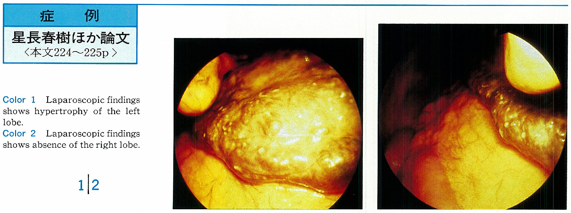
    1996 年 49 巻 p. 224-225
    発行日: 1996/12/02
    公開日: 2015/03/20
    ジャーナル フリー
    A very rare case of hypogenesis of the right lobe of the liver is reported.
    A 51-year-old male was admitted to our hospital because of acute hepatitis B. Abdominal US and CT scanning showed aplasia of the right lobe with compensatory hypertrophy of the left lobe and the caudate lobe. The gallbladder was situated posterior to the hypertrophied left hepatic lobe. The right portal branch were not observed on abdominal angiography. On laparoscopy, the right lobe of the liver was also found to be absent and the left lobe of the liver showed compensatory enlargement. Based on these findings, this patient was diagnosed as having a hypogenesis of the right lobe of the liver associated with hepatitis B.
    Only thirteen cases of the right hepatic lobe agenesis have been reported since 1987 in Japan. Although this anomaly is considered to be extremely uncommon, it will be more frequently in the future with advances in noninvasive imaging diagnosis.
  • 狩野 聡子, 小林 出, 山田 俊彦, 阿部 毅彦, 石原 弘, 市川 武, 田村 茂生
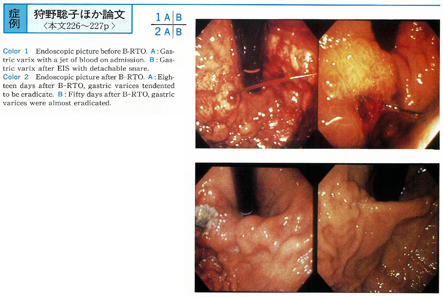
    1996 年 49 巻 p. 226-227
    発行日: 1996/12/02
    公開日: 2015/03/20
    ジャーナル フリー
    A 51-year-old male patient was admited to our hospital because of a large amount of hematemesis. Large cardio solitary varices with a jet of blood were seen via endoscopy on admission. We performed endoscopic injection sclerotherapy (EIS) , and succeeded in stopping the hemorrhage. After the therapy the patient fell into bleeding shock for a time. But 10 days after, gastric varices bled again, therefore we performed EIS and used detachable snare, resulted in the hemorrhage.
    But because of enlarged gastric varices, we were afraid of the varices rupture for the second time. Next, since angiographic examination disclosed a gastric renal shunt, we tried B-RTO. The gastric renal shunt was occluded with a balloon and we performed injection of 24ml 5% EOI through the balloon catheter. Gastric varices were almost eradicated on endoscopic examination 50 days after B-RTO.
    B-RTO is assessed to be a useful non-invasive procedure but it is difficult to treat gastric varices with bleeding, therefore we need EIS or detachable snare a state of emergency bleeding. By both therapies use jointly, we are possible to meet with good results.
  • 齋藤 光浩, 中島 洋, 佐藤 悦久, 川村 直弘, 今村 真紀子, 徳植 秀樹, 横山 孝典, 高須 政夫, 宇都宮 潔, 高橋 信一, ...
    1996 年 49 巻 p. 228-229
    発行日: 1996/12/02
    公開日: 2015/03/20
    ジャーナル フリー
    Accessory liver lobe is attached to main liver, and different from ectopic liver which is detached from main liver. Accessory liver lobes are incidentally noted at operation or necropsy in most cases. We reported 7 cases with accessory liver lobe diagnosed by laparoscopy.
    The cases were of 22 to 63 years old, including 6 females and one male. The biopsy diagnosis of main liver was chronic hepatitis in 4 cases, acute hepatitis, drug-indused hepatitis and autoimmune hepatitis in 1 case each. These accessory liver lobes were attached to quadrate lobe (S4) in 4 cases, and left liver lobe (S3) in 3 cases. Accessory liver lobes measured 3mm minimum and 10mm maximum in size. In shape, they were tongue-like in 3 cases, semioval and hemispherical in 2 cases each. All of them could not be demonstrated by ultrasonography and CT, and were asymptomatic.
  • 畑 真, 中川 浩之, 長谷 祐治, 織畑 道宏, 佐々木 秀雄, 城所 仂
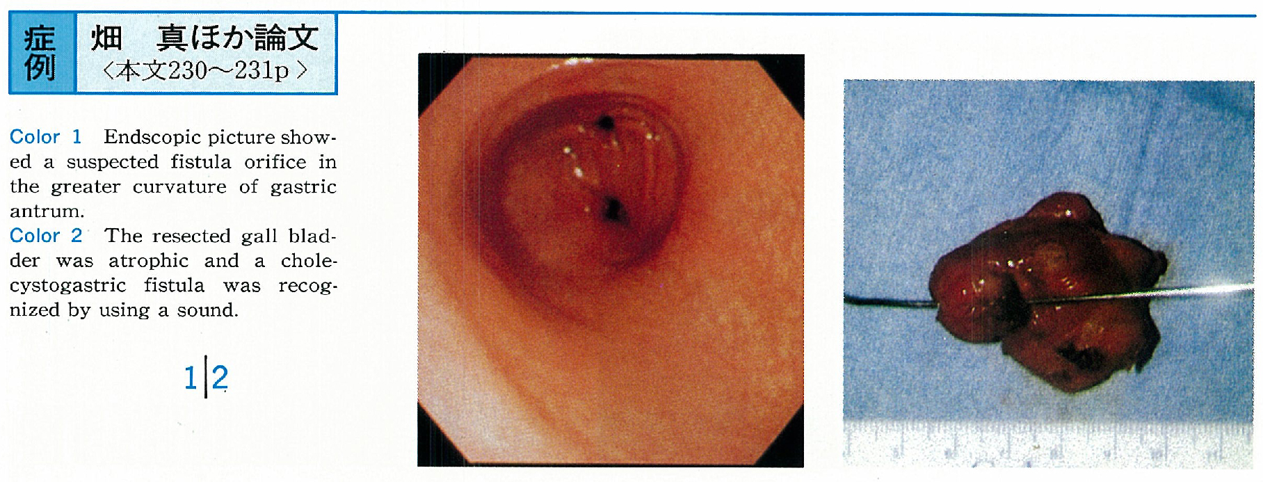
    1996 年 49 巻 p. 230-231
    発行日: 1996/12/02
    公開日: 2015/03/20
    ジャーナル フリー
    The patient, a-57-year-old female who had a history of cholecystitis about 25 years before, was seen in our hospital with a chief complaint of right hypochondralgia in September 1994.
    Abdominal plain X-ray revealed pneumobilia and she was admitted with a diagnosis of enterobiliary fistula. Pneumobilia and atrophic gall bladder were detected by CT and Ultrasonography. The orifice, which was considered to be a cholecystogastric fistula, was found in the antrum of the stomach by gastroendoscopy, but the fistula wasn't clearly visualized although endoscopic fistulography was tried. The fistula wasn't visualized by endoscopic retrograde cholangiography (ERC) and percutaneus transhepatic cholangiography (PTC) .
    Surgical operation was performed with a diagnosis of cholecystitis, cholangitis and enterobiliary fistula. During the operation, the gall bladder was remarkablly contracted and adhered to the antrum of the stomach with a fistula formation. Cholecystectomy and closure of the fistula was performed. The cholecystogastric fistula was recognized by using a sound. Histologically, chronic cholecystitis pattern was demonstrated. It has been well known that cholecystogastric fistula is rare among the sponstaneous internal biliary fistulae.
    We found 38 cases in the Japanese literature. Surgical therapy at an early stage is recommended even for asymptomatic patients in principle.
  • 佐藤 康永, 木田 光広, 日高 央, 菅野 聡, 松田 摩也, 今泉 弘, 西元寺 克禮, 仙石 紀彦, 飯野 善一郎, 吉田 宗紀
    1996 年 49 巻 p. 232-233
    発行日: 1996/12/02
    公開日: 2015/03/20
    ジャーナル フリー
    A 53-year-old male presented with right upper quadrant pain. Two gallstones were visualized on ultrasonography. He was treated by laparoscopic cholecystectomy. On the first postoperative day (POD) , substantial biliary leakage from the abdominal drain occured. Computed tomography and ultrasonography showed subphrenic fluid collection.
    Endoscopic retrograde cholangiography was performed on the 10th POD. It showed extravasation of contrast from the posterior branch. Then a 7 Fr gauge endoprosthesis was placed in, after that bile drainage decreased, and followed ultrasonography showed decreasing of the fluid collection. One month later, the stent was removed and disclosure of the biliary leakage was confirmed by ERC. He have asymptomatic now.
    We conclude that endoscopic retrograde biliary drainage is one of the choice for treating the biliary leakage following laparoscopic cholecystetomy.
  • 町田 彰男, 新谷 隆, 平塚 研之, 李 雨元, 成田 和広, 相田 貞継, 清水 喜徳, 村上 雅彦, 草野 満夫, 諸星 利男
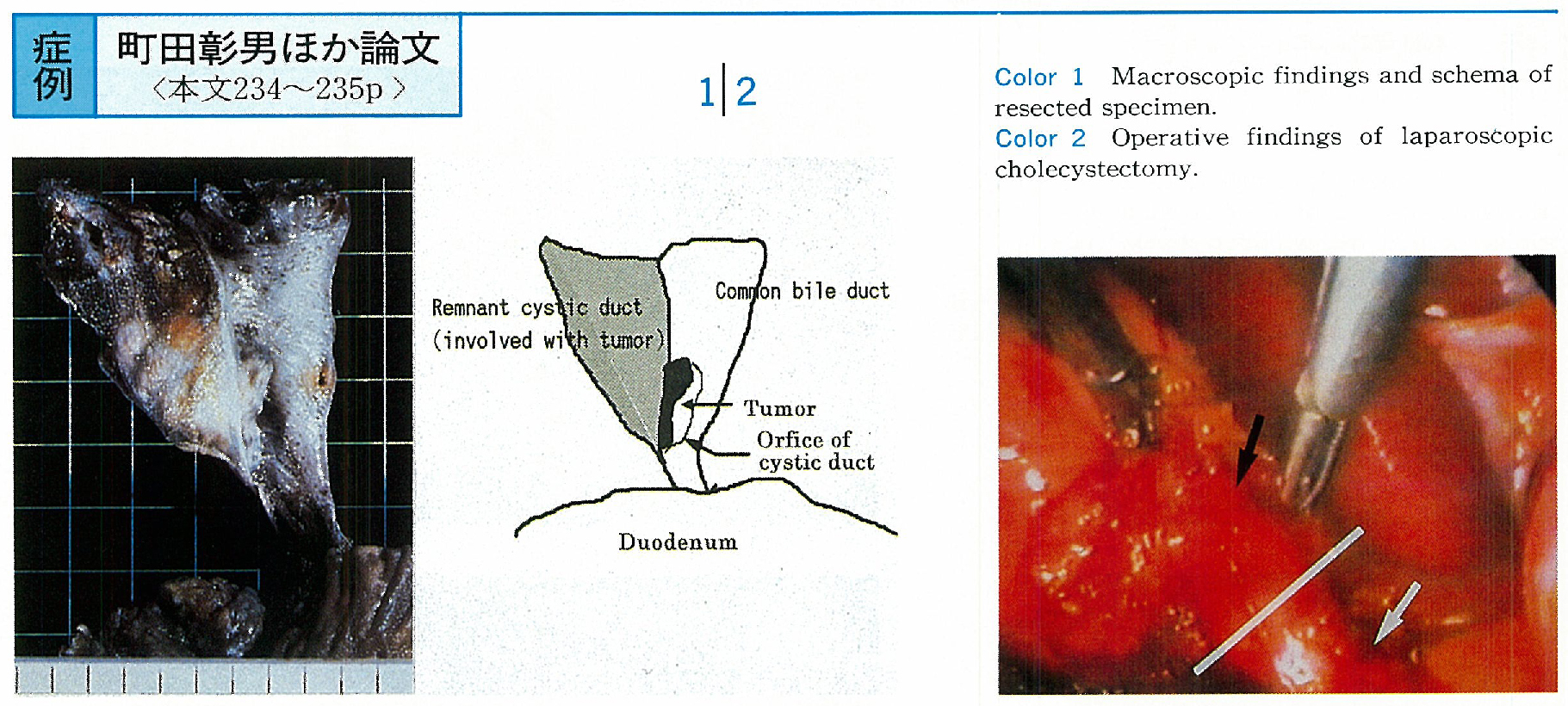
    1996 年 49 巻 p. 234-235
    発行日: 1996/12/02
    公開日: 2015/03/20
    ジャーナル フリー
    A 67-year-old man was admitted to our hospital for obstructive jaundice. Before four years, he had a laparoscopic cholecystectomy for acute cholecystitis by limy bile. Abdominal computed tomography and ultrasonography revealed obstruction of superior common bile duct by a tumor. Following PTCD and cholangiography showed same findings and cholangiocarcinoma was diagnosed by cytology from bile juice.
    Pancreaticoduodenectomy was performed. Histological examination of resected specimen revealed papillary adenocarcinoma in remnant cystic duct with the invasion limited to the muscularis propria, without lymph node metastasis. We reported a very interesting case of remnant cystic duct cancer after cholecystectomy.
  • 土谷 まり子, 柳沢 明子, 板橋 聖子, 北村 容子, 有田 由美子, 渡辺 七六, 前田 淳, 重本 六男, 山下 克子, 横山 泉, ...
    1996 年 49 巻 p. 236-237
    発行日: 1996/12/02
    公開日: 2015/03/20
    ジャーナル フリー
    A 43-year-old man was admitted with a painfull pancreatic cyst. He had a long history of alcohol abuse and had presented with acute alcoholic pancreatitis four years previously. After that episode, alcohol consunption was repeatedly associated with pain in the right iliac fossa.
    Ultrasound and CT scanning of the abdomen revealed a cystic structure in the anterior aspect of the pancreatic body. ERCP showed dilatation and tortuosity of the main pancreatic duct. Based on the ERCP findings the clinical diagnosis was chronic pancreatitis complicated by pseudocyst. The objective findings, inflammatory reaction, etc, resolved in response to medical treatment.
    However, the cyst was sometimes symptomatic and giant in size (8×4.5cm) . It showed no tendency to shrink and a drainage procedure was required. Although we initially attempted laparoscopic cyst-gastrostomy, the wall of cyst was free in the abdominal space and had no point of attachment to the digestive duct. We therefore performed surgical cyst-gastrostomy, which resulted remarkable reduction in the size of cyst. There are several approaches to the treatment of symptomatic pancreatic pseudocyst, in addition to the surgery, such as laparoscopy, etc.
  • 真田 淳, 荻原 正示, 武井 和夫, 水村 泰夫, 水口 泰宏, 武田 一弥, 三輪 一彦, 真神 易, 堀部 俊哉, 角谷 宏, 新戸 ...
    1996 年 49 巻 p. 238-239
    発行日: 1996/12/02
    公開日: 2015/03/20
    ジャーナル フリー
    A 60-year-old female took medical checkup and examination of upper GI tract revealed irregularities of outlines of stomach, suggestive of compression by extramural mass. Further examination was performed at our hospital.
    Ultrasonography revealed an echogenic tumor picture and a small cyst in the tail of pancreas. CT and endoscopic ultrasonography showed intratumor honeycomb appearance. Deviation of the main pancreatic duct adjacent to the tumor was demonstrated by ERP. The angiography showed a hypervascular area at the tumor and an intermission of pancreatic magna artery.
    This patient underwent distal pancreatectomy. Histological dignosis was serous cystadenoma, and tumor showed microcystic pattern, single lining epithelial cells without cellular atypism, and two intracystic hemorrhages in the tumor.
第62回日本消化器内視鏡学会関東地方会 後抄録
特別講演
シンポジウム
ワークショップ
スペシャルフォーラム
ビデオセッション
一般演題
feedback
Top