消化器内視鏡の進歩:Progress of Digestive Endoscopy
Online ISSN : 2189-0021
Print ISSN : 0389-9403
50 巻
選択された号の論文の76件中1~50を表示しています
掲載論文カラー写真集
臨床研究
  • 小山 博, 成木 行彦, 西野 執, 笹島 雅彦, 神田 映子, 栗田 俊夫, 北條 裕, 有木 寿史, 米谷 隆, 山田 秀一, 中谷 尚 ...
    1997 年 50 巻 p. 147-151
    発行日: 1997/06/06
    公開日: 2015/02/17
    ジャーナル フリー
     われわれは,Helicobacter pyloriHP)陽性胃潰瘍30例,十二指腸潰瘍23例,胃十二指腸潰瘍11例,慢性胃炎38例に対して,amoxicillin(AMPC)またはclarithromycin(CAM)にて除菌治療を行い,胃内視鏡所見と胃粘膜組織所見を治療前後で比較検討をした。方法は除菌治療前,除菌終了4週間目の胃内視鏡検査施行時に胃体上部,体下部,前庭部の各大弯側からそれぞれ2カ所ずつ生検し,培養および組織学的検査をした。13C-urea breath test(UBT)は13C標識の尿素(Isotech社)100mgを無菌水30mlに溶解し,空腹時に投与し,さらに無菌水30mlで2回口腔内を濯いだ。前,5,10,20,30,60分後に呼気約2.5lを採取した。13CO2はレーザ分光計で測定した。UBTは除菌前,除菌終了直後,2週後,4週後,12週後に測定した。慢性胃炎に対してはSydney systemで評価した。HP除菌治療はA群55例にlansoprazole(LAZ)30mg/日,CAM 600mg/日,ecabet Na(E-Na)2.0g/日,B群47例にLAZ 30mg/日,AMPC 2000mg/日,E-Na 2.0g/日を2週間投与した。全体の除菌率は72/102(70.5%)で,A群では30/55(54.5%)認められ,B群では42/47(89.3%)認められた。治療前の内視鏡所見では,びらん性胃炎は除菌有効例が無効例に比べてgradeが軽い傾向が認められ,治療後の有効例ではほとんどびらんの改善を認めた。発赤した胃炎では,除菌有効例はほとんど治療後改善を認め,胃粘膜組織像では好中球や単核球浸潤の改善を認めた。UBTは安全で簡便な検査法であり,HP除菌治療の効果判定に有用であると思われた。
  • 松久 威史, 羽山 享宏, 飯田 章太郎, 新井 朱音, 草間 泉, 遠藤 まゆみ, 吉原 一郎, 井上 泰夫, 森 洋, 山田 宣孝
    1997 年 50 巻 p. 152-156
    発行日: 1997/06/06
    公開日: 2015/02/17
    ジャーナル フリー
     消化性潰瘍を中心としたH.pylori感染症例(181例)の除菌成績について検討した。除菌による血清ガストリン値,ペプシノゲン(PG)値の変化についても観察した。除菌の判定は除菌療法開始6週間後に実施し,生検組織の病理学的検査,13C尿素呼気試験(UBT)により行った。病理学的診断,UBTともに陰性で,背景胃粘膜の活動性胃炎が消失しているものを除菌成功例とした。生検組織上わずかに菌が残存している際や,菌はみられないが活動性胃炎が残存している場合,尿素呼気試験が陰性化していない場合は,同一薬剤による追加除菌を行った。菌数,組織学的活動性に全くあるいはほとんど変化がみられない症例では,他剤による再除菌を試みた。種々のregimenを検討した結果,proton pump inhibitor,tinidazoleにamoxicillinまたはclarithromycinを加えた1週間除菌療法が高い除菌率を示した(各々76.9%,90.0%)。追加除菌,再除菌を含め181例中77.3%において除菌が確認された。除菌成功例における血清ガストリン値は,除菌6週間後に有意に低下していた(p<0.05)。血清PGⅡ値は除菌成功例,不成功例とも6週間後に低下していたが,成功例においてより低値であった。PGⅠ/Ⅱ比も除菌前に比し上昇しているが,成功例においてその上昇傾向が目立つ。これは,不成功例の中に再除菌症例と追加除菌症例が含まれているためと思われた。血清ガストリン値,血清PGⅡ値,PGⅠ/Ⅱ比は除菌判定の指標として有用と考える。
  • 五十嵐 直喜, 熊井 浩一郎, 一色 聡一郎, 石川 洋一郎, 横山 剛義, 藤田 晃司, 石川 秀樹, 大谷 吉秀, 久保田 哲朗, 北島 ...
    1997 年 50 巻 p. 157-159
    発行日: 1997/06/06
    公開日: 2015/02/17
    ジャーナル フリー
     胃生検でGroup Ⅲとなった場合の臨床上取扱の観点から,胃腺腫の肉眼病型としては圧倒的に多い扁平隆起性病変について,内視鏡的および病理組織学的診断について検討した。1990年1月から1994年12月までの5年間に胃高分化型腺癌(tub1)m癌および胃腺腫(adenoma)に対して施行された胃切除28例,または内視鏡下粘膜切除(EMR)73例によって得られた切除標本の肉眼所見および病理組織学的所見と,術前の内視鏡所見および生検組織診断結果を対比検討した。治療前後の病理組織学的所見が一致したものはGroup Ⅴ→tub1が36例(35%),Group Ⅲ→adenomaは33例(33%)であった。一方,不一致症例はGroup Ⅲ→tub1が32例(32%)であった。その内視鏡所見はびらんを伴わないものが多く,治療後の切除標本の腫瘍径では20mm以下が多かった。なおGroup Ⅴ→adenoma症例はなかった。
  • 藤田 晃司, 大谷 吉秀, 石川 洋一郎, 一色 聡一郎, 横山 剛義, 五十嵐 直喜, 石川 秀樹, 石塚 裕人, 林 憲孝, 大上 正裕 ...
    1997 年 50 巻 p. 160-163
    発行日: 1997/06/06
    公開日: 2015/02/17
    ジャーナル フリー
     最近6年間に当院内視鏡センターで内視鏡的粘膜切除(EMR)を施行した胃癌症例は135症例である。そのうち切除標本の病理組織学的所見により胃切除が施行された19例(14%)を対象とし,EMR標本と切除胃標本の比較から癌遺残とリンパ節の転移の状況を検討した。症例は男性17例,女性2例であった。年齢は35-80歳(平均61.4歳)であった。肉眼型はⅠ型1例,Ⅱa型7例,Ⅱc型9例,Ⅱa+Ⅱb 1例,Ⅱa+Ⅱc 1例であった。AまたはM領域で小弯または後壁が78.9%を占めた。腫瘍長径は5-35mm(平均13.4mm),組織型はtub19例,tub29例,por11例であった。EMR切除標本の深達度はm癌9例,sm癌10例であった。追加EMRを施行した症例が3例あり,そのうち2例はレーザー照射を追加していた。術式は幽門側胃切除14例,胃全摘3例,噴門側胃切除1例,部分切除1例であった。手術が追加された理由としては,EMR切除断端陽性7例,EMR切除断端不明瞭2例,EMR切除断端陽性かつsm浸潤10例であった。m癌9例中7例(79%),sm癌10例中8例(80%)に癌遺残を認めた。sm癌で癌遺残を認め,総胆管結石を併発したため,2年後に胃切除を行った1例は深達度ss,n2の進行癌となっていたが,他の18例にはリンパ節転移を認めなかった。EMR後に断端陽性の場合,高率に癌遺残を認め,胃切除が必要と考えられた。
  • 富樫 一智, 小西 文雄, 石塚 恒夫, 仙波 真吾, 柏木 宏, 金澤 暁太郎
    1997 年 50 巻 p. 164-167
    発行日: 1997/06/06
    公開日: 2015/02/17
    ジャーナル フリー
     われわれは,通常の内視鏡的摘除法では不完全切除となったり,内視鏡的摘除の完全性に関する組織学的判定が不十分となる可能性がある病変に対して,内視鏡的粘膜切除(以下EMR)を実施してきた。本論文では,われわれが施行した478回のEMR症例を対象として検討した。EMRの方法は,生食の注入方法(注入部位・注入回数・注入速度)を,病変の形態・大きさ・存在部位に応じて決定した。15mm以下の病変に対してはミニ楕円型スネアを用いた。EMR後の粘膜欠損部のクリップによる閉鎖術は,出血傾向がある場合には原則的に施行した。結果として,478回のEMR後に穿孔例はなかったが,直後の出血は16例,翌日以降の出血は3例に認められた。直後に出血した16例はいずれも直ちに内視鏡的に止血可能であり,その後に再出血した症例はなかった。後出血した3例中2例では,検査時に明かな出血がなかったが,1例では静脈性出血が認められクリップにより止血された。EMR後の粘膜欠損部のクリップによる閉鎖が実施された20例中1例においても後出血がみられた。EMR後の局所再発は分割切除となった1病変に認められ,粘膜下層浸潤癌のため外科的追加切除された4病変においても,局所に腫瘍腺管の残存は認められなかった。以上より,EMRに伴う合併症の頻度は他の内視鏡的摘除法と同等であり,局所の完全切除を得るためにはEMRを実施することが適切であろうと考えられた。
  • 六倉 俊哉, 三輪 博久
    1997 年 50 巻 p. 168-172
    発行日: 1997/06/06
    公開日: 2015/02/17
    ジャーナル フリー
     早期胃癌および早期食道癌に対する内視鏡的粘膜切除術においては,透明キャップを用いた陰圧吸引下における内視鏡的粘膜切除術(endoscopic mucosal resection using a transparent cap,以下EMR-C)が普及しつつある。しかし,大腸病変に対しては,大腸壁が薄いため穿孔の危険があるとされ,一般には普及していない。われわれは1995年以降,大腸腫瘍性病変27病変(このうち早期大腸癌12病変)に対しEMR-Cを施行してきており,①病変の粘膜下層に10-20mlという大量の生理的食塩水を注入し大きく膨張させることにより,病変を筋層より十分に剥離させる,②切除の途中でスネアを一時ゆるめることにより,絞扼されている可能性のある筋層を解除する,などの工夫を行うことにより,大腸病変に対しても安全にEMR-Cを施行しうると考えている。そして断端が病変近傍の場合には,顕微鏡的遺残病変を消滅させるため,高周波凝固による焼灼術を追加して行っており,早期大腸癌12症例中1例のみが,病変が広範で切除不可能と判断されたため手術を要したが,その他の11症例においては遺残,再発を認めていない。本法は,直腸病変に対しては穿孔の危険がないので安心して施行しうる。また,襞の裏側の病変などの正面視が困難な病変の切除にも適している。さらに,側方進展を示す病変に対しても,分割切除の手技が容易に行えることより非常に有用である。切除後には送気を行わず,クリップによる縫縮術を速やかに施行することにより穿孔,出血に対する予防を行っており,合併症は1例も経験していない。本法は早期大腸癌に対する治療法として,特に側方進展を示す病変に対し,極めて有用な治療法と考えられる。
  • 神澤 輝実, 江川 直人, 榊 信廣, 石渡 淳一
    1997 年 50 巻 p. 173-176
    発行日: 1997/06/06
    公開日: 2015/02/17
    ジャーナル フリー
     膵頭部に異常がない背側膵管像の得られた膵管非癒合12例を対象とし,副乳頭開口部から40mmまでの背側膵管から派生する下頭枝について検討した。全長10mm以上の下頭枝を全例で認め,長さ20mm以上の長い下頭枝が9例9本,10-19mmの短い下頭枝が7例8本であった。長い下頭枝が1本のみ認められた症例が5例,長い下頭枝と短い下頭枝が各1本ずつが4例,短い下頭枝のみが3例であった。長い下頭枝の派生部位は副乳頭開口部より20mmから26mm,短い下頭枝は7mmから15mmの部位が多かった。主乳頭開口部から副膵管合流部までの主膵管の距離が長く,副膵管が上流背側膵管と直線化を呈する70例の副膵管からは,長い下頭枝が59例70本,短い下頭枝が42例53本認められ,長い下頭枝の派生部位は副乳頭開口部より13mmから25mm,短い下頭枝では7mmから14mmの部位に多かった。膵管非癒合の背側膵管と,上流背側膵管と直線化を呈する副膵管は,走行形態のみならず,同部より派生する下頭枝の状況も類似しており,ともに胎生期の背側膵原基の主導管がそのまま残存したと考えられた。
症例
  • 城田 繁, 卜部 元道, 溝渕 昇, 諌山 冬実, 坂本 一博, 榊原 宣
    1997 年 50 巻 p. 177-180
    発行日: 1997/06/06
    公開日: 2015/02/17
    ジャーナル フリー
     症例は62歳,男性。1996年3月嚥下時つかえ感が出現した。上部消化管造影X線検査では,胸部下部食道に長径4.5cmの辺縁不整な隆起性病変を認めた。上部消化管内視鏡検査では,切歯列より38cmの部位から噴門へ向かって,約4cmに及ぶルゴール不染帯を呈する全周性の粘膜の発赤を認めた。その内部には,12時方向を中心とした約半周性の立ち上がり急峻な隆起性病変を認めた。生検の結果は腺癌であった。以上より食道癌と診断し,5月28日胸部下部・腹部食道噴門側胃切除術を施行した。病理組織学的検査で腫瘍は大きさ5×3.3cm,sm,n(-),V0,ly2の腺癌であり,Barrett食道より発生したものと考えられた。腫瘍周囲には異型上皮を認なかった。Barrett上皮部では,抗p53免疫組織染色で陽性細胞を認めず,抗PCNA labelling index(LI)は42%と,正常粘膜と比較して強い増殖活性が示唆された。
  • 西巻 学, 谷 穣, 三治 哲哉, 緑川 昌子, 半田 豊, 森田 重文, 大野 博之, 吉田 肇, 鶴井 光治, 三坂 亮一, 川口 実, ...
    1997 年 50 巻 p. 181-184
    発行日: 1997/06/06
    公開日: 2015/02/17
    ジャーナル フリー
     患者は51歳の男性。検診にて胃粘膜下腫瘍を指摘され,精査目的で入院となった。胃透視および上部内視鏡検査にて,胃前庭部大弯側に径3cm大の表面平滑で,立ち上りのなだらかなbridging foldを伴う同色調の粘膜下腫瘍を認めた。同腫瘍は超音波内視鏡上,固有筋層と連続し,境界明瞭な径2.0×1.6cm大の腫瘤で,エコーレベルはやや低く,内部エコーは不均一で,低エコーのスポットを認めた。腹部CT上では径2cm大の造影剤で濃染される壁外発育性の腫瘤を呈した。悪性腫瘍も否定できないため,腹腔鏡下胃楔状切除術を施行した。病理組織学的には腫瘍は血管腔の増生を伴い,その周囲に核異型のみられない類円形の腫瘍細胞が,シート状あるいは蜂巣状に増殖していた。電顕上では平滑筋細胞と類似の構造を呈し,glomus腫瘍の所見と一致していた。以上より胃glomus腫瘍と診断した。
  • 飯塚 雄介, 光永 篤, 内山 めぐみ, 内田 耕司, 根本 行仁, 中村 真一, 村田 洋子, 鈴木 茂, 林 直諒
    1997 年 50 巻 p. 185-187
    発行日: 1997/06/06
    公開日: 2015/02/17
    ジャーナル フリー
     症例は65歳,女性。1991年より当科にて高ガストリン血症を伴うA型胃炎として定期的に経過観察されていた。1996年4月の胃内視鏡検査にて胃体上部前壁および中部前壁に,粘膜下腫瘍様の山田Ⅰ型の小隆起性病変を認めた。血中ガストリン値は4,432pg/mlと著明な高値を示し,抗胃壁細胞抗体も陽性であった。超音波内視鏡検査では,主として胃壁第3層内に直径4×2mmの低エコー腫瘤像として認められた。同部の組織生検像にて,粘膜上皮深部に索状充実性に配列する腫瘍細胞を認め,これらはGlimerius染色,Chromogranin-A染色陽性であり,胃カルチノイドと診断された。諸検査にてリンパ節転移,肝転移は認められなかった。A型胃炎による高ガストリン血症は胃カルチノイドの発生原因の1つと考えられており,A型胃炎を胃カルチノイドの発生母地として定期的に経過観察することは,その早期発見のために非常に重要であると考えられた。
  • 杉坂 宏明, 成宮 徳親, 杉本 泉, 門脇 泰憲, 丸山 達志, 宮島 浩人, 常喜 真理, 石戸 浩之, 井上 冬彦, 中田 哲也, 永 ...
    1997 年 50 巻 p. 188-191
    発行日: 1997/06/06
    公開日: 2015/02/17
    ジャーナル フリー
     症例は60歳,男性。肝障害の精査目的で入院した。腹腔鏡下肝生検の結果,肝臓の炎症細胞浸潤は軽度であったが,検診目的で行った上部内視鏡検査で凹凸不整な粘膜がみられ,組織学的検索からMALTリンパ腫の診断が確定した。超音波内視鏡では第2層と第3層の著明な肥厚が,赤外線電子内視鏡では腫瘍辺縁に点在する円形のpooling像が認められた。以上からsmに浸潤する病変と考えられ,胃全摘術を行った。切除標本の検索でsm3に浸潤する病変であった。本例では赤外線電子内視鏡による粘膜下層血管の観察が,腫瘍のsm浸潤を判定する一助になると考えられた。
  • 鹿間 伸明, 大井田 正人, 今泉 弘, 木田 芳樹, 田辺 聡, 木田 光広, 小泉 和三郎, 西元寺 克禮
    1997 年 50 巻 p. 192-196
    発行日: 1997/06/06
    公開日: 2015/02/17
    ジャーナル フリー
     症例は45歳,女性。1995年6月近医にて内視鏡検査を行い,胃癌を疑われたが放置。同年10月本院受診。内視鏡検査では,体下部小弯の瘢痕しか指摘しえなかった。再検時,体下部大弯前壁に日の丸状紅斑様所見に類似したⅡc型胃癌を認めた。生検では未分化型腺癌であった。毛細血管拡張症と本症例の鑑別点を探るため,画像解析を用い発赤部と褪色部それぞれの粘膜血液量比(関心領域の粘膜血液量/健常部)を検討した。本症例の発赤部の粘膜血液量比は1.37と,発赤型の微小胃癌の1.40と近似していたが,毛細血管拡張症の発赤部では,1.91と,他の2つに比して高い粘膜血液量を有していた。褪色部では,それぞれの間に明らかな差は認められなかった。以上の結果より,発赤部の色調の強さが鑑別診断点となりうるものと考えられた。
  • 岩崎 良三, 澤口 朗, 山田 俊夫, 板津 智子, 長田 太郎, 中島 美香子, 田中 大, 大蔵 隆一, 三輪 洋人, 荻原 達雄, 佐 ...
    1997 年 50 巻 p. 196-199
    発行日: 1997/06/06
    公開日: 2015/02/17
    ジャーナル フリー
     症例は72歳,女性。1996年6月,上腹部不快感が出現し,近医受診。胃内視鏡検査で胃角部に隆起性病変を認め,生検組織でGroup Ⅲと診断されたため,精査目的にて当科入院。胃内視鏡検査で胃角部小弯に約10cm大の桑実状の柔らかい隆起性病変を認め,前庭部後壁には約2cm大の褪色調扁平隆起を認めた。生検結果はいずれもGroupⅢであった。胃X線検査で胃角部の隆起病変は多数の結節状の透亮像からなる,いわゆる“soap-bubble appearance”を呈していた。胃内視鏡検査所見もvillous tumorの特徴を示し,その癌化率の高さと腫瘍の大きさから癌である可能性が高いと考えられ,胃亜全摘術を施行した。切除胃肉眼所見で10×5×3cm大の分葉粗大顆粒状の隆起性病変と2×1.5cm大の扁平隆起を認めた。組織学的に前者はm層に限局した絨毛状構造をとる高分化型腺癌,後者は粘膜層に限局した管状腺癌であった。胃における絨毛状発育を呈する腫瘍はまれであり,若干の文献的考察を加えて報告する。
  • 大久保 明子, 今枝 博之, 都築 義和, 宮口 信吾, 永田 篤文, 竹内 一郎, 岩男 泰
    1997 年 50 巻 p. 200-202
    発行日: 1997/06/06
    公開日: 2015/02/17
    ジャーナル フリー
     症例は61歳,女性。先天性股関節脱臼による股関節痛のため,1977年頃よりindomethacin坐薬を常用していた。1996年3月下旬より右下腹部痛と下血を認め,当科に入院した。大腸内視鏡検査と小腸透視で終末回腸の全周にわたり多発したびらん,小潰瘍と周囲粘膜の浮腫,血管不透見像を認めた。indomethacinの中止により症状は軽快した。しかし,股関節痛のため再び服用を始めたところ腹痛が出現し,再入院した。消化管穿孔と診断し,緊急手術を行い,回盲部より約30cm口側の回腸に3カ所の穿孔部位を認め,回腸部分切除術を施行した。病理組織学的には多発したUl ⅡからⅣの潰瘍と穿孔を認め,リンパ球を中心とした炎症細胞浸潤と肉芽組織がみられた。NSAIDs起因性回腸潰瘍はまれで,穿孔例は少なく,示唆に富む症例と考え報告する。
  • 熊野 雄一, 金子 和弘, 三田村 圭二, 村上 雅彦, 草野 満夫, 半沢 雅人, 加藤 和夫, 竹内 治男
    1997 年 50 巻 p. 203-205
    発行日: 1997/06/06
    公開日: 2015/02/17
    ジャーナル フリー
     症例は49歳,男性。横行結腸ポリープの内視鏡的切除術の経過観察のため受診。大腸内視鏡検査により,S状結腸に径10mm大のⅡa+Ⅱc様の病変を認め,注腸検査では同部位に扁平な隆起性病変と壁の変形があり,深達度はsm以深と診断し,手術を施行した。組織学的には低分化型腺癌で,深達度は漿膜下層であった。また,肉眼的に病変と推定される領域を明らかに越えた部位に,組織学的に漿膜下層への浸潤が認められた。本症例は早期癌様の肉眼形態を呈していたが,側方および深部への浸潤傾向が強く,興味ある発育様式を示しており,当院で経験した低分化型腺癌を加えての検討と文献的考察を行い報告する。
  • 太田 優子, 藤崎 順子, 一之瀬 方紀子, 池上 雅博, 足利 健, 増田 勝紀, 大政 良二, 秋庭 宏紀, 奥脇 秀一郎, 新井 弥生 ...
    1997 年 50 巻 p. 206-209
    発行日: 1997/06/06
    公開日: 2015/02/17
    ジャーナル フリー
     症例は69歳,男性。1996年5月17日に突然の下血が出現し,近医を受診したところ,直腸診にて腫瘤を認め,その精査・加療目的にて当院へ紹介入院となった。大腸内視鏡検査では歯状線直上より,直腸前壁に正常粘膜に被われた大きさ6cmの隆起性病変を認め,中央には深い潰瘍を伴っていた。生検の結果,直腸平滑筋肉腫と診断した。骨盤内CT,骨盤内MRI,腹部超音波では腫瘍と前立腺との境界は不明瞭であったが,さらに超音波内視鏡を施行したところ,腫瘍は固有筋層より連続し,前立腺と接していたが,その間に脂肪層を連続して追うことができ,浸潤はないと診断した。手術所見でも同様であった。直腸平滑筋肉腫は比較的まれな疾患であるが,超音波内視鏡がその治療方針に有用であった1例を経験したので報告した。
  • 岩瀬 滋, 斉藤 紀文, 坪井 秀夫, 杉森 一哉, 梅沢 正, 小金井 裕之, 山内 裕代, 永瀬 肇, 島田 誠, 富永 静男, 鈴木 ...
    1997 年 50 巻 p. 210-213
    発行日: 1997/06/06
    公開日: 2015/02/17
    ジャーナル フリー
     症例は52歳男性。平成5年2月頃より月に数回程度出現する粘血便と腹痛を主訴に,平成5年8月当科受診。大腸内視鏡検査にて直腸に軽度の発赤,浮腫を認め直腸炎と診断した。生検では非特異性慢性炎症であった。以後も時々極少量の血便が続いていたが,平成8年5月13日,突然強度の腹痛と血便が出現し,緊急入院となった。内視鏡検査では,直腸に限局して壊死物質様の白苔と蒼白な粘膜を認めた。生検では,粘膜の梗塞様壊死が認められた。便培養や腹部血管造影では異常所見はなく,血管炎を伴う基礎疾患も存在しなかった。絶食,ステロイド治療により約2カ月で軽快した。広範な粘膜壊死を生じた原因不明の虚血性直腸炎と考えられるまれな1例を経験したので,若干の文献的考察を加えて報告する。
  • 上田 純二, 横田 敏弘, 細川 浩一, 近藤 仁, 木場 崇剛, 岩渕 正広, 横山 正, 白尾 国昭, 斉藤 大三, 藤田 伸, 石川 ...
    1997 年 50 巻 p. 214-217
    発行日: 1997/06/06
    公開日: 2015/02/17
    ジャーナル フリー
     症例は62歳,女性。平成7年9月頃よりめまい,脱力感,粘液性下痢が出現したため近医を受診した。脱水症と診断され,点滴治療を受けたが軽快せず,また大腸内視鏡検査で直腸に腫瘤が発見されたため,精査加療目的で当院に紹介された。当院受診時,下痢に伴う脱水および著明な血清電解質異常(Na 137mEq/l,K 2.2mEq/l,Cl 82mEq/l)が認められた。内視鏡検査で直腸に巨大な絨毛腫瘍が認められ,また生検組織診で中等度異型を伴う絨毛腺腫と診断されたため,肛門括約筋を温存した外科的切除が施行された。腫瘍は大きさが17×9.5cmで,表面は絨毛状を呈し,組織学的には粘膜内に限局した高分化型腺癌であった。術後,血清電解質は正常化した。本邦における電解質異常をきたした大腸絨毛腫瘍の報告はこれまでに25例にすぎず,まれな症例と考えられたため報告した。
  • 川村 直弘, 浅葉 宣之, 小山 元一, 佐藤 悦久, 矢島 佐江子, 齋藤 光浩, 今村 真紀子, 徳植 秀樹, 横山 孝典, 高須 政夫 ...
    1997 年 50 巻 p. 218-221
    発行日: 1997/06/06
    公開日: 2015/02/17
    ジャーナル フリー
     症例は27歳,女性。妊娠28週目に黄疸と高度の肝障害にて1992年7月30日入院した。入院時HCV抗体陽性であったが,急性妊娠性脂肪肝(AFLP)の疑いで帝王切開を施行し,その際の肝生検で広範な肝細胞壊死と脂肪沈着を認め,AFLPと診断した。血漿補充療法などにて救命した。第61病日に施行した腹腔鏡所見は,高度の凹凸不整像と陥凹部に赤色紋理を認めた。組織学的には脂肪沈着は認めず,広範な肝細胞壊死と門脈域の高度な炎症性細胞浸潤を認めた。以後,肝機能検査の間欠的な変動を認めたため,1996年9月第2回腹腔鏡下肝生検を施行した。前回に比し凹凸の程度は著明に改善し,小陥凹の多発を認めた。赤色紋理は著明に退色縮小し,一部に認められるのみであった。組織学的には,C型慢性肝炎(F1/A1)であった。HCV抗体陽性AFLPの腹腔鏡像の報告はなく,経時的に肝表面および組織像を観察しえた1例を経験したので報告した。
臨床研究
  • 祖山 暁子, 佐藤 慎一, 西村 元伸, 栗山 根廣, 藤井 隆之, 北澤 恵美子, 山田 英夫, 後藤 剛貞, 松下 一之, 松本 康久, ...
    1997 年 50 巻 p. 222-223
    発行日: 1997/06/06
    公開日: 2015/02/17
    ジャーナル フリー
    From 1977 to 1996 we treated endoscopically 19 cases of esophageal foreign body at Sakura National Hospital without any complication. Nineteen were 12 males and 7 females and 2 to 79 years in age. Objects of foreign bodies were a PTP in 7 cases, foods in 5, a coin in 4 and others. All foreign bodies in childhood were a coin. For the removal of pointed objects such as a PTP or denture, use of an overtube, a transparent hood and a rigid esophagoscope is important.
  • 白石 好, 小澤 壯治, 安藤 暢敏, 石 志紘, 中村 榮一, 竹内 裕也, 小柳 和夫, 林 浩二, 田中 大, 古川 秋生, 熊井 浩 ...
    1997 年 50 巻 p. 224-225
    発行日: 1997/06/06
    公開日: 2015/02/17
    ジャーナル フリー
    The flexible self-expandable metallicstent (Ultra flex esophageal setent system) was implanted in 5 patients with malignant stricture of the esophagus, in whom 4 patients had advanced esophageal cancer and 1 patient anastmotic stricture after total gastrectomy for advanced gastric cancer. All patients were able to swallow liquid diet and/or gruel after stent implantation, and 3 of 5 had no complaints. Two patients had fever over 38.5℃ or pain which required analgesics, but improved within 3 days.
    We conclude that flexible self-expandable metallicstent was effective for improvement of quality of life for patients with malignant stricture of the esophagus because of better improvement of stricture and less complication.
  • 蜂巣 忠, 山田 英夫, 後藤 剛貞, 松下 一之, 松本 康久, 楠目 健一, 北澤 恵美子, 佐藤 慎一, 西村 元伸, 栗山 根廣, ...
    1997 年 50 巻 p. 226-227
    発行日: 1997/06/06
    公開日: 2015/02/17
    ジャーナル フリー
    The endoscopic clip is made from austenite of stainless steel which consist of Fe, Ni, and Cr. The austenite does not have magnetism its-self, however, slight magnetism is produced in the plastic workings during manufacturing of the clips. And there were no reports with respect to the affect of endoscopic clip on MRI. Therefore, the experimental studies on physical injury and imaging artifact of the endoscopic clip associated with MRI were carried out.
    The materials in this studies were 12 endoscopic clips (MD-850) which grasped a chunk of fresh beef, and a MRI equipment used was the Gyroscan, ACS-II (1.5T) . Scanning condition was spine-echo (T1-weighted image, T2-weighted image, proton density-weighted image) and field-echo similar to clinical usage. Total time of the scanning was one hour. All clips remained on the beef throughout the scanning, and there were no physical injuries around them. However, the imaging artifact of the endoscopic clip became bigger in such an order as, T1-weighted image, proton density-weighted image, T2-weighted image and field-echo. We conclude that MRI examination being carried out with the remaining endoscopic clips is a safe procedure.
  • 吉本 一哉, 酒井 義浩, 須田 浩晃, 角田 二郎, 神津 隆弘, 松村 修志, 中島 俊一, 石井 俊也, 小林 博之, 掛村 忠義, ...
    1997 年 50 巻 p. 228-229
    発行日: 1997/06/06
    公開日: 2015/02/17
    ジャーナル フリー
    Three dimensional endoscopic ultrasonpgraphy (3D-EUS) using ultrasonic probe is gradually performed according to the development of 3-D ultrasound system. This system is able to visualize the ultrasound image of radial, linear and 3-D image also able to calculate the mass volume of the lesion. We examined the accuracy and usefulness of this mass volume calculating system.
    In the latest about 6 month, 8 cases of colorectal tumor were examined using this system made by Olympus Co. Experimental examination of mass volume calculation using phantom so called“konnyaku”also performed. Procedure of mass volume calculation were operated in the control panel of the 3D-EUS system. Precise tracing of the shape of obtained tumor image were repeated 5 times and reconstructed tumor image were made by several tumor imaged slice of 40 slices of radial image obtained by 3D-EUS. Mass volume was immediately calculated by this system and indicated in the TV monitor. Five types of phantom (round shape, cubic shape) and eight clinical cases (leiomyoma 2, lymphangioma 2, carcinoid 1, malignant lymphoma 1, colon cancer 2) were calculated the mass volume 5 times respectively. These experimental phantoms were also measured the mass volume precisely another procedure.
    The data of phantom volume were generally calculated larger than precise measured volume (range about 9% to 27%) . Concerning to the phantom shape, round shaped phantom revealed more accurate data than cubic shaped one. Data of colorectal tumor volume were calculated ranged from 0.09cm of small carcinoid to 3.85cm of advanced cancer. Mass volume calculation using ultrasound system were tried in various organs but the obtained data was not accurate because of operating soft was not sufficient. This 3D-EUS system is easily obtained the data of mass volume and operating time is very quick. This procedure will be more useful for the following up of the tumor mass volume. Further improvement and convenience of this system is much expected.
  • 片桐 正人, 蜂矢 朗彦, 松崎 浩司, 織田 良夫, 斉藤 秀, 栗田 俊夫, 三浦 富宏, 米谷 隆, 近藤 栄作, 飯田 和成, 成木 ...
    1997 年 50 巻 p. 230-231
    発行日: 1997/06/06
    公開日: 2015/02/17
    ジャーナル フリー
    It is thought to be important to observe as many collateral veins as possible before performing B-RTO in our experience. This study was conducted to evaluate (1) whether all inlets and outlets of gastric varices may be visualized at both percutaneous transhepatic portography (PTP) and balloon-occuluded retrograde transvenography (B-RTV) , (2) the influence of collateral veins other than left infraphrenic vein on B-RTO.
    The subjects consisted of 5 cirrhotic patients with gastric varices who underwent both PTP and B-RTV before B-RTO.
    The inlets, which were visualized at PTP, were observed in 3 cases at B-RTV, while the other outlets were observed at B-RTV after PTP in 4 of 5 cases. B-RTO was performed successfully in 3 cases who were able to block all the outlets using sklerosant and microcoils. On the other hand, B-RTO was unsuccessful in 2 cases who had multiple major collaterals which were not shown at PTP and were too extensive to be blocked.
    It seems to be so difficult to observe all the inlets and outlets of gastric varices at either PTP or B-RTV suggesting that both procedures should be performed before B-RTO, which may determine the application of B-RTO.
  • 影山 裕, 佐野 秀弥, 本橋 修, 高木 精一, 幾世橋 篤
    1997 年 50 巻 p. 232-233
    発行日: 1997/06/06
    公開日: 2015/02/17
    ジャーナル フリー
    In order to elucidate the clinical characteristics of the MALT lymphoma of the stomach this study was performed. The subjects were 9 patients operated in a period between November 1986 and December 1995. The results were the following : (1) The patients with the superficial spreading type were 7 of 9 patients with MALT lymphoma. (2) The endoscopic findings were composed of granular or nodular mucosa, and erosions or ulcers. (3) Endoscopic ultrasonography was useful for the diagnosis of the extent of tumor infiltration under the mucosa.
  • 塩田 滋朗, 岡田 利邦, 佐伯 伊知郎, 芦川 和広, 郷 佳克, 中村 尚志, 中井 呈子, 東 馨, 山田 耕三, 細井 董三, 山村 ...
    1997 年 50 巻 p. 234-235
    発行日: 1997/06/06
    公開日: 2015/02/17
    ジャーナル フリー
    Stomach cancers in high position are more likely to be detected at rather advanced stages. Early stomach cancers in the cardia and its vicinity, in spite of the difficulty in the early detection and relatively poor prognosis, if detected at the earlier stages, could be well indicated to EMR or surgical partial resection. Therefore, early carcinomas (14 cases, 16 lesions) were analysed in comparison with those in other gland regions in order to elucidate the following two points : (1) Probability of less interventional treatment. (2) Endoscopic findings concerned on the early detection.
    To sum up, (1) EMR is to be indicated mainly at the stage of m. Very occasionally, however, is sm to be allowed under a given condition (age, general weakness, etc) . (2) m cancer involved is featured by blurred redness surrounded by subtle elevation, while sm cancer reveals erosion easy to haemorrhage indicative of fragility of the mucous membrane and sometimes accompanied by whitish coating. Moreover, lesions revealing tumor formation have all invaded sm.
    Early detection in here is still at issue despite the recent advances of endoscopic diagnosis.
  • 遠藤 まゆみ, 羽山 享宏, 井上 泰夫, 松久 威史, 山田 宣孝
    1997 年 50 巻 p. 236-237
    発行日: 1997/06/06
    公開日: 2015/02/17
    ジャーナル フリー
    A relation among Helicobacter pylori (H. pylori) infection, histological chronic active gastritis with biopsy specimen and endoscopic gastric mucosa in duodenal ulcer were studied.
    1) Many cases were infected with H. pylori. An infection rate occupied 88.9%.
    2) A frequency of histological chronic active gastritis in H. pylori infected cases in the greater curvature of lower antrum showed over 90%, which was same in the greater curvature and lesser cuevature of body.
    3) We got no relation between infection rate and gastric mucosa in duodenal ulcer. It was different from gastric ulcer and normal control group. In these groups, a positive rate rose with an increase of atrophic grade, and decreased it in severe atrophic group.
  • 国東 幹夫, 木田 光広, 渡辺 摩也, 菅野 聡, 木田 芳樹, 今泉 弘, 大井田 正人, 西元寺 克礼
    1997 年 50 巻 p. 238-239
    発行日: 1997/06/06
    公開日: 2015/02/17
    ジャーナル フリー
    Staritz et al made the first report about endoscopic papillary balloon dilation (EPBD) for treating bile duct stones in 1983. However EPBD has not gained wide acceptance. Because following study reported that EPBD complications such as pancreatitis were higher than that of endoscopic sphyncterotomy (EST) . However EPBD is recently begining to employ again in treating small bile duct stones instead of EST.
    We have introduced EPBD in 17 patients (EPBD group) with bile duct stones from May, 1996. Duodenal papilla were dilated with 8ATM and 8mm balloon for 2 minutes with endoscopic and radiologic guidance. And we have compared efficacy and complications of EPBD with 66 patients (EST group) treated by EST. Maximum size of bile duct stone in EPBD group and EST group is nearly of the same such as 12.8±6.8mm and 14.1±6.9mm. Using EPBD, it is also practicable to remove large bile duct stone after applicating mechanical lithotripsy. There was no perforated cases in EPBD and EST group. Hemorrhage requiring hemostasis, hyperamylasemia and acute pancreatitis is 0%, 17.6%, 0% in EPBD group, and 3.0%, 37.9%, 4.5% in EST group, respectively. That is, efficacy and complications of EPBD is equal to or slightly superior to that of EST.
    We concluded that EPBD is one of the best options for treating bile duct stones and will be emploied frequently in the near future.
症例
  • 関 英一郎, 権田 厚文, 藤井 佑二, 勝浦 康光, 大坊 昌史, 酒井 康孝, 岡田 豪, 大内 一智, 飯田 敦子, 桜井 秀樹, 冨 ...
    1997 年 50 巻 p. 240-241
    発行日: 1997/06/06
    公開日: 2015/02/17
    ジャーナル フリー
    A 51-year-old female was admitted to our hospital with dysphagia. Double contrast esophagram shows a filling defect with smooth contour and sharp margins at the lower part of the esophagus. Endoscopic examination confirmed the protruded polyp (Yamada III type) with an erosion on its top at the lower esophagus. Endoscopic ultrasonography demonstrated a low echoic solid lesion at the submucosal layer. The polyp was considered a hemangioma of the esophagus. We could resect the polyp by endoscopic polypectomy with a detachable snear without bleeding and perforation. Pathological diagnosis of the resected material was cavernous hemangioma of the esophagus.
    Some endoscopic and clinical features and therapeutic choice of esophageal hemangioma were summarized based on 73 cases (including our own case) reported in Japan.
    The detachable snear was very useful for the prevention of hemorrhage in resecting and the fixing of the protruded lesion at the lower esophagus.
  • 草野 昌男, 永野 圭一郎, 篠原 正夫, 梅沢 理依, 牧野 博之, 石井 耕司, 岡島 存男, 山室 渡, 住野 泰清
    1997 年 50 巻 p. 242-243
    発行日: 1997/06/06
    公開日: 2015/02/17
    ジャーナル フリー
    A 53-year-old female with dysphagia was admitted. A barium meal and endoscopic examination revealed a protruding tumor that arose from the lower thoracic esophagus with a longitudinal diameter of 2cm. The tumor was covered with normal esophageal mucosa and stained by iodine. Endoscopic ultrasonography revealed high echoic tumor with acoustic shadow in the submucosal layer. Computed tomography showed fatty density (-70HU) which appeared to be an intraluminal position. Therefore, the tumor was diagnosed as lipoma of esophagus and removed via endoscopy. The tumor was a soft consistent mass enclosed with a thin capsule and measuring 2.5×1.6×1.2cm. The pathological finding showed a benign esophageal lipoma consisting of mature adipose tissue.
    Benign tumors of esophagus are relatively rare in comparison with malignant tumors. The majority of benign tumors of esophagus are leiomyoma, but lipoma is rare that is account for about 2%. Only thirty-two cases of lipoma have been reported in Japan. Endoscopic resection is the best therapy for esophageal lipoma, if the site, size and stalk are satisfied with indication.
  • 後藤 亨, 花田 晃一, 堀本 満智子, 田中 正仁, 志和 忠志, 栃久保 修, 石井 當男
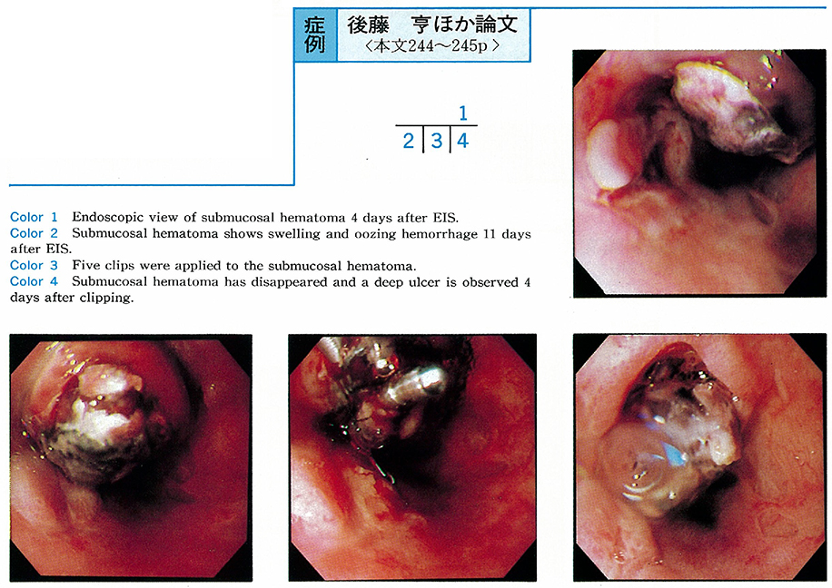
    1997 年 50 巻 p. 244-245
    発行日: 1997/06/06
    公開日: 2015/02/17
    ジャーナル フリー
    A 64-year-old woman was diagnosed as having liver cirrhosis by biopsy 3 years previously. Esophageal varices were noted the previous year. She was treated by endoscopic variceal ligation (EVL) and EIS, but the varices recurred. Therefore, we performed endscopic injection sclerotherapy ligation (EISL) . EISL was successful and 1 week after EISL, we performed EIS by extra-vascular injection of 4ml of Aethoxysklerol.
    At 4 days after EIS, a submucosal hematoma was observed. She was treated by conservative measures, but submucosal hematoma had increased in size at 11 days. So, we tried clipping the surface of the hematoma. The hematoma had disappeared 4 days after clipping, but then a deep ulcer appeared. After one month, the ulcer was completely healed and the appearance of the varices was F0.
    We reported one successful case of clipping to treat submucosal hematoma after EIS.
  • 中野 茂, 蜂矢 朗彦, 近藤 栄作, 栗田 俊夫, 小山 博, 神田 映子, 片桐 正人, 米谷 隆, 山田 秀一, 西野 執, 大塚 幸 ...
    1997 年 50 巻 p. 246-247
    発行日: 1997/06/06
    公開日: 2015/02/17
    ジャーナル フリー
    A 64-year-old female with liver cirrhosis was admitted to our hospital because of recurring esophageal varices.
    Endoscopy showed linear esophageal varices with communicating vessels to the varices on the lower esophagus as well as a slightly dilated cardiac plexus. Hepatopetal flow of paraesophageal veins via cardiac plexus was observed and esophageal variceal flow remained unclear as a result of percutaneous transhepatic portography.
    Endoscopic ligations were performed on the cardiac region to reduce blood flow toward the esophageal varices, at this time a developed esophageal varices was observed, while paraesophageal veins showed no change by EUS. Finally, EVL was performed using a spiral pattern to eradicate the varices.
    These results above suggest that esophageal varices were upward flowing rather than downward flowing, and had grester flow than that of paraesophageal veins in this case.
  • 斉藤 秀, 山田 秀一, 西野 執, 織田 良夫, 片桐 正人, 神田 映子, 栗田 俊夫, 北條 裕, 近藤 栄作, 中谷 尚登, 長山 ...
    1997 年 50 巻 p. 248-249
    発行日: 1997/06/06
    公開日: 2015/02/17
    ジャーナル フリー
    On rare occasions hemorrhagic shock develops in patients with Mallory-Weiss syndrome. We encountered a patient with Mallory-Weiss syndrome presenting with hemorrhagic shock. Hemostasis was successfully achieved with O-rings.
    The patient was a 68-year-old male who admitted our department with hematemesis after drinking a bottle of beer. When he arrived this hospital, his blood pressure was 70/40mmHg and he had a soft pulse. He had severe anemia. Emergency endoscopy following gastric lavage revealed three lacerations in the esophagogastric junction, two on the lesser curvature and one on the greater curvature. Ligation therapy with O-ring was perfomed and hemostasis was achieved completely.
    Although ulcers remained, hemorrhage did not recur to the patient. The patient was discharged without further incident. O-rings are widely used in the treatment of esophagogastric varices. We reported a case of Mallory-Weiss syndrome successfuly treated with O-ring.
  • 阿部 誠司, 竹下 俊隆, 溝上 裕士, 白石 貴久, 柏原 光介, 大坪 十四哉, 奈良坂 俊明, 松岡 健, 草間 博
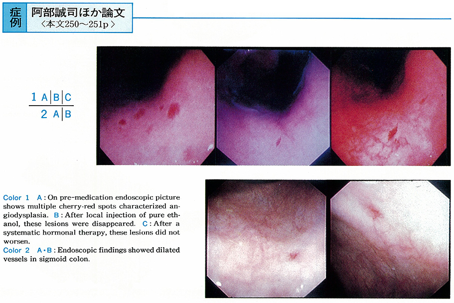
    1997 年 50 巻 p. 250-251
    発行日: 1997/06/06
    公開日: 2015/02/17
    ジャーナル フリー
    A 71-year-old woman was hospitalized due to appetite loss and abdominal pain. Laboratory examination revealed anemia and fecal positive occult blood. Blood loss from angiodysplasia was diagnosed by endoscopically characteristic multiple gastric cherry-red spots.
    These lesions were treated endoscopically by pure ethanol injections. Treatment was successful and anemia was improved. Further more, estriol therapy was added with 2mg per day. Patient recovered completely. Local injection of pure ethanol and systemic hormonal therapy is effective treatment for bleeding from gastric angiodysplasia.
  • 野口 典男, 本田 徹, 佐伯 伊知郎, 斎藤 直也, 竹下 公矢, 遠藤 光夫
    1997 年 50 巻 p. 252-253
    発行日: 1997/06/06
    公開日: 2015/02/17
    ジャーナル フリー
    A 53-year-old man complained of bloody vomiting and tarry stool on May 18th, 1996. About 5 days ago, he ate Shimesaba (raw mackerel) and so on. Endoscopic examination revealed an open and irregular ulcer with clots on the greater curvature aspect of the fornix. At that area, a codworm penetrating the gastric mucosa was found and was removed endoscopically using biopsy forceps under the direct vision.
    Histopathologically, the larva was identified as larva of Anisakis. On May 23rd, 1996, he complained of those symptoms again and was admitted to the hospital in pre-shock state. An endoscopic examination was performed immediately. There were massive blood and clots in the stomach and a ruptured vessel at the bottom of the ulcer was found. Endoscopic clippings hemostasis was performed at the vessel twice. About a month later, gastric endoscopy revealed that the ulcer was healed.
    This case was thought to be very rare that an Anisakis penetrated the gastric wall to form an ulcer with massive bleeding.
  • 栗山 根廣, 佐藤 慎一, 祖山 暁子, 西村 元伸, 楠目 健一, 松本 康久, 松下 一之, 山田 英夫, 後藤 剛貞, 北澤 恵美子, ...
    1997 年 50 巻 p. 254-255
    発行日: 1997/06/06
    公開日: 2015/02/17
    ジャーナル フリー
    A patient of 46-year-old male under hemodialysis owing to chronic renal failure (CRF) admitted to the hospital due to massive hematemesis and decreasing in blood pressure. An emergent endoscopy was carried out, and bleeding from a gastric elevated erosion in the antrum was found. By applying five times of clipping to the lesion, complete hemostasis was achieved.
    Erosive lesions of the upper gastrointestinal tract occurred with CRF, are known to be refractory to medical treatment. According to our data, the hemorrhage from gastric elevated erosion in CRF patient was one of the major causes of gastrointestinal hemorrhage. Usually the gastric erosions were located in the antrum and the source of active hemorrhage from them was single. Moreover, that hemorrhage tended to occur in the patients with long-term hemodialysis. We concluded that when the hemorrhage from the gastric erosion is difficult to control only by medications, it is necessary to add an endoscopic hemostasis such as clipping which does not damage the gastric mucosa so much.
  • 渋谷 進, 河島 孝彦, 飯田 浩行, 青柳 啓之, 井坂 直哉, 足立 信也, 高瀬 靖広, 近森 文夫
    1997 年 50 巻 p. 256-257
    発行日: 1997/06/06
    公開日: 2015/02/17
    ジャーナル フリー
    We introduced endoscopic variceal ligation (EVL) with a giant ligator and a three-shooter for solitary gastric varix. We had 2 died cases with one autopsied case after EVL for gastric varix. Aim of this study is to determine histological changes after EVL and the indication of EVL.
    Each case was elective and emergency cases. EVL was performed for gastric varix. They had hematemesis two days after EVL and were died. It has been reported that the borderline cases (Child C terminal) for esophageal endoscopic injection sclerotherapy, had 2 out of 3 conditions ; less than albumin 2.5g/dl, more than total bilirubin 5.0mg/dl and delirium. Two reported cases was Child C terminal. First case was autospied. Histopathological findings of the autopsied specimen showed that ligated gastric varix was ulcerated without bleeding, varix between the ligated ones was thrombosed.
    Therefore, it is thought that the varix is histologically vanished after ulceration or thrombosis after EVL and the indication of EVL for gastric varix must be strictly limited than that of esophageal injection sclerotherapy in the viewpoint of liver function.
  • 星田 有人, 篠木 啓, 山手 祐一郎, 竹内 司, 山門 進, 永井 俊彦
    1997 年 50 巻 p. 258-259
    発行日: 1997/06/06
    公開日: 2015/02/17
    ジャーナル フリー
    A 70-year-old female patient with liver cirrhosis (unknown etiology) was diagnosed as hemorrhagic portal hypertensive gastropathy by upper GI endoscopy and admitted to our hospital 4 times because of iron deficiency anemia since January 1991.
    In April, 1996 (76yr) , She complained progressive dyspnea on physical exertion and fainting. Laboratory data revealed severe iron deficiency anemia. The upper GI endoscopy (June 19th) showed multiple protruding red marks with oozing type hemorrhage at cardia and antrum in the stomach. Propranolol administration was started to relieve portal hypertension at the dose of 30mgTID from July 2nd. On the 5th day, heart rate decreased 60 to 42 bpm (30%) , blood pressure 120/60 to 82/64mmHg and her symptom was getting worse. So the dose of propranolol was reduced to 20mgBID from July 15th. Heart rate decrement was 60 to 50 bpm (16.7%) , blood pressure 100/60mmHg and her symptom was improved. The upper GI endoscopic findings of PHG were improved and active hemorrhage was not observed. On hemorrhagic PHG in the elderly, adequate low dose medical treatment was suggested to be useful and safe.
  • 植田 健治, 中田 薫, 原田 容治, 鶴井 茂, 齋藤 利彦
    1997 年 50 巻 p. 260-261
    発行日: 1997/06/06
    公開日: 2015/02/17
    ジャーナル フリー
    The patient was a 54-year-old man who presented because of epigastric discomfort. Gastric X-ray examination and endoscopic examination of the upper gastrointestinal tract revealed a tumor of 2cm by 2cm with central depression. The patient was suspected of having a submucosal tumor. Routine biopsy yielded only normal gastric mucosal tissue so that no tumor cell was identified.
    An artificial ulcer was produceds by microwave irradiation and another biopsy was taken, with no success in the identification of tumor cells. Considering the size of the lesion and the X-ray and endoscopic findings, however, a malignant tumor could not be completely ruled out. As a result of partial gastrectomy, a definitive diagnosis of heterotopic cystic malformation of the stomach was made.
  • 佐伯 伊知郎, 竹下 公矢, 谷 雅夫, 本田 徹, 斉藤 直也, 遠藤 光夫
    1997 年 50 巻 p. 262-263
    発行日: 1997/06/06
    公開日: 2015/02/17
    ジャーナル フリー
    Case 1 : A 65-year-old male was admitted to our hospital for the operation of urinary bladder tumor. Preoperative upper gastrointestinal endoscopy revealed a protruding lesion with an ulcer in the antrum of the stomach. It was 25mm in size and the endoscopic examination using an ultrasonic probe showed homogeneous hypoechoic mass invading to the bottom of the third layer (submucosa) of the gastric wall. Partial gastrectomy was performed and the lesion was diagnosed as inflammatory fibroid polyp (IFP) histologically.
    Case 2 : A 37-year-old male was pointed out a submucosal tumor 10mm in size in the antrum of the stomach by standard endoscopy. Endoscopic ultrasonography showed homogeneous hypoechoic mass in the second and middle-third layer of the gastric wall. It was resected endoscopically and diagnosed as IFP histologically.
    It was difficult to diagnose gastric IFP endoscopically, but endoultrasonographic examination was useful in the diagnosis and treatment of these two cases.
  • 山根 建樹, 古谷 徹, 石井 隆幸, 中村 眞, 小沼 康男, 川村 忠夫, 河原 秀次郎, 宮沢 善夫
    1997 年 50 巻 p. 264-265
    発行日: 1997/06/06
    公開日: 2015/02/17
    ジャーナル フリー
    A 49-year-old male visited our hospital with a complaint of epigastric discomfort. A depressed lesion was detected in the greater curvarure of his stomach by endoscopic examination. We suspected that it was gastric early cancer, type IIc and biopsy specimen of it showed group IV (well differentiated adenocarcinoma suspected) . But specimen obtained by repeated biopsy showed group III (adenoma) .
    So we performed endoscopic mucosal resection with ligating device (EMR-L) and recognized it as adenoma. The diagnosis of depressed adenoma of the stomach is very difficult because it is similar to IIc. The method of EMR-L was useful in diagnosis of this case.
  • 松本 政雄, 井野元 勤, 森下 慎二, 中村 昭子, 新村 和平, 佐藤 芳之, 池田 有成, 井上 泰
    1997 年 50 巻 p. 266-267
    発行日: 1997/06/06
    公開日: 2015/02/17
    ジャーナル フリー
    Recently, it has been reported that Helicobacter pylori (H. pylori) infection refered to the development of gastric mucosa-associated lymphoid tissue (MALT) lymphoma, because MALT lymphoma was improved by eradication of H. pylori.
    This patient was a 56-years-old man. Barium meal examination showed a small depressed lesion in the middle body of the stomach. Endoscopic examination revealed a scattered irregular depressed lesion in the same location. Histological study of the biopsy specimen revealed proliferation of atypical lymphoid cells and lympho-epithelial lesions in the gastric mucosa, and the diagnosis of gastric MALT lymphoma was suggested.
    H. pylori was detected by both culture and histological findings. So, we tried antibiotic therapy for H. pylori by 30mg of lansoprazole, 1500mg of amoxicillin, and 400mg of clarithromycin for 2 weeks. Six months after the therapy, eradication of H. pylori was ascertained and the MALT lymphoma lesion had disappeared endoscopically and histologically. It is suggested that H. pylori infection play a role in the pathgenesis of gastric MALT lymphoma, and that eradication of H. pylori is the first choice treatment for gastric MALT lymphoma.
  • 奥村 俊明, 井上 詠, 林 丈晴, 池澤 智明, 雨宮 哲朗, 高石 雅章, 笠谷 知宏, 仁科 牧子, 西田 次郎, 海老原 洋子, 水 ...
    1997 年 50 巻 p. 268-269
    発行日: 1997/06/06
    公開日: 2015/02/17
    ジャーナル フリー
    We experienced a case with gastric leiomyosarcoma resected by laparoscopic procedure. A 37-year-old woman was referred to the hospital because of an elevated lesion of the stomach. We performed barium meal examination as well as esophagogastroduodenoscopy, indicating submucosal tumor in the posterior wall of the upper body of the stomach. The tumor was further clarified by endoscopic ultrasonography and abdominal CT, suggesting leiomyosarcoma.
    The tumor was resected by laparoscopic procedure with a sufficient surgical margin, and finally diagnosed gastric leiomyosarcoma. The patient was discharged on 9th day after the operation. The laparoscopic local resection is thought to be an useful technique for submucosal tumor.
  • 三治 哲哉, 坂井 康明, 西巻 学, 谷 穣, 緑川 昌子, 半田 豊, 森田 重文, 大野 博之, 吉田 肇, 鶴井 光治, 三坂 亮一 ...
    1997 年 50 巻 p. 270-271
    発行日: 1997/06/06
    公開日: 2015/02/17
    ジャーナル フリー
    A 51-year-old female with no complaint underwent medical examination in 1993, and gastric X-ray revealed multiple small gastric polyps on the greater curvature from upper to middle corpus. Endoscopy showed multiple appearance of small gastric polyps in that area, corresponded to Yamada II type and had same color tone as peripheral mucosa. Histopathological finding of polyps demonstrated fundic gland polyps.
    Follow-up endoscopy was performed once a year, and findings in August 1995 indicated disappearance of polyps and sessile type elevated lesion with redness, 2mm in diameter, on the greater curvature of angulus. The lesion became larger in size (10mm in diameter) and showed sub-pedunculated type in June 1996. The results of biopsy led us to diagnose the lesion as papillary adenocarcinoma.
    Endoscopic mucosal resection was performed in July 1996 and histopathological fingings revealed depth of invasion was m and no malignant cells was recognized in the cut end. From the evidence in the sets of examination and from a survey of the literature, we concluded this case as rare.
  • 山村 冬彦, 山田 浩隆, 三上 哲弥, 金子 和弘, 秋田 泰, 吉川 望海, 三田村 圭二, 太田 秀一
    1997 年 50 巻 p. 272-273
    発行日: 1997/06/06
    公開日: 2015/02/17
    ジャーナル フリー
    The patient was a 62-year-old woman. In 1984, she was pointed out to have a 5mm-sized polyp in the upper body of stomach and it was diagnosed as hyperplastic polyp by a biopsied specimen. The polype enlarged to 10mm in diameter over the next 12 years and its endoscopic appearance changed to lobulated and variegated one.
    Since histological findings of biopsied specimens in 1996 showed adenocarcinoma, polypectomy was performed. Microscopic study of the resected specimen demonstrated well-defferentiated adenocarcinoma located on the surface of hyperplastic polyp. This is a very rare case of gastric hyperplastic polyp accompanied by the malignant transformation recognized by serial endoscopy during follow-up for 12 years.
    This case suggests that enlargement and lobulating tendency could be considered as sings of malignant transformation of hyperplastic polyps. Diagnostic reliability of biopsy could not be always perfect in case of malignant transfomation of gastric hyperplastic polyp, therefore, endoscopic follow-up should be performed continuously. When the signs indicated above are observed, endoscopic polypectomy should be applied regardless of negativeresults of biopsied specimens.
  • 林 毅, 高橋 泰人, 小林 裕太郎, 鈴木 啓弘, 山口 開, 巴 雅威, 長田 達郎, 遠山 裕樹, 吉田 秀樹, 草刈 幸次, 柴山 ...
    1997 年 50 巻 p. 274-275
    発行日: 1997/06/06
    公開日: 2015/02/17
    ジャーナル フリー
    Upper GI endoscopy was performed on a 75-year-old male patient complaining of abdominal discomfort on March 22. A polyp 2cm in diameter with the smooth surface, Type III (Yamada's criteria) was detected at the lesser curvature of the pyloric antrum, and was diagnosed as adenoma.
    After endoscopic polypectomy on may 30, an enlarged polyp (3cm) with lobulation was observed. Histological findings revealed adenoma including hyperplasia of the lacunar epithelium and intestinal epithelial metaplasia. Finally, the polyp was diagnosed as focal cancer in adenoma with high grade atypical cells and structure.
    This is an important case to study differentiated adenocarcinoma of the stomach.
  • 神田 映子, 中野 茂, 栗田 俊夫, 小山 博, 近藤 栄作, 山田 秀一, 岩崎 格, 瓜田 純久, 小島 保明, 吉永 淑子, 尾崎 ...
    1997 年 50 巻 p. 276-277
    発行日: 1997/06/06
    公開日: 2015/02/17
    ジャーナル フリー
    A 43-year-old female patient had epigastralgia and had been pointed out SMT like elevated lesion on the anterior wall of the antrum in February 1995. Follow up study had done in April 1995, and diagnosed poorly differentiated adenocarcinoma by biopsy. On ultrasonic endoscopy, the invasive depth of the tumor showed in the muscle layer. Gastrectomy was carried out.
    There are more than 60 cases reported as SMT tumors in the stomach. We found this case as one of striking resemblance to gastric cancer and concidered about the some features of this type cancer in the stomach.
  • 海野 潤, 石河 利隆, 奥山 尚, 荒井 咲子, 横山 喜恵, 林田 憲正, 田所 昌夫
    1997 年 50 巻 p. 278-279
    発行日: 1997/06/06
    公開日: 2015/02/17
    ジャーナル フリー
    An 81-year-old man visited our hospital because of loss of appitite and general fatigue. A panendoscopic examination revealed a round ulcerated lesion on the posterior wall of the gastric antrum and a pathological examination of the biopsied specimen showed a poorly differentiated adenocarcinoma. Since multiple liver tumors were detected by an abdominal CT scan and an elevated value of serum AFP, 273ng/ml, was noticed, a liver tumor biopsy was done after admission. As it was the same histology as that of the gastric carcinoma, an AFP producing gastric carcinoma with liver metastasis was highly suspected.
    Ten units of OK-432 and 250mg of 5-furuolouracil (5-FU) were injected concomitantly at the site of the gastric carcinoma through a fine needle via a panendoscopy twice with a week's interval. Ten days after the local injection, 750mg of 5-FU and 120mg of methotrexete were given intravenously. On that day the patient suddenly exhibited a fever, a chill, jaundice and hematuria. The next day he died of disceminated intervascular coagulation and multiple organ failure.
    At the autopsy, a lot of odorous gas was retained intraperitoneum and subcutaneously. There were multiple cavities in the organs such as the liver, kidney, heart and so on. A microscopic examination showed multiple rod shaped organisms on the sides of the cavities and in the vein. All these findings indicated the sepsis of gas producing organisms.
  • 吉田 由紀子, 永尾 重昭, 青野 茂昭, 岡田 千津子, 畠中 賢司, 加藤 真吾, 山口 勉, 黒木 雅彦, 岩下 悦郎, 岩井 淳浩, ...
    1997 年 50 巻 p. 280-281
    発行日: 1997/06/06
    公開日: 2015/02/17
    ジャーナル フリー
    A 44-year-old man visited our hospital due to the epigastralgia. An endoscopic examination revealed an active gastric ulcer in the antrum of the stomoch and a healing ulcer in the duodenal bulb. Despite that either H2 receptor antagonists or proton pump inhibitors had been administered, none of them had been effective enough for ulcers to be cured. An endoscopic examination after 17 months of the conventional therapy revealed the gastroduodenal fistula, so-called the double pylorus.
    Because he was found to be positive for Helicobacter pylori by the culture of gastric biopsy specimen as well as pathologically, he received an eradication therapy of H. pylori with omeprazole 40mg plus amoxiciline 1500mg for 14 consecutive days. The therapy brought both ulcers to be cured with red scars and set the patient free from the symptom although the ulcers were recurrent after 12 weeks. Due to his request, recradication therapy was done with omeprazole 40mg, amoxicilin 1,500mg plus clarithromycin 400mg for 14 consecutive and had brought the ulcers to be cured with white scars and negative for H. pylori. The ulcers have never been recurrent and remained scars since re-eradication therapy.
    This case is considered rare and worth to report because it had been able to observe that the intractable gastroduodenal ulcers had become the gastroduodenal fistula“the double pylorus”and the eradication of H. pylori had been effective for preventing the intractable ulcers from recurring.
feedback
Top