Journal of Reproduction and Development
Online ISSN : 1348-4400
Print ISSN : 0916-8818
ISSN-L : 0916-8818
Volume 63, Issue 6
Displaying 1-12 of 12 articles from this issue
SRD Innovative Technology Award 2016
  • Yutaka HASHIYADA
    Article type: SRD Innovative Technology Award 2016
    2017 Volume 63 Issue 6 Pages 527-538
    Published: 2017
    Released on J-STAGE: December 15, 2017
    Advance online publication: October 15, 2017
    JOURNAL FREE ACCESS

    Production of sires with high breeding potential is indispensable for prompt and reliable breeding using their semen in the cattle industry. Currently, in Japan, we aim to further the production of Japanese black sires via a new breeding system that uses genetically homologous monozygotic twins so that better growth performance and carcass traits can be translated to the increased production of beef with higher economic value. Several studies have reported that monozygotic twins are produced by embryo bisection. On the other hand, with the evolution and stabilization of in vitro fertilization technology, it has become possible to produce multiple monozygotic twin calves from blastomeres separated from a cleavage-stage embryo. This review attempts to clarify breeding practices through revalidation of the factors that affect the production efficiency of monozygotic twin calves by embryo bisection. Furthermore, the establishment of a system for monozygotic twin embryo production via the simplified technique of blastomere separation is reviewed while showing data from our previously performed studies.

    Download PDF (5179K)
Original Article
  • Ayumi HASEGAWA, Keiji MOCHIDA, Narumi OGONUKI, Michiko HIROSE, Toshiko ...
    Article type: Original Article
    2017 Volume 63 Issue 6 Pages 539-545
    Published: 2017
    Released on J-STAGE: December 15, 2017
    Advance online publication: August 20, 2017
    JOURNAL FREE ACCESS

    In embryo transfer experiments in mice, pseudopregnant females as recipients are prepared by sterile mating with vasectomized males. Because only females at the proestrus stage accept males, such females are selected from a stock of animals based on the appearance of their external genital tract. Therefore, the efficiency of preparing pseudopregnant females largely depends on the size of female colonies and the skill of the operators who select females for sterile mating. In this study, we examined whether the efficiency of preparing pseudopregnant females could be improved by applying an estrous cycle synchronization method by progesterone (P4) pretreatment, which significantly enhances the superovulation outcome in mice. We confirmed that after two daily injections of P4 (designated Days 1 and 2) in randomly selected females, the estrous cycles of most females (about 85%) were synchronized at metestrus on Day 3. When P4-treated females were paired with vasectomized males for 4 days (Days 4–8), a vaginal plug was found in 63% (20/32) of the females on Day 7. After the transfer of vitrified-warmed embryos into their oviducts, 52% (73/140) of the embryos successfully developed into offspring, the rate being comparable to that of the conventional embryo transfer procedure. Similarly, 77% (24/31) of females became pregnant by fertile mating with intact males for 3 days, which allowed the scheduled preparation of foster mothers. Thus, our estrous cycle synchronization method may omit the conventional experience-based process of visually observing the vagina to choose females for embryo transfer. Furthermore, it is expected that the size of female stocks for recipients can be reduced to less than 20%, which could be a great advantage for facilities/laboratories undertaking mouse-assisted reproductive technology.

    Download PDF (809K)
  • Chao WANG, Ruiming ZHANG, Le ZHOU, Jintian HE, Qiang HUANG, Farman A S ...
    Article type: Original Article
    2017 Volume 63 Issue 6 Pages 547-554
    Published: 2017
    Released on J-STAGE: December 15, 2017
    Advance online publication: August 31, 2017
    JOURNAL FREE ACCESS

    Intrauterine growth retardation (IUGR) impairs fetal intestinal development, and is associated with high perinatal morbidity and mortality. However, the mechanism underlying this intestinal injury is largely unknown. We aimed to investigate this mechanism through analysis of intestinal autophagy and related signaling pathways in a rat model of IUGR. Normal weight (NW) and IUGR fetuses were obtained from primiparous rats via ad libitum food intake and 50% food restriction, respectively. Maternal serum parameters, fetal body weight, organ weights, and fetal blood glucose were determined. Intestinal apoptosis, autophagy, and the mechanistic target of rapamycin (mTOR) signaling pathway were analyzed. The results indicated that maternal 50% food restriction reduced maternal serum glucose, bilirubin, and total cholesterol and produced IUGR fetuses, which had decreased body weight; blood glucose; and weights of the small intestine, stomach, spleen, pancreas, and kidney. Decreased Bcl-2 and increased Casp9 mRNA expression was observed in IUGR fetal intestines. Analysis of intestinal autophagy showed that the mRNA expression of WIPI1, MAP1LC3B, Atg5, and Atg14 was also increased, while the protein levels of p62 were decreased in IUGR fetuses. Compared to NW fetuses, IUGR fetuses showed decreased mTOR protein levels and enhanced mRNA expression of ULK1 and Beclin1 in the small intestine. In summary, the results indicated that maternal 50% food restriction on gestational days 10–21 reduced maternal serum glucose, bilirubin, and total cholesterol contents, and produced IUGR fetuses that had low blood glucose and reduced small intestine weight. Intestinal injury of IUGR fetuses caused by maternal food restriction might be due to enhanced apoptosis and autophagy via the mTOR signaling pathway.

    Download PDF (882K)
  • Jeong Hyo LEE, Jeong-Woong PARK, Si Won KIM, Joonghoon PARK, Tae Sub P ...
    Article type: Original Article
    2017 Volume 63 Issue 6 Pages 555-562
    Published: 2017
    Released on J-STAGE: December 15, 2017
    Advance online publication: September 01, 2017
    JOURNAL FREE ACCESS

    In mammals, germ cells originate outside of the developing gonads and follow a unique migration pattern through the embryonic tissue toward the genital ridges. Many studies have attempted to identify critical receptors and factors involved in germ cell migration. However, relatively few reports exist on germ cell receptors and chemokines that are involved in germ cell migration in avian species. In the present study, we investigated the specific migratory function of C-X-C chemokine receptor type 4 (CXCR4) in chicken primordial germ cells (PGCs). We induced loss-of-function via a frameshift mutation in the CXCR4 gene in chicken PGCs using clustered regularly interspaced short palindromic repeat-CRISPR-associated protein 9 (CRISPR/Cas9) genome editing. The migratory capacity of CXCR4 knockout PGCs was significantly reduced in vivo after transplantation into recipient embryos. However, CXCR4-expressing somatic cell lines, such as chicken DT40 and DF1, failed to migrate into the developing gonads, suggesting that another key factor(s) is necessary for targeting and settlement of PGCs into the genital ridges. In conclusion, we show that CXCR4 plays a critical role in the migration of chicken germ cells.

    Download PDF (4326K)
  • Po-Liang CHENG, Hui-Ru WU, Cheng-Yan LI, Chih-Feng CHEN, Hsu-Chen CHEN ...
    Article type: Original Article
    2017 Volume 63 Issue 6 Pages 563-570
    Published: 2017
    Released on J-STAGE: December 15, 2017
    Advance online publication: September 08, 2017
    JOURNAL FREE ACCESS
    Supplementary material

    Previous studies have shown that grafted neonatal chicken testicular tissue can develop and produce functional sperm; however, it was unclear whether regenerative processes or proportional growth caused the re-appearance of spermatogenic tissue. We dissociated testicular tissues, performed subcutaneous auto-transplantation of the re-aggregated cells to castrated cockerels, and monitored the post-surgery development of these transplanted aggregates. We found that these transplanted cell aggregates experienced compensatory growth in the form of a 300-fold increase in size, rather than the 30-fold increase observed in normal testis development. Further, these dissociated testicular cell aggregates restored seminiferous tubule structure and were able to produce testosterone and motile sperm. Therefore, we concluded that the dissociated testicular cells from 11-week-old cockerels retained a strong regenerative potential, as they exhibited compensatory growth, restored destroyed structure, and sustained spermatogenesis.

    Download PDF (3553K)
  • Hiroaki OKAMURA, Takashi YAMAMURA, Yoshihiro WAKABAYASHI
    Article type: Original Article
    2017 Volume 63 Issue 6 Pages 571-580
    Published: 2017
    Released on J-STAGE: December 15, 2017
    Advance online publication: November 07, 2017
    JOURNAL FREE ACCESS
    Supplementary material

    A population of neurons in the arcuate nucleus (ARC) coexpresses kisspeptin, neurokinin B (NKB), and dynorphin, and therefore they are referred to as KNDy neurons. It has been suggested that KNDy neurons participate in several brain functions, including the control of reproduction. The present study aimed to advance our understanding of the anatomy of the KNDy neural system. We first produced an antiserum against goat kisspeptin. After confirming its specificity, the antiserum was used to histochemically detect kisspeptin-positive signals. Using the colocalization of kisspeptin and NKB immunoreactivity as a marker for KNDy neurons, we mapped distributions of their cell somata and fibers in the whole brain (except the cerebellum) of ovariectomized (OVX) goats. KNDy neuronal somata were distributed throughout the ARC, and were particularly abundant in its caudal aspect. KNDy neuronal fibers projected into several areas within the septo-preoptic-hypothalamic continuum, such as the ARC, median eminence, medial preoptic nucleus, and bed nucleus of the stria terminalis. Kisspeptin immunoreactivity was not found outside of the continuum. We then addressed to the hypothesis that substance P (SP) is also involved in the KNDy neural system. Double-labeling immunohistochemistry for kisspeptin and SP revealed that KNDy neurons did not coexpress SP, but nearly all of the KNDy neuronal somata were surrounded by fibers containing SP in the OVX goats. The present results demonstrate anatomical evidence for a robust association between the KNDy and SP neural systems.

    Download PDF (2721K)
  • Eunhye KIM, Seon-Ung HWANG, Junchul David YOON, Eui-Bae JEUNG, Eunsong ...
    Article type: Original Article
    2017 Volume 63 Issue 6 Pages 581-590
    Published: 2017
    Released on J-STAGE: December 15, 2017
    Advance online publication: October 06, 2017
    JOURNAL FREE ACCESS
    Supplementary material

    Compared with the in vivo environment, porcine in vitro embryo-culture systems are suboptimal, as they induce oxidative stress via the accumulation of reactive oxygen species (ROS). High ROS levels during early embryonic development cause negative effects, such as apoptosis. In this study, we examined the effects of the antioxidant carboxyethylgermanium sesquioxide (Ge-132) during in vitro culture (IVC) on embryonic development in porcine in vitro fertilization (IVF) embryos. Zygotes were treated with different concentrations of Ge-132 (0, 100, 200 and 400 μg/ml). All of the Ge-132 treatment groups displayed greater total cell numbers after IVC (98.1, 98.5 and 103.4, respectively) compared with the control group (73.9). The 200 μg/ml Ge-132 treatment group exhibited significantly increased intracellular GSH levels compared with the control group, whereas the ROS generation levels decreased in Ge-132 dose-dependent manner (P < 0.05). The mRNA expression levels of the KEAP1 gene and proapoptotic genes BAX and CASPASE3 were lower in the Ge-132 treated blastocysts compared with the control group (P < 0.05). The percentages of apoptotic and necrotic cells in the Ge-132 treated embryos on day 2 (48 h) were significantly lower than the untreated embryos (9.1 vs. 17.1% and 0 vs. 2.7%, respectively). In the day 7 blastocysts, the percentages of apoptotic cells in 200 µg/ml Ge-132 treated group were lower compared to controls (1.6 vs. 2.5%). More KEAP1 protein was found to be localized in cytoplasm of the 200 μg/ml Ge-132 treated blastocysts, whereas KEAP1 protein was predominantly nuclei in the control blastocysts. These results indicate that the developmental competence of embryos cultured under Ge-132 treatment may be associated with KEAP1 signaling cascades involved in oxidative stress and apoptosis during porcine preimplantation embryo development.

    Download PDF (1466K)
  • Takayuki YAMOCHI, Shu HASHIMOTO, Masaya YAMANAKA, Yoshiharu NAKAOKA, Y ...
    Article type: Original Article
    2017 Volume 63 Issue 6 Pages 591-595
    Published: 2017
    Released on J-STAGE: December 15, 2017
    Advance online publication: October 12, 2017
    JOURNAL FREE ACCESS

    To determine the optimum culture duration for porcine growing oocytes (GOs) to attain maturation competence, we examined the meiotic competence, chromatin configuration, and fertilization ability of porcine oocytes obtained from early antral follicles and cultured for 10–16 days. The survival rate of oocytes after 10 days of culture (62.8%) was similar to that of oocytes after 12 days of culture (55%) and significantly higher than that of oocytes cultured for 14 and 16 days (52.9 and 24.3%, respectively). No significant difference was observed in the diameter of ooplasm from oocytes cultured for different durations (117.4–118.3 μm). The maturation rates of surviving oocytes after 10 and 16 days of culture (38.3 and 22.7%, respectively) were significantly lower than those of oocytes cultured for 12 and 14 days, and their in vivo counterparts (52.8–62.4%). The number of oocytes with surrounded-nucleolus chromatin was significantly lower in the 10-day culture group (78.4%) as compared with 14-day culture and in vivo counterpart groups (93.6 and 95.1%, respectively). After in vitro maturation and intracytoplasmic sperm injection, no significant difference was observed in the rate of fertilization among oocytes cultured for 12 and 14 days, and their in vivo counterparts (40.5–47.2%). Thus, porcine GOs required at least 12 days to acquire meiotic and fertilization competence, and the culture duration to maximize the number of mature oocytes ranged from 12 to 14 days.

    Download PDF (744K)
  • Toshiaki SUMIYOSHI, Natsumi ENDO, Tomomi TANAKA, Hideo KAMOMAE
    Article type: Original Article
    2017 Volume 63 Issue 6 Pages 597-604
    Published: 2017
    Released on J-STAGE: December 15, 2017
    Advance online publication: October 27, 2017
    JOURNAL FREE ACCESS

    Relaxation of the intravaginal part of the uterus is obvious around 6 to 18 h before ovulation, and this is considered the optimal time for artificial insemination (AI), as demonstrated in recent studies. Estrous signs have been suggested as useful criteria for determining the optimal time for AI. Therefore, this study evaluated the usefulness of estrous signs, particularly the relaxation of the intravaginal part of the uterus, as criteria for determining the optimal time for AI. A Total of 100 lactating Holstein-Friesian cows kept in tie-stall barns were investigated. AI was carried out based on the criterion for the optimal time for AI (optimal group), and earlier (early group) and later (late group) than the optimal time for AI, determined on the basis of estrous signs. After AI, ovulation was assessed by rectal palpation and ultrasonographic observation at 6-h intervals. For 87.5% (35/40) of cows in the optimal group, AI was carried out 24-6 h before ovulation, which was previously accepted as the optimal time for AI. AI was carried out earlier (early group) and later (late group) than optimal time for AI in 62.1% (18/29) and 71.0% (22/31) of cows, respectively. The conception rate for the optimal group was 60.0%, and this conception rate was higher than that for the early group (44.8%) and late group (32.2%), without significance. Further, the conception rate of the optimal group was significantly higher than the sum of the conception rates of the early and late groups (38.3%; 23/60) (P < 0.05). These results indicate that the criteria postulated, relaxation of the intravaginal part of the uterus and other estrous signs are useful in determining the optimal time for AI. Furthermore, these estrous signs enable the estimations of stages in the periovulatory period.

    Download PDF (1003K)
Technology Report
  • Akihiko OHTA, Yuichiro TSUNODA, Yoshihiko TAMURA, Kayoko IINO, Naoto N ...
    Article type: Technology Report
    2017 Volume 63 Issue 6 Pages 605-609
    Published: 2017
    Released on J-STAGE: December 15, 2017
    Advance online publication: October 13, 2017
    JOURNAL FREE ACCESS
    Supplementary material

    The gonadotropins, luteinizing hormone (LH) and follicle-stimulating hormone (FSH), are important hormones in vertebrate reproduction. The isolation of gonadotropins from the pituitary gland is sub-optimal, as the cross-contamination of one hormone with another is common and often results in the variation in the measured activity of LH and FSH. The production of recombinant hormones is, therefore, a viable approach to solve this problem. This study aimed to express recombinant rat, mouse, and mastomys FSH and LH in Chinese hamster ovary (CHO) cells. Their common α-subunits along with their hormone-specific β-subunits were encoded in a single mammalian expression vector. FSH from all three species was expressed, whereas expression was achieved only for the mouse LH. Immunohistochemistry for rat alpha subunit of glycoprotein hormone (αGSU) and LHβ and FSHβ subunits confirmed the production of the dimeric hormone in CHO cells. The recombinant rodent gonadotropins were confirmed to be biologically active; estradiol production was increased by recombinant FSH in granulosa cells, while recombinant LH increased testosterone production in Leydig cells.

    Download PDF (1158K)
  • Masumi HIRABAYASHI, Hiromasa HARA, Teppei GOTO, Akiko TAKIZAWA, Melind ...
    Article type: Technology Report
    2017 Volume 63 Issue 6 Pages 611-616
    Published: 2017
    Released on J-STAGE: December 15, 2017
    Advance online publication: August 19, 2017
    JOURNAL FREE ACCESS

    The present study was conducted to establish haploid embryonic stem (ES) cell lines using fluorescent marker-carrying rats. In the first series, 7 ES cell lines were established from 26 androgenetic haploid blastocysts. However, only 1 ES cell line (ahES-2) was found to contain haploid cells (1n = 20 + X) by fluorescence-activated cell sorting (FACS) and karyotypic analyses. No chimeras were detected among the 10 fetuses and 41 offspring derived from blastocyst injection with the FACS-purified haploid cells. In the second series, 2 ES cell lines containing haploid cells (13% in phES-1 and 1% in phES-2) were established from 2 parthenogenetic haploid blastocysts. Only the phES-2 cell population was purified by repeated FACS to obtain 33% haploid cells. Following blastocyst injection with the FACS-purified haploid cells, no chimera was observed among the 11 fetuses; however, 1 chimeric male was found among the 47 offspring. Although haploid rat ES cell lines can be established from both blastocyst sources, FACS purification may be necessary for maintenance and chimera production.

    Download PDF (1607K)
  • Minami W OKUYAMA, Tomochika SUGIURA, Masaharu MORIYOSHI, Kazuto YAMASH ...
    Article type: Technology Report
    2017 Volume 63 Issue 6 Pages 617-622
    Published: 2017
    Released on J-STAGE: December 15, 2017
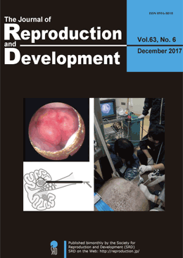
    Advance online publication: October 14, 2017
    JOURNAL FREE ACCESS
    Supplementary material

    For examining pig ovaries, which have complex structures, laparoscopy is a useful technique, but requires general anesthesia; therefore, it cannot be performed repeatedly within a short period of time. We report a transvaginal endoscopy-based technique for conducting ovarian examinations without general anesthesia. Sows were sedated in pig stalls. Using a colonoscope, the vaginal wall was punctured with a trocar. To avoid the trocar being caught in the broad ligament of the uterus or the connective tissue around the vagina, the trocar was inserted close to the external uterine os and between the 2:00 and 3:00 or the 9:00 and 10:00 positions (in a clockwise direction). Then, a urethroscope was inserted into the abdomen, and an examination was carried out after the ovaries had been moved towards the urethroscope camera via rectal palpation. This less invasive procedure may allow repeated examinations and will increase our understanding of ovarian dynamics in pigs.

    Editor's pick

    Cover Story : 
    Okuyama et al. reported a transvaginal endoscopy-based technique for conducting ovarian examination in sows in a standing position. Sows were sedated in pig stalls, and their vaginal walls were punctured. Subsequently, a urethroscope was inserted into their abdomen, and an examination was conducted after their ovaries had moved towards the urethroscope camera via rectal palpation (Okuyama MW, et al. A transvaginal endoscopy-based technique for performing ovarian examinations in sows. pp. 617–622). This less invasive procedure without the use of general anesthesia may allow repeated ovarian examinations and increase our understanding of the ovarian dynamics in pigs.

    Download PDF (1819K)
feedback
Top