Journal of Reproduction and Development
Online ISSN : 1348-4400
Print ISSN : 0916-8818
ISSN-L : 0916-8818
Volume 66, Issue 2
Displaying 1-13 of 13 articles from this issue
Original Article
  • Ryutaro MORIYAMA, Koichi IWAMOTO, Teruki HAGIWARA, Saishu YOSHIDA, Tak ...
    Article type: Original Article
    2020 Volume 66 Issue 2 Pages 97-104
    Published: 2020
    Released on J-STAGE: April 10, 2020
    Advance online publication: December 09, 2019
    JOURNAL OPEN ACCESS

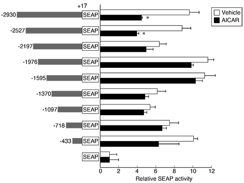
    Malnutrition is one of the factors that induces reproductive disorders. However, the underlying biological processes are unclear. AMP-activated protein kinase (AMPK) is an enzyme that plays crucial role as a cellular energy sensor. In the present study, we examined the effects of AMPK activation on the transcription of the murine gonadotropin subunit genes Cga, Lhb, and Fshb, and the gonadotropin-releasing hormone receptor Gnrh-r. Real-time PCR and transcription assay using LβT2 cells demonstrated that 5-amino-imidazole carboxamide riboside (AICAR), a cell-permeable AMP analog, repressed the expression of Lhb. Next, we examined deletion mutants of the upstream region of Lhb and found that the upstream regulatory region of Lhb (–2527 to –2198 b) was responsible for the repression by AICAR. Furthermore, putative transcription factors (SP1, STAT5a, and TEF) that might mediate transcriptional control of the Lhb repression induced by AICAR were identified. In addition, it was confirmed that both AICAR and a competitive inhibitor of glucose metabolism, 2-deoxy-D-glucose, induced AMPK phosphorylation in LβT2 cells. Therefore, the upstream region of Lhb is one of the target sites for glucoprivation inducing AMPK activation. In addition, AMPK plays a role in repressing Lhb expression through the distal –2527 to –2198 b region.

    Download PDF (1697K)
  • Kouji KOMATSU, Satoru MASUBUCHI
    Article type: Original Article
    2020 Volume 66 Issue 2 Pages 105-113
    Published: 2020
    Released on J-STAGE: April 10, 2020
    Advance online publication: December 29, 2019
    JOURNAL OPEN ACCESS

    The controlled activation of dormant primordial follicles is important for the maintenance of periodic ovulation. Previous reports have clearly identified the signaling pathway in granulosa cells and oocytes that controls the activation of primordial follicles; however, the exact cue for the in vivo activation of dormant primordial follicles is yet to be elucidated. In this study, we found that almost all activated primordial follicles made contact with blood vessels. Based on this result, we speculated that the contact between primordial follicles and blood vessels may provide a cue for the activation of dormant primordial follicles. To confirm this hypothesis, we attempted to activate dormant primordial follicles within the ovaries by inducing angiogenesis through the use of biodegradable gels containing recombinant vascular endothelial growth factor and in cultured ovarian tissues by increasing the serum concentration within the culture medium. The activation of dormant primordial follicles was promoted in both experiments, and our results indicated that an increase in the supply of the serum component, from new blood vessels formed via angiogenesis, to the dormant primordial follicles is the cue for their in vivo activation. In the ovaries, angiogenesis often occurs during every estrous cycle, and it is therefore likely that angiogenesis is the crucial event that influences the activation of primordial follicles.

    Editor's pick

    Cover Story:
    Most primordial follicles present in ovaries are dormant and only a few of them are activated in every estrus cycle. However, the mechanism controlling the activation of dormant primordial follicles in vivo remains unclear. In this study, Komatsu et al. found that almost all the activated primordial follicles (black arrows) made contact with blood vessels (red arrows) in mouse ovaries (Komatsu et al. Increased supply from blood vessels promotes the activation of dormant primordial follicles in mouse ovaries. pp. 105–113). To confirm the hypothesis that angiogenesis is crucial for activation of the dormant primordial follicles in vivo, Komatsu et al. induced angiogenesis using recombinant VEGF. They found that the activation of dormant primordial follicles was promoted by an increase in the number of blood vessels in the ovaries. Furthermore, the number of activated follicles increased in cultured ovarian tissues depending on the serum concentration in the medium. These results confirm that the supply of serum components through new blood vessels formed via angiogenesis is a cue for the activation of dormant primordial follicles in the ovaries.

    Download PDF (4871K)
  • Tamás SOMFAI, Hiep Thi NGUYEN, Men Thi NGUYEN, Thanh Quang DANG-NGUYEN ...
    Article type: Original Article
    2020 Volume 66 Issue 2 Pages 115-123
    Published: 2020
    Released on J-STAGE: April 10, 2020
    Advance online publication: January 27, 2020
    JOURNAL OPEN ACCESS
    Supplementary material

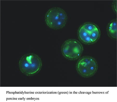
    The aim of the present study was to clarify whether or not our vitrification procedure at the germinal vesicle (GV)-stage triggers the apoptotic cascade in oocytes and subsequent embryos. Immature porcine cumulus-oocyte complexes were either vitrified and warmed (vitrified group) or subjected to cryoprotectant agents (CPA group) or cultured without any treatment (control). Oocytes of all treatment groups were subjected to in vitro maturation (IVM), fertilization, and embryo culture. Apoptosis was assayed in live oocytes at the end of IVM culture and in cleavage-stage embryos after in vitro fertilization (IVF). We detected similar frequencies of DNA fragmentation, levels of caspase activity, phosphatidylserine externalization, and mRNA levels for pro-apoptotic Bax and CASP3 genes in oocytes at the end of IVM and in early embryos among all groups. However, in the vitrified group, the anti-apoptotic Bcl-XL gene was upregulated in 4–8 cell embryos, which caused an 8-fold significant increase in the Bcl-XL/Bax mRNA ratio compared with the control and CPA groups (P < 0.05). In conclusion, vitrification of porcine oocytes at the GV stage by our method did not trigger the apoptotic cascade in oocytes and subsequent embryos but triggered the upregulation of the anti-apoptotic Bcl-XL gene in embryos.

    Download PDF (1069K)
  • Kei HORIHATA, Naoko INOUE, Yoshihisa UENOYAMA, Kei-ichiro MAEDA, Hirok ...
    Article type: Original Article
    2020 Volume 66 Issue 2 Pages 125-133
    Published: 2020
    Released on J-STAGE: April 10, 2020
    Advance online publication: January 19, 2020
    JOURNAL OPEN ACCESS

    Kisspeptin, encoded by Kiss1, is essential for reproduction in mammals. Kiss1 expression is regulated by estrogen via histone acetylation in the Kiss1 promotor region. Thus, elucidation of histone modification factor(s) involved in the regulation of Kiss1 expression is required to gain further understanding of the mechanisms of its control. The RNA-seq analysis of isolated kisspeptin neurons, obtained from the arcuate nucleus (ARC) of female rats, revealed that Rbbp7, encoding retinoblastoma binding protein 7 (RBBP7), a member of histone modification and chromatin remodeling complexes, is highly expressed in the ARC kisspeptin neurons. Thus, the present study aimed to investigate whether RBBP7 is involved in Kiss1 expression. Histological analysis using in situ hybridization (ISH) revealed that Rbbp7 expression was located in several hypothalamic nuclei, including the ARC and the anteroventral periventricular nucleus (AVPV), where kisspeptin neurons are located. Double ISH for Rbbp7 and Kiss1 showed that a majority of kisspeptin neurons (more than 85%) expressed Rbbp7 mRNA in both the ARC and the AVPV of female rats. Further, Rbbp7 mRNA knockdown significantly decreased in vitro expression of Kiss1 in a mouse immortalized kisspeptin neuronal cell line (mHypoA-55). Estrogen treatment significantly decreased and increased Kiss1 mRNA levels in the ARC and AVPV of ovariectomized female rats, respectively, but failed to affect Rbbp7 mRNA levels in both the nuclei. Taken together, these findings suggest that RBBP7 is involved in the upregulation of Kiss1 expression in kisspeptin neurons of rodents in an estrogen-independent manner.

    Download PDF (5324K)
  • Sho NAKAMURA, Kohei NODA, Masafumi MIWA, Shiori MINABE, Teruki HAGIWAR ...
    Article type: Original Article
    2020 Volume 66 Issue 2 Pages 135-141
    Published: 2020
    Released on J-STAGE: April 10, 2020
    Advance online publication: December 27, 2019
    JOURNAL OPEN ACCESS

    Negative energy balance in domestic animals suppresses their reproductive function. These animals commonly use long-chain fatty acids (LCFAs) from adipocytes as an energy source under states of malnutrition. The G-protein coupled receptor, GPR120, is a specific receptor for LCFAs, but its role in reproductive function remains unknown in domestic animals. The purpose of this study was to examine whether GPR120 is involved in the reproductive system of cattle. GPR120 mRNA expression was evaluated in brain, pituitary, and ovarian tissue samples by RT-PCR. GPR120 gene expression was detected with high intensity only in the anterior pituitary sample, and GPR120-immunoreactive cells were found in the anterior pituitary gland. Double immunohistochemistry of GPR120 in the anterior pituitary hormone-producing cells, such as gonadotropes, thyrotropes, lactotropes, somatotropes, and corticotropes, was performed to clarify the distribution of GPR120 in the anterior pituitary gland of ovariectomized heifers. Luteinizing hormone β subunit (LHβ)- and follicle-stimulating hormone β subunit (FSHβ)-immunoreactive cells demonstrated GPR120 immunoreactivity at 80.7% and 85.9%, respectively. Thyrotropes, lactotropes, somatotropes, and corticotropes coexpressed GPR120 at 21.1%, 5.4%, 13.6%, and 14.5%, respectively. In conclusion, the present study suggests that GPR120 in the anterior pituitary gland might mediate LCFA signaling to regulate gonadotrope functions, such as hormone secretion or production, in cattle.

    Download PDF (1789K)
  • Chikaya DEURA, Ryutaro MORIYAMA
    Article type: Original Article
    2020 Volume 66 Issue 2 Pages 143-148
    Published: 2020
    Released on J-STAGE: April 10, 2020
    Advance online publication: December 29, 2019
    JOURNAL OPEN ACCESS

    High-fat diet (HFD) is associated with the regulation of reproductive functions. This study aimed to investigate the effects of short-term HFD on the mRNA expression levels of follicle-stimulating hormone β subunit (FSHβ), luteinizing hormone β subunit (LHβ), gonadotropin-releasing hormone receptor, and long-chain fatty acid receptor, GPR120, in the matured male mouse pituitary gland. Adult male mice were fed either control chow or HFD for 1, 2, 5, 10, 30 and 150 days. Fshb and Gpr120 mRNA expression levels in the pituitary glands were significantly increased during 2 to 30 days of HFD feeding. Gnrh-r mRNA in the 30 days HFD fed group and body weight in the 30 and 150 days HFD fed groups were higher than control. However, there were no significant differences in plasma non-esterified fatty acids or glucose levels during the 150 days of HFD feeding. These results suggest that male mice feeding a short-term HFD induces FSHβ synthesis and GPR120 expression in their pituitary gonadotropes.

    Download PDF (790K)
  • Dagvajamts BADRAKH, Yojiro YANAGAWA, Masashi NAGANO, Seiji KATAGIRI
    Article type: Original Article
    2020 Volume 66 Issue 2 Pages 149-154
    Published: 2020
    Released on J-STAGE: April 10, 2020
    Advance online publication: January 28, 2020
    JOURNAL OPEN ACCESS

    Epidermal growth factor (EGF) concentrations in the uterus show two peaks on days 2–4 and 13–14 during the estrous cycle in fertile cows. Loss of the two peaks has been linked to reduced fertility in repeat breeder cows. This study aimed to examine the effect of seminal plasma (SP) on normalizing endometrial EGF concentrations and restoring fertility in repeat breeder cows with low EGF concentrations on day 3. In study 1, we examined the effect of the deposition sites (the vagina and uterus) of SP on the endometrial EGF concentrations in repeat breeder cows. SP infusion into the vagina, but not uterus, on the first day of the estrus cycle (day 0) normalized the endometrial EGF concentrations (≥ 4.7 ng/g tissue weight) on day 3. In study 2, the effect of SP volume (0.5 and 10 ml of SP and 0.5 ml of SP diluted to 10 ml) on EGF concentrations was examined. All groups with SP infusion had increased EGF concentrations on day 3, and cows with 10 ml of SP and 0.5 ml of SP diluted to 10 ml showed the highest levels of EGF concentrations. In study 3, we examined the effect of SP infusion on fertility. SP infusion normalized two peaks of endometrial EGF concentrations in about 60% of repeat breeder cows and produced more pregnancies than the controls (44.4 vs. 19.4%). Therefore, we concluded that SP may contain an activity to normalize the EGF profile and restore fertility in repeat breeder cows with altered EGF profiles.

    Download PDF (640K)
  • Hiroka OKAJI, Kenta TETSUKA, Ren WATANABE, Satoshi KISHIGAMI
    Article type: Original Article
    2020 Volume 66 Issue 2 Pages 155-161
    Published: 2020
    Released on J-STAGE: April 10, 2020
    Advance online publication: January 24, 2020
    JOURNAL OPEN ACCESS

    The extracellular matrix between the oocyte and zona pellucida (ZP) plays an important role in mammalian fertilization and preserves the specific environment of the perivitelline space (PVS) during the development of a preimplantation embryo after fertilization. In this study, we applied a highly sensitive luminescent protein dye, LumiteinTM, to observe the hydrophobic status of proteins in oocytes and preimplantation embryos. LumiteinTM is widely used for detecting denatured proteins after sodium dodecyl sulfate-polyacrylamide gel electrophoresis. LumiteinTM fluorescence was detected primarily in the PVS and degenerated first polar body of fresh normal metaphase II (MII) oocytes but much less within the ZP and ooplasm, which suggested a hydrophobic PVS environment in the MII oocytes. Unexpectedly, abnormally-shaped fresh or aged oocytes showed stronger fluorescence in the PVS, which reflected oocyte quality. Interestingly, 10 h after fertilization, the fluorescent signal in the PVS temporarily increased in a patched pattern that appeared and then disappeared by the two-cell stage. After the two-cell stage, the decreased fluorescent signal was maintained throughout the development of the preimplantation embryo. These results suggest new protein dynamics in the PVS during the one-cell stage of the oocyte. Thus, cellular imaging of oocytes and preimplantation embryos using LumiteinTM provides new information on protein dynamics.

    Download PDF (2735K)
  • Seiki HARAGUCHI, Thanh Quang DANG-NGUYEN, David WELLS, Daiichiro FUCHI ...
    Article type: Original Article
    2020 Volume 66 Issue 2 Pages 163-174
    Published: 2020
    Released on J-STAGE: April 10, 2020
    Advance online publication: January 26, 2020
    JOURNAL OPEN ACCESS

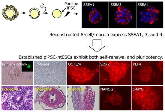
    We investigated whether sequential reprogramming via porcine induced pluripotent stem cells (piPSCs) or exposure to oocyte cytoplasm following nuclear transfer could generate nuclear transfer-derived ESCs (piPSCs-ntESCs). Nuclear transfer embryos were reconstructed with piPSCs possessing a ZsGreen fluorescent marker for expression of exogenous Nanog and Lin28. Reconstructed oocytes developed to morphologically normal 8-cell/morulae (35/93, 37.6%) and blastocysts (12/93, 12.9%). Although most green fluorescent protein-positive blastocysts showed efficient outgrowth (8/10, 80%), none formed primary colonies and all cultures degenerated. Conversely, 15% of fluorescent positive 8-cell/morula stage embryos showed outgrowth (6/40), with three forming primary colonies (7.5%). All three were expanded and maintained as piPSC-ntESC lines. These cell lines expressed stem cell marker genes and proteins. Despite inactivation of one X chromosome, all piPSC-ntESC lines formed teratomas comprising derivatives from all three embryonic germ layers. Strong SSEA1, 3, and 4 expression was detected at the 8-cell/morula stage in embryos reconstructed from both piPSCs and porcine embryonic fibroblasts (PEFs). SSEA3 was notably absent from IVF controls at pre-implantation embryo stages. Finally, we attempted to establish ntESCs from 8-cell/morulae reconstructed with PEFs using the same culture conditions as those for piPSC-ntESC derivation. Although eight primary colonies arose from 107 embryos (7.5%), they all degenerated after the first passage culture. Early and sustained expression of key reprogramming regulatory factors may be critical for pluripotent stem cell derivation to derive piPSC-ntESCs from 8-cell/morula stages, while the expression of SSEAs may be involved in the initial stem cell colony formation phases.

    Download PDF (8336K)
  • Shiori MUSHA, Saishu YOSHIDA, Syo MURAKAMI, Ryotaro KOJIMA, Masahito D ...
    Article type: Original Article
    2020 Volume 66 Issue 2 Pages 175-180
    Published: 2020
    Released on J-STAGE: April 10, 2020
    Advance online publication: January 19, 2020
    JOURNAL OPEN ACCESS

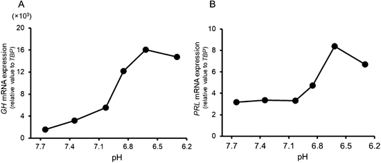
    Hormone-secreting pituitary adenomas show unregulated hormonal hypersecretion and cause hyperpituitarism. However, the mechanism of the unregulated hormone production and secretion has not yet been fully elucidated. Solid tumors show reduced extracellular pH, partly due to lactate secretion from anaerobic glycolysis. It is known that extracellular acidification affects hormone secretion. However, whether and how the extracellular acidification influences the unregulated hormone production and secretion remain unknown. In the present study, we found that GPR4, a proton-sensing G protein-coupled receptor, was highly expressed in MtT/S cells, a growth hormone-producing and prolactin-producing pituitary tumor cell line. When we reduced the extracellular pH, growth hormone and prolactin mRNA expressions increased in the cells. Both increased expressions were partially suppressed by a GPR4 antagonist. We also found that extracellular acidification enhanced growth hormone-releasing factor-induced growth hormone secretion from MtT/S cells. These results suggest that GPR4 may play a role in hypersecretion of the hormone from hormone-producing pituitary tumors. A GPR4 antagonist will be a useful tool for preventing the hypersecretion.

    Download PDF (751K)
  • Rebecca LAPP, Volker RÖTTGEN, Torsten VIERGUTZ, Joachim M. WEITZEL, An ...
    Article type: Technology Report
    2020 Volume 66 Issue 2 Pages 181-188
    Published: 2020
    Released on J-STAGE: April 10, 2020
    Advance online publication: January 27, 2020
    JOURNAL OPEN ACCESS

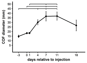
    The aim of this study was to establish a model to induce cystic ovarian follicles (COFs) in cattle using the cyclooxygenase inhibitor, indomethacin. Eighteen Holstein-Frisian cattle were synchronized with prostaglandin F2alpha (PGF2α) and gonadotropin-releasing hormone (GnRH). Ultrasound-guided transvaginal intrafollicular injections were performed in 23 preovulatory follicles with different concentrations of indomethacin 16 h after GnRH administration. An injection of 0.2 ml 35 µM indomethacin solution (resulting in a final concentration of 8 µg/ml in the follicular fluid) was the minimal dosage leading to COF formation. The induced COFs reached a maximum mean diameter of 36.9 ± 4.5 mm eleven days after injection. The estrous cycle was extended to 25–39 days. Luteinization was first observed 4 days after injection, accompanied by a slight increase in plasma progesterone concentration. The bioactivity of indomethacin was demonstrated by the decrease of prostaglandin E2 in the follicular fluid of three animals. The method presented here is minimally invasive and allows for the generation of defined COFs for further investigations.

    Download PDF (916K)
Technology Report
  • Kenzo UCHIKURA, Rumiko YAMAMOTO, Shigeyuki TAJIMA, Masahiro SUZUKI, Ay ...
    Article type: Technology Report
    2020 Volume 66 Issue 2 Pages 189-192
    Published: 2020
    Released on J-STAGE: April 10, 2020
    Advance online publication: January 16, 2020
    JOURNAL OPEN ACCESS

    The effects of a single subcutaneous or intramuscular injection of follicle-stimulating hormone (FSH) on follicular growth and expression of estrous behavior and its single subcutaneous administration on the number of corpora lutea (CL) and embryos were investigated in pigs. All four sows that were subcutaneously administered 5 AU FSH expressed normal estrus and had no ovarian cysts. Two of the four sows that were administered 5 AU FSH intramuscularly did not exhibit estrus, and another sow had a short estrus period. All four sows had ovarian cysts. The mean numbers of CL, embryos, and blastocysts following the subcutaneous administration of 5 AU FSH (16.8, 16.0, and 13.8, respectively) did not differ significantly from those for the control animals treated intramuscularly with 1000 IU equine chorionic gonadotropin (18.5, 16.5, and 14.3, respectively). In conclusion, embryo recovery was possible using a single subcutaneous administration of FSH.

    Download PDF (1174K)
  • M A HANNAN, Shingo HANEDA, Kaishi MURATA, Shiori TAKEUCHI, Soon Hon CH ...
    Article type: Technology Report
    2020 Volume 66 Issue 2 Pages 193-197
    Published: 2020
    Released on J-STAGE: April 10, 2020
    Advance online publication: January 26, 2020
    JOURNAL OPEN ACCESS

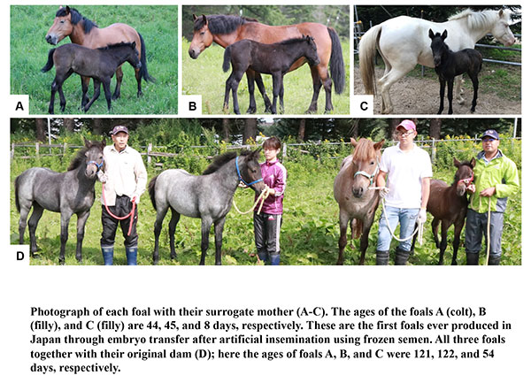
    Until now, there have been no reports of foals born through embryo transfer after artificial insemination using frozen semen in Japan. The aims of this study were to develop a riding crossbred horse and evaluate the prospects of embryo transfer technology in multiplying horse population. In both donor and recipient mares, luteolysis was induced by the administration of 0.1 mg Cloprostenol to synchronize the onset of estrus, and ovulation was induced by administering 2000 IU human chorionic gonadotropin (hCG) or 0.75 mg Deslorelin. Frozen semen from an Irish Connemara pony stallion was used to breed a Hokkaido native pony mare by deep-horn artificial insemination (dose, 400 × 106 sperm). A non-surgical technique was used to collect embryos from the donor mare at day 7 post-ovulation and transfer them transcervically into the uterus of recipient mares (n = 4) immediately after collection. Weekly blood samples were collected from the recipients throughout pregnancy. A total of four embryos were recovered from seven collection attempts (57% recovery) from a donor mare in a single breeding season. Three of the four transferred embryos maintained successful pregnancy and delivered a healthy live foal (75% birth). A normal progesterone profile was observed throughout gestation in recipient mares. In conclusion, for the first time, to the best of our knowledge, this study describes the birth of foals through non-surgical transcervical embryo transfer in Japan after artificial insemination using frozen semen. We expect that this new crossbreed (Connemara pony × Hokkaido native pony) will be a good riding breed.

    Download PDF (1610K)
feedback
Top