Journal of Reproduction and Development
Online ISSN : 1348-4400
Print ISSN : 0916-8818
ISSN-L : 0916-8818
Volume 65, Issue 4
Displaying 1-14 of 14 articles from this issue
SRD Young Investigator Award 2018
  • Kanako MOROHAKU
    Article type: SRD Young Investigator Award 2018
    2019 Volume 65 Issue 4 Pages 281-287
    Published: 2019
    Released on J-STAGE: August 09, 2019
    Advance online publication: April 20, 2019
    JOURNAL FREE ACCESS

    Eggs are female germ cells that are required for producing offspring through sexual reproduction. In mammals, eggs are produced in the ovary and ovulated into the oviduct. It is well known that over 99% of eggs are degenerated without ovulation, so that many studies have attempted in vitro folliculogenesis to produce many eggs in different species for a few decades. Although many methods have been developed, a success of in vitro egg production with the resultant live birth of offspring has been limited, especially in livestock animals. More recently, we have succeeded in producing live pups derived from in vitro/ex vivo egg production in mice. This review aims to introduce our recent findings with a brief history of in vitro/ex vivo culture systems for follicles and ovaries.

    Editor's pick

    Cover Story:
    In vitro/ex vivo egg production has been widely studied in various mammalian species over half of the century to utilize the majority of the immature oocytes stocked in the female ovaries. Recently, the first successful protocol of in vitro oogenesis from primordial germ cells (PGC) has been established by Morohaku K, et al., resulting in the live birth of offspring in mice. The protocol consists of two vital steps; 1) ex vivo organ culture of mouse PGC ovaries to complete the process of follicle formation, with successful incorporation of antagonists for the existing estrogen receptors, and 2) in vitro follicle culture of the growing follicles isolated from the cultured ovaries. The review in this issue introduces the current findings and aspects governing in vitro oogenesis, with a brief history (Morohaku K. A way for in vitro/ex vivo egg production in mammals. pp. 281–287).

    Download PDF (4306K)
  • Ryotaro MIURA
    Article type: SRD Young Investigator Award 2018
    2019 Volume 65 Issue 4 Pages 289-295
    Published: 2019
    Released on J-STAGE: August 09, 2019
    Advance online publication: May 13, 2019
    JOURNAL FREE ACCESS

    The first follicular wave emerges soon after ovulation, and its dominant follicle (DF) develops during the first 8–10 days of the estrous cycle in cattle. And, the first-wave DF is a non-ovulatory follicle, because it develops during the first half of the estrous cycle simultaneously with the corpus luteum (CL), which produces and secretes progesterone. Regarding the characteristics of development and the mechanisms of deviation in the DF during the follicular wave, the first-wave DF has been well studied. However, the characteristics of the first-wave DF, such as growth, blood flow in the follicular wall, concentration of sex steroid hormones in the peripheral blood and follicular fluid, amounts of mRNA in granulosa cells, as well as the characteristics of the CL formed after the first-wave DF and the influence of the first-wave DF on fertility (conception rate), have not been well studied. Additionally, the first-wave DF synthesizes and secretes 17β-estradiol (E2), and plasma E2 concentration increases during the early stage of the estrous cycle. Consequently, there is a possibility that the first-wave DF might affect the fertility in cattle. In this review, to provide the new perspective on reproductive physiology in cattle, characteristics of the first-wave DF were examined in detail and its characteristics were compared with that of the second-wave DF. In addition, the locational effects of the first-wave DF and CL on conception rate are discussed.

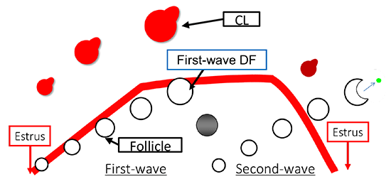
    Download PDF (1113K)
Original Article
  • Kumiko NAKATA, Kaoru YOSHIDA, Naoki YAMASHITA
    Article type: Original Article
    2019 Volume 65 Issue 4 Pages 297-304
    Published: 2019
    Released on J-STAGE: August 09, 2019
    Advance online publication: April 11, 2019
    JOURNAL FREE ACCESS

    The aim of this study was to develop a new container for cryopreservation of a limited number of spermatozoa. To evaluate the efficacy and safety of this new container, we performed preclinical evaluations using human sperm or mouse oocytes and sperm. First, using human sperm that was frozen and then thawed, we demonstrated that the sperm recovery rate using the new container was 96.7% (58/60), which was significantly higher (P < 0.05) than the recovery rate of 21.2% (11/52) when using the Cryotop®. Sperm motility rates were 19.2% (10/52) using the Cryotop® and 35.0% (21/60) using the new container. Second, murine epididymal spermatozoa were divided into three groups: fresh spermatozoa, spermatozoa frozen using a straw, and spermatozoa frozen using the new container. Sperm motility, sperm membrane and DNA integrity, in vitro development of fertilized eggs, and offspring development after embryo transfer were assessed. The motility of freeze-thawed sperm was lower in spermatozoa that were frozen using the new container than in fresh spermatozoa or those that were frozen using a straw. After intracytoplasmic sperm injection, the survival rate was 96.7% (145/150), the 2-cell development rate was 90.3% (131/145), and the blastocyst development rate was 77.2% (112/145), when using the new container. There were no differences in the sperm membrane, DNA integrity, or in the embryo development rates to the blastocyst stage among the different frozen groups. Six offspring were derived from spermatozoa freeze-thawed in the new container, and they developed normally. Thus, the new container allows easy handling of a small number of sperms and minimizes sperm loss during cryopreservation.

    Download PDF (1161K)
  • Kumiko TAKEDA, Eiji KOBAYASHI, Kagetomo NISHINO, Akira IMAI, Hiromichi ...
    Article type: Original Article
    2019 Volume 65 Issue 4 Pages 305-312
    Published: 2019
    Released on J-STAGE: August 09, 2019
    Advance online publication: May 02, 2019
    JOURNAL FREE ACCESS

    Age-associated methylation changes in genomic DNA have been recently reported in spermatozoa, and these changes can contribute to decline in fertility. In a previous study, we analyzed the genome-wide DNA methylation profiles of bull spermatozoa using a human DNA methylation microarray and identified one CpG site (CpG-1) that potentially reflects age-related methylation changes. In the present study, cryopreserved semen samples from a Japanese Black bull were collected at five different ages, which were referred to as JD1-5: 14, 19, 28, 54, and 162 months, respectively, and were used for genome-wide DNA methylation analysis and in vitro fertilization (IVF). Distinct age-related changes in methylation profiles were observed, and 77 CpG sites were found to be differently methylated between young and adult samples (JD1-2 vs. JD4-5). Using combined bisulfite restriction analysis (COBRA), nine CpG sites (including CpG-1) were confirmed to exhibit significant differences in their age-dependent methylation levels. Eight CpG sites showed an age-dependent increase in their methylation levels, whereas only one site showed age-dependent hypomethylation; in particular, these changes in methylation levels occurred rapidly at a young age. COBRA revealed low methylation levels in some CpG regions in the majority of the IVF blastocyst-stage embryos derived from spermatozoa at JD2-5. Interestingly, bulls with different ages did not show differences in their methylation levels. In conclusion, our findings indicated that methylation levels at nine CpG sites in spermatozoa changed with increasing age and that some CpG regions were demethylated after fertilization. Further studies are required to determine whether age-dependent different methylation levels in bull spermatozoa can affect fertility.

    Download PDF (1047K)
  • Hanako BAI, Talukder Md Abdus SHABUR, Hiroki KUNII, Tsukino ITOH, Mana ...
    Article type: Original Article
    2019 Volume 65 Issue 4 Pages 313-318
    Published: 2019
    Released on J-STAGE: August 09, 2019
    Advance online publication: May 02, 2019
    JOURNAL FREE ACCESS

    Calving is a critical but stressful event required for milk production in dairy cows. In the present study, we investigated the immune status of peripheral blood mononuclear cells (PBMCs) isolated from periparturient cows to better understand and, thus, possibly prevent stress during the periparturient period. To evaluate the immune response of PBMCs, we assessed their proliferation with or without a mitogen (concanavalin A, ConA). Blood samples were collected 24 h before and after calving and 1 week after calving. The proliferation of non-treated cells remained unchanged throughout the examination period. The immune response of PBMCs isolated from the cows before calving was relatively low, even after ConA stimulation; however, the immune response of PBMCs collected at both time points after calving was significantly higher than those of non-stimulated controls. Next, we examined the expression patterns of T cell related and inflammatory cytokine genes in PBMCs. We found that the mRNA expression levels of both CD4 and CD8 showed decreasing trends after calving. The expression of the Th1 cell marker gene IFNG also decreased after calving. The mRNA expression level of the inflammatory cytokine gene TNFA increased after parturition. Overall, our results suggest that the PBMC immune response was weakened in cows before delivery and part of the expression of the immune cell-related genes in these cells is altered 24 h before and after calving.

    Download PDF (917K)
  • Kazuki TAKAHASHI, Pablo J. ROSS, Ken SAWAI
    Article type: Original Article
    2019 Volume 65 Issue 4 Pages 319-326
    Published: 2019
    Released on J-STAGE: August 09, 2019
    Advance online publication: April 25, 2019
    JOURNAL FREE ACCESS

    Zinc finger and SCAN domain containing 4 (Zscan4) is a gene that is specifically expressed during zygotic genome activation (ZGA) in mouse preimplantation embryos, and a reduction of Zscan4 transcripts leads to developmental failure. In mouse embryonic stem cells (ESCs), Zscan4 is expressed transiently in as little as 1–5% of the cell population. Zscan4 has also been shown to enhance the efficiency of mouse induced pluripotent stem cells (iPSCs) generation and their quality. Although ZSCAN4 plays important roles in murine embryos and stem cells, its expression and role in bovine embryos is unknown. This study examines ZSCAN4 transcripts in bovine embryos at various developmental stages and attempts to elucidate the functions of ZSCAN4 during bovine preimplantation development. ZSCAN4 transcripts were found to be upregulated at the 8- and 16-cell stages. We next attempted ZSCAN4 downregulation in bovine early embryos by RNA interference and evaluated developmental competency and transcripts levels of genes involved in ZGA and iPSCs generation. Although the bovine embryos injected with ZSCAN4-siRNA could develop to the 8-cell stage, very few were developing beyond the 16-cell stage. PIWIL2 expression was reduced in ZSCAN4 downregulated embryos. It is possible that ZSCAN4 downregulated embryos fail to regulate gene expression during ZGA. Our results indicate that ZSCAN4 is an important factor for the preimplantation development of bovine embryos.

    Download PDF (1189K)
  • Shouhei KURATA, Yuuki HIRADATE, Kohei UMEZU, Kenshiro HARA, Kentaro TA ...
    Article type: Original Article
    2019 Volume 65 Issue 4 Pages 327-334
    Published: 2019
    Released on J-STAGE: August 09, 2019
    Advance online publication: June 10, 2019
    JOURNAL FREE ACCESS

    In mammals, ejaculated sperm acquire their fertilizing ability during migration through the female reproductive tract, which secretes several factors that contribute to sperm capacitation. Gamma-aminobutyric acid (GABA) is a well-known neurotransmitter in the central nervous system, but additionally enhances the sperm acrosome reaction in the rat and cow. However, the detailed effects of GABA concentration on sperm function remain unclear. In this study, we detected the presence of the GABA type A receptor (GABA A) in mouse epididymal sperm by western blot analysis and in the sperm acrosome by immunocytochemistry. We also investigated the effects of GABA on sperm fertilizing ability. We found that GABA facilitated the tyrosine phosphorylation of sperm proteins, which is an index of sperm capacitation. GABA also promoted the acrosome reaction, which was suppressed by a selective GABA A receptor antagonist. We then found that the effective GABA concentration for the acrosome reaction corresponds to sperm concentration, but we did not detect any marked effect of GABA on sperm motility using a computer-assisted sperm analysis system. Using immunohistochemistry, we also detected GABA expression in the epithelia of the mouse uterus and oviduct. Furthermore, we found that the mRNA levels of glutamate decarboxylase (Gad), which generates GABA from L-glutamate, were higher in the oviduct than in the uterus, and that Gad mRNA levels were higher at estrus than at the diestrus stage. These results indicate that the GABA concentration can act as a modulator of the acrosome reaction and sperm capacitation in the female reproductive tract.

    Download PDF (1447K)
  • Olga AMELKINA, Prattana TANYAPANYACHON, Ampika THONGPHAKDEE, Kaywalee ...
    Article type: Original Article
    2019 Volume 65 Issue 4 Pages 335-343
    Published: 2019
    Released on J-STAGE: August 09, 2019
    Advance online publication: May 30, 2019
    JOURNAL FREE ACCESS

    In recent years, the Kiss1 gene has been reported in a number of vertebrate species, and a substantial dataset has been acquired to demonstrate the critical role of kisspeptins in the reproductive system; yet limited information is available for carnivores. In the present study, we identified and characterized feline Kiss1 by isolating and cloning its full-length cDNA in the domestic cat hypothalamus and caracal testis, using the method of rapid amplification of cDNA ends. Additionally, we isolated and cloned the 3′ end of Kiss1 cDNA, containing kisspeptin-10 (Kp10), from the ovaries of a clouded leopard and Siberian tiger. Nucleotide sequencing revealed that domestic cat Kiss1 cDNA is of 711 base pairs and caracal Kiss1 cDNA is of 792 base pairs, both having an open reading frame of 450 base pairs, encoding a precursor protein Kiss1 of 149 amino acids. The core sequence of the feline kisspeptin Kp10 was found to be identical in all species analyzed here and is highly conserved in other vertebrate species. Using an anti-Kp10 antibody, we found the immunoreactive kisspeptin to be localized in the periventricular and infundibular nuclei of the cat hypothalamus. The results show that kisspeptin is highly conserved among different feline families, and its immunoreactive distribution in the hypothalamus may indicate its physiological function in the domestic cat.

    Download PDF (1640K)
  • Tomoko OBUCHI, Masahiro OSADA, Takeyuki OZAWA, Hiroshi NAKAGAWA, Michi ...
    Article type: Original Article
    2019 Volume 65 Issue 4 Pages 345-352
    Published: 2019
    Released on J-STAGE: August 09, 2019
    Advance online publication: June 10, 2019
    JOURNAL FREE ACCESS

    This study was conducted to evaluate and compare the economic benefits of different embryo sexing methods, based on the cost per female dairy calf produced. Female calves were produced from four kinds of female embryos: (1) those collected from superstimulated donors at 7–8 days after artificial insemination (AI) with X-sorted semen; (2) those sex-determined by loop-mediated isothermal amplification assay of a biopsy sample of embryos collected from superstimulated donors after AI with conventional unsorted semen; (3) those obtained by invitro embryo production (IVEP), using X-sorted semen and in vitro-matured oocytes collected from donors by ovum pick-up (OPU); and (4) those obtained by IVEP, using X-sorted semen and oocytes collected by OPU after dominant follicle ablation and follicle growth stimulation of the donors. The respective productivities of female calves per technical service and the total production cost per female calf of each sexing method were compared. The production cost per female calf (66,537 JPY), as calculated from the number of female calves per service (1.30), pregnancy rate of transfer (42.9%), rate of female calves obtained (92.9%), and total cost of the method (56,643 JPY plus embryo transfer fee), was less for IVEP with X-sorted semen and follicular growth-stimulated (FGS) oocytes than for the other groups (P < 0.05). The results demonstrate that embryo production with X-sorted semen and FGS oocytes provides a more efficient method for producing female calves than the other embryo sexing methods.

    Download PDF (653K)
  • Daiyu ITO, Sayaka WAKAYAMA, Yuko KAMADA, Ikue SHIBASAKI, Satoshi KAMIM ...
    Article type: Original Article
    2019 Volume 65 Issue 4 Pages 353-359
    Published: 2019
    Released on J-STAGE: August 09, 2019
    Advance online publication: May 23, 2019
    JOURNAL FREE ACCESS

    Freeze-drying of spermatozoa is a convenient and safe method to preserve mammalian genetic material without the use of liquid nitrogen or a deep freezer. However, freeze-dried spermatozoa (FD sperm) are not frequently used because of the low success rate of offspring after intracytoplasmic spermatozoa injection (ICSI). In this study, we determined the optimal concentration and a point of action of trehalose as a protectant for the preservation of FD sperm from different mouse strains at room temperature (RT). Although trehalose demonstrated no potential to protect the FD sperm of ICR mice against the freeze-drying procedure itself, the blastocyst rate was significantly improved when FD sperm was preserved for more than 1 month at RT (56–63% vs. 29% without trehalose). The optimal concentration of trehalose was 0.5 M. Importantly, remarkable results were obtained when spermatozoa of inbred mouse strains (C57BL/6N, C3H/He, and 129/Sv) were used, and many offspring were obtained when FD sperm that was preserved for 3 months at RT (26–28% vs. 6–11% of without trehalose) was used. However, when DNA damage in FD sperm was examined by gamma-H2Ax assays, it was found that trehalose failed to protect the FD sperm from DNA damage. These results suggest that trehalose has the potential to protect other sperm factors rather than sperm DNA during preservation at RT for longer periods and trehalose is more effective for inbred mouse strains.

    Download PDF (1251K)
  • Natsuko EMURA, Kazuki TAKAHASHI, Yuriko SAITO, Ken SAWAI
    Article type: Original Article
    2019 Volume 65 Issue 4 Pages 361-368
    Published: 2019
    Released on J-STAGE: August 09, 2019
    Advance online publication: May 25, 2019
    JOURNAL FREE ACCESS

    TEA domain family transcription factor 4 (Tead4) is known to be important for the trophectoderm (TE) segregation in murine embryos. However, the role of TEAD4 in early development of porcine embryos is still unknown. We examined TEAD4 expression patterns and attempted to determine the functions of TEAD4 during porcine preimplantation development using RNA interference. TEAD4 mRNA was upregulated from the 2–4-cell to 8–16-cell stages and then decreased to the blastocyst stage. Nuclear localization of TEAD4 protein was detected at the 16-cell stage, as well as at subsequent developmental stages. In porcine embryos injected with TEAD4 siRNA, transformation from morula to blastocyst was inhibited. Although TEAD4 downregulation did not affect the expression levels of POU class 5 homeobox 1 (OCT-4), transcription of SRY-related HMG-box gene 2 (SOX2) was detected at high level in TEAD4-downregulated embryos. It is possible that TEAD4 contributes to blastocyst formation in porcine embryos through downregulation of SOX2 expression. Collectively, our results indicate that TEAD4 is an important factor for the preimplantation development of porcine embryos.

    Download PDF (2497K)
Technology Report
  • Yasuo FUSHIMI, Danielle MONNIAUX, Mitsuhiro TAKAGI
    Article type: Technology Report
    2019 Volume 65 Issue 4 Pages 369-374
    Published: 2019
    Released on J-STAGE: August 09, 2019
    Advance online publication: March 30, 2019
    JOURNAL FREE ACCESS

    In this study, we evaluated the efficiency of a single measurement of plasma anti-Müllerian hormone (AMH) concentration in heifers in determining the number of oocytes recoverable by ovum pick-up (OPU), and compared AMH concentrations among sister heifers from the same parents. For this, blood samples from 50 embryo-transfer-derived female Japanese Black (JB) heifers (mean: 8.7 age in months) were collected and plasma AMH concentration was measured. At 13–15 months of age, both the number of follicles (2–9 mm) and the number of collected oocytes after OPU were counted and compared. Results indicated that the heifers with the highest AMH concentration had the highest number of follicles in their ovaries and gave the highest number of collected oocytes with OPU, thereby indicating that a single measurement of plasma AMH concentration is informative for the selection of OPU-donor heifers in herd breeding programs. The practice of performing a single AMH measurement may accelerate the intensive breeding of JB herds.

    Download PDF (1067K)
  • Hidenori SHIBAHARA, Yasuhisa MUNAKATA, Ai ISHIGURO, Koumei SHIRASUNA, ...
    Article type: Technology Report
    2019 Volume 65 Issue 4 Pages 375-379
    Published: 2019
    Released on J-STAGE: August 09, 2019
    Advance online publication: April 14, 2019
    JOURNAL FREE ACCESS

    This study compared the effects of different volumes of culture medium for the in vitro growth of oocytes derived from porcine early antral follicles (EAFs). Oocyte granulosa cell complexes (OGCs) were collected from EAFs (0.5–0.7 mm in diameter) and individually cultured for 14 days. When OGCs were cultured in 1 ml of medium with or without polyacrylamide gel (PAG), the presence of PAG supported granulosa cell (GC) proliferation and oocyte growth. When OGCs were cultured in 0.2 or 1 ml of medium on PAG, the number of GC in the OGC culture and the developmental ability of the oocytes cultured in vitro were significantly higher for the 1 ml of culture medium group than for the 0.2 ml group. In conclusion, a combination of a large volume of culture medium with PAG improved the growth and developmental ability of the oocytes cultured in vitro, which were comparable to the oocytes collected from large antral follicles.

    Download PDF (807K)
  • Shiori MUSHA, Syo MURAKAMI, Ryotaro KOJIMA, Hideaki TOMURA
    Article type: Technology Report
    2019 Volume 65 Issue 4 Pages 381-388
    Published: 2019
    Released on J-STAGE: August 09, 2019
    Advance online publication: April 20, 2019
    JOURNAL FREE ACCESS

    Cyclic adenosine monophosphate (cAMP) plays a pivotal role in gonadotrope responses in the pituitary. Gonadotropin-releasing hormone (GnRH) mediated synthesis and secretion of follicle-stimulating hormone (FSH) and luteinizing hormone (LH) are regulated by both the Gs/cAMP and Gq/Ca2+ signaling pathways. Pituitary adenylate cyclase-activating polypeptide (PACAP) also regulates GnRH responsiveness in gonadotropes through the PACAP receptor, which activates the Gs/cAMP signaling pathway. Therefore, measuring intracellular cAMP levels is important for elucidating the molecular mechanisms of FSH and LH synthesis and secretion in gonadotropes. The GloSensor cAMP assay is useful for detecting cAMP levels in intact, living cells. In this study, we found that increased GloSensor luminescence intensity did not correlate with cAMP accumulation in LβT2 cells under low pH conditions. This result indicates that cell type and condition must be considered when using GloSensor cAMP.

    Download PDF (904K)
feedback
Top