Journal of Reproduction and Development
Online ISSN : 1348-4400
Print ISSN : 0916-8818
ISSN-L : 0916-8818
Volume 69, Issue 3
Displaying 1-7 of 7 articles from this issue
SRD Outstanding Research Award 2022
  • Kimiko INOUE
    Article type: SRD Outstanding Research Award 2022
    2023 Volume 69 Issue 3 Pages 129-138
    Published: 2023
    Released on J-STAGE: June 06, 2023
    Advance online publication: March 16, 2023
    JOURNAL OPEN ACCESS

    Somatic cell nuclear transfer (SCNT) is the only reproductive technology used to produce individuals from somatic cells by transferring them to enucleated oocytes. Although more than 25 years have passed since the first mammalian SCNT was reported in sheep, problems such as low birth rates and morphological abnormalities have persisted and limited its practical applications. The mouse is the ideal laboratory animal to unveil these questions due to its established reproductive technologies and extensive knowledge base of its genome and various strains. We investigated the causes of incomplete reprogramming after nuclear transfer of donor somatic cells and found that the loss of imprint in some placenta-specific imprinted genes could induce non-random SCNT abnormalities. By ameliorating aberrantly expressed imprinted genes, we succeeded in increasing the low birth rate and improving morphological abnormalities observed in SCNT fetuses. Furthermore, we sought appropriate mouse strains and cell types as nuclear donors to increase their developmental efficiencies and expand their applications in various fields. Peripheral blood cells are useful as ethical and economical cell species because they can be collected easily, even though SCNT embryos derived from hematopoietic cells show poor developmental abilities after reconstruction. Additionally, it is possible to obtain mice that are reactive to specific antigens of interest by using lymphocytes. Although there are still many limitations to the practical use of SCNT, its utilization is steadily expanding.

    Download PDF (4494K)
Review
  • Ryuki SHIMADA, Kei-ichiro ISHIGURO
    Article type: Review
    2023 Volume 69 Issue 3 Pages 139-146
    Published: 2023
    Released on J-STAGE: June 06, 2023
    Advance online publication: March 17, 2023
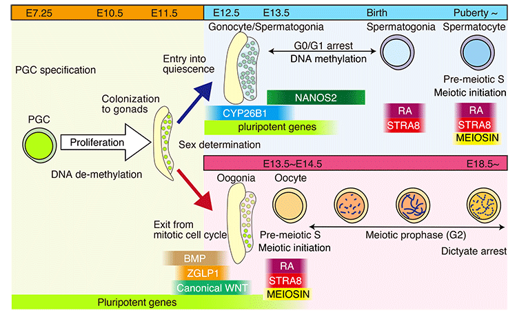
    JOURNAL OPEN ACCESS

    In mouse fetal gonads, germ cell development is accompanied by changes in cell cycle mode in response to external signals and intrinsic mechanisms of cells. During fetal development, male germ cells undergo G0/G1 arrest, while female germ cells exit the mitotic cell cycle and enter meiosis. In fetal testes, NANOS2 and CYP26B1 force germ cells to stay in G0/G1 arrest phase, preventing them from entering the meiotic cell cycle. In the fetal ovary, external signals, such as RA, BMP, and WNT, promote the competency of female germ cells to enter the meiotic cell cycle. MEIOSIN and STRA8 ensure the establishment of the meiotic cell cycle by activating meiotic genes, such that meiotic entry coincides with the S phase. This review discusses germ cell development from the viewpoint of cell cycle regulation and highlights the mechanism of the entry of germ cells into meiosis.

    Editor's pick

    Cover Story:
    The development of germ cells is accompanied by alterations in the cell cycle in response to external signals and intrinsic cellular mechanisms. During fetal development, male germ cells undergo G0/G1 arrest, whereas female germ cells exit the mitotic phase of the cell cycle and enter meiosis. The NANOS2 and CYP26B1 proteins in the fetal testes cause the germ cells to remain in G0/G1 arrest, which prevents them from entering the meiotic cell cycle. External signals such as RA, BMP, and WNT promote the female germ cells in the fetal ovaries to enter the meiotic phase of the cell cycle. MEIOSIN and STRA8 are transiently co-expressed in the pre-leptotene phase in spermatocytes and oocytes. The MEIOSIN-STRA8 complex ensures the establishment of the meiotic phase by activating meiotic genes in such a manner that the entry into meiosis coincides with the S phase of the cell cycle. This review discusses the development of germ cells from the viewpoint of cell cycle regulation and highlights the mechanism by which germ cells enter the meiotic phase of the cell cycle (Shimada and Ishiguro. Cell cycle regulation for meiosis in mammalian germ cells, pp. 139–146).

    Download PDF (5886K)
Original Article
  • Risa SUZUKI, Masakatsu FUJINOKI
    Article type: Original Article
    2023 Volume 69 Issue 3 Pages 147-153
    Published: 2023
    Released on J-STAGE: June 06, 2023
    Advance online publication: March 18, 2023
    JOURNAL OPEN ACCESS

    Progesterone (P) enhances spermatozoal hyperactivation, a capacitation event. Hyperactivation is associated with successful in vitro fertilization (IVF). In this study, we examined the effects of P on hyperactivation and IVF in mice. P enhanced spermatozoal hyperactivation and increased IVF success rate in a dose-dependent manner. Moreover, P affected spermatozoal hyperactivation and IVF through the membrane progesterone receptor of the spermatozoal head. These results show that P regulates spermatozoal capacitation and fertilization in mice. The concentration of P changes during the estrous cycle, indicating that spermatozoa are capacitated in response to the oviductal environment and subsequently fertilize the oocyte.

    Download PDF (1629K)
  • Lanfang MA, Shuo WANG, Jun YANG, Weicheng TANG, Zhangying WU, Lili CAO ...
    Article type: Original Article
    2023 Volume 69 Issue 3 Pages 154-162
    Published: 2023
    Released on J-STAGE: June 06, 2023
    Advance online publication: April 18, 2023
    JOURNAL OPEN ACCESS

    MicroRNA (miR)-145 is enriched in the follicular granulosa cells (GCs) of 3-week-old mice. Downregulating miR-145 inhibits the proliferation and differentiation of GCs and induces evident changes in their cytoskeleton. In this study, we examined how miR-145 induces cytoskeletal changes in mouse GCs and its potential mechanism in regulating GC steroidogenesis. We found that actin related protein 2/3 complex subunit 5 (Arpc5) is a target of miR-145. The miR-145 antagomir increased ARPC5 expression but not β-ACTIN, β-TUBULIN, and PAXILLIN expression. Arpc5 overexpression inhibited GC proliferation, differentiation, and progesterone synthesis. Furthermore, the expression of progesterone synthesis-associated enzymes was downregulated in the Arpc5 overexpression group, and the GC cytoskeleton exhibited evident changes. We conclude that Arpc5, a new target of miR-145, regulates primary GC proliferation and progesterone production by regulating the cytoskeleton remodeling.

    Download PDF (2594K)
  • Satoshi NAGASHIMA, Takuma KIMURA, Ryota TERASHIMA, Makoto SUGIYAMA, Ke ...
    Article type: Original Article
    2023 Volume 69 Issue 3 Pages 163-169
    Published: 2023
    Released on J-STAGE: June 06, 2023
    Advance online publication: April 13, 2023
    JOURNAL OPEN ACCESS

    Lysophosphatidic acid (LPA) has been implicated in the uterine endometrial functions of implantation and decidualization; however, not much is known about its myometrial contractile function. Herein we characterized the uterotonic effects of LPA in non-pregnant (estrus) and peri-parturient rats in vitro. LPA dose-dependently (0.01–10 μM) stimulated the amplitude and integral, but not the frequency, of the uterine strip contraction of estrous rats. The stimulatory effect of LPA was enhanced 1 day before parturition but was lost 1 day postpartum. LPA did not cause the de novo synthesis of prostaglandin (PG) F2α but stimulated contractions cooperatively with the PG. LPA-induced contractions were significantly inhibited by an LPA1/2/3 antagonist in the uteri of estrous rats but not in term rats. This study characterized the uterotonic effect of a natural LPA that occurs at physiological concentrations, changes with reproductive states, and is independent of mediation by the newly synthesized PG.

    Download PDF (1472K)
  • Natsuko MIYAMOTO, Akihiro OHYA, DURITAHALA, Mitsuhiro SAKASE, Hiroshi ...
    Article type: Original Article
    2023 Volume 69 Issue 3 Pages 170-177
    Published: 2023
    Released on J-STAGE: June 06, 2023
    Advance online publication: April 18, 2023
    JOURNAL OPEN ACCESS
    Supplementary material

    This study aimed to characterize calyculin A (CL-A)-induced and thimerosal-induced hyperactivation of cryopreserved bovine spermatozoa. Hyperactivation was effectively induced by treating with 10 nM CL-A for 60 min in the presence of cyclic AMP analogs, extracellular Ca2+, and albumin or with 12.5 µM thimerosal briefly in the absence of these capacitation-supporting factors. Majority of the spermatozoa exhibiting CL-A-induced hyperactivation were characterized by the 3-dimensional helical movement with head rotation, higher degree of flagellar curvature, and faster beating of the flagella than those exhibiting thimerosal-induced hyperactivation of the 2-dimensional planar movement without head rotation. The CL-A-induced hyperactivation was linked to the activation of cAMP/protein phosphorylation-dependent signaling cascades and to the decreased activity of glycogen synthase kinase-3α (GSK-3α). In contrast, the thimerosal-induced hyperactivation was suppressed by pretreatment with CL-A and cyclic AMP analogs in the absence of CaCl2 to activate cAMP/protein phosphorylation-dependent signaling cascades. Additionally, the intracellular Ca2+ level in live sperm flagella was significantly higher in the CL-A-treated samples than in the thimerosal-treated samples. These results indicate that CL-A-induced hyperactivation of cryopreserved bovine spermatozoa is an extracellular Ca2+-dependent type with the 3-dimensional helical movement, which can be regulated not only by the activation of cAMP/protein phosphorylation-dependent signaling cascades, leading to a large enhancement of the intracellular Ca2+ level, but also by the reduction in GSK-3α activity. Considering the different characteristics of thimerosal-induced hyperactivation, our results suggest that the diversity of sperm hyperactivation arises from different combinations of flagellar bending and head rotation.

    Download PDF (5324K)
  • Satoshi FUNAYA, Yuan WANG, Masataka G. SUZUKI, Masahito IKAWA, Fugaku ...
    Article type: Original Article
    2023 Volume 69 Issue 3 Pages 178-182
    Published: 2023
    Released on J-STAGE: June 06, 2023
    Advance online publication: April 17, 2023
    JOURNAL OPEN ACCESS

    Linker histone variants regulate higher-order chromatin structure and various cellular processes. It has been suggested that linker histone variant H1a loosens chromatin structure and activates transcription. However, its role in early mouse development remains to be elucidated. We investigated the functions of H1a during preimplantation development using H1a gene-deleted mice. Although H1a homozygous knockout (KO) mice were born without any abnormalities, the number of offspring were reduced when the mothers but not fathers were homozygous KO animals. Maternal H1a KO compromised development during the morula and blastocyst stages, but not differentiation of the inner cell mass or trophectoderm. Thus, maternal linker histone H1a is important in early development.

    Download PDF (1673K)
feedback
Top