Journal of Reproduction and Development
Online ISSN : 1348-4400
Print ISSN : 0916-8818
ISSN-L : 0916-8818
Volume 64, Issue 4
Displaying 1-11 of 11 articles from this issue
Review
  • Saffet OZTURK, Fatma UYSAL
    Article type: Review
    2018 Volume 64 Issue 4 Pages 289-296
    Published: 2018
    Released on J-STAGE: August 20, 2018
    Advance online publication: May 18, 2018
    JOURNAL FREE ACCESS

    Spermatogenesis is briefly defined as the production of mature spermatozoa from spermatogonial stem cells at the end of a strictly regulated process. It is well known that, to a large extent, transcriptional activity ceases at mid-spermiogenesis. Several mRNAs transcribed during early stages of spermatogenesis are stored as ribonucleoproteins (RNPs). During the later stages, translational control of these mRNAs is mainly carried out in a time dependent-manner by poly(A)-binding proteins (PABPs) in cooperation with other RNA-binding proteins and translation-related factors. Conserved PABPs specifically bind to poly(A) tails at the 3′ ends of mRNAs to regulate their translational activity in spermatogenic cells. Studies in this field have revealed that PABPs, particularly poly(A)-binding protein cytoplasmic 1 (Pabpc1), Pabpc2, and the embryonic poly(A)-binding protein (Epab), play roles in the translational regulation of mRNAs required at later stages of spermatogenesis. In this review article, we evaluated the spatial and temporal expression patterns and potential functions of these PABPs in spermatogenic cells during spermatogenesis. The probable relationship between alterations in PABP expression and the development of male infertility is also reviewed.

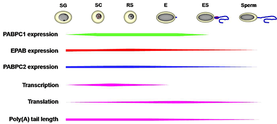
    Download PDF (1198K)
Original Article
  • Shinnosuke HONDA, Yuka MIKI, Yuya MIYAMOTO, Yu KAWAHARA, Satoshi TSUKA ...
    Article type: Original Article
    2018 Volume 64 Issue 4 Pages 297-301
    Published: 2018
    Released on J-STAGE: August 20, 2018
    Advance online publication: May 03, 2018
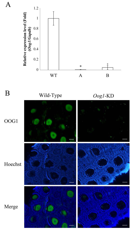
    JOURNAL FREE ACCESS

    Oog1, an oocyte-specific gene that encodes a protein of 425 amino acids, is present in five copies on mouse chromosomes 4 and 12. In mouse oocytes, Oog1 mRNA expression begins at embryonic day 15.5 and almost disappears by the late two-cell stage. Meanwhile, OOG1 protein is detectable in oocytes in ovarian cysts and disappears by the four-cell stage; the protein is transported to the nucleus in late one-cell to early two-cell stage embryos. In this study, we examined the role of Oog1 during oogenesis in mice. Oog1 RNAi-transgenic mice were generated by expressing double-stranded hairpin Oog1 RNA, which is processed into siRNAs targeting Oog1 mRNA. Quantitative RT-PCR revealed that the amount of Oog1 mRNA was dramatically reduced in oocytes obtained from Oog1-knockdown mice, whereas the abundance of spermatogenesis-associated transcripts (Klhl10, Tekt2, Tdrd6, and Tnp2) was increased in Oog1 knockdown ovaries. Tdrd6 is involved in the formation of the chromatoid body, Tnp2 contributes to the formation of sperm heads, Tekt2 is required for the formation of ciliary and flagellar microtubules, and Klhl10 plays a key role in the elongated sperm differentiation. These results indicate that Oog1 down-regulates the expression of spermatogenesis-associated genes in female germ cells, allowing them to develop normally into oocytes.

    Editor's pick

    Cover Story: Oog1, an oocyte-specific gene, encodes the protein belonging to the leucine-rich repeat (LRR) superfamily. LRR is a motif involved in protein-protein interactions. Complete knockout of Oog1 is challenging because five copies of the Oog1 gene are present on chromosomes 4 and 12. Honda et al. generated Oog1 RNA interference (RNAi)-transgenic mice to investigate the effects of Oog1 knockdown on gene expression in the oocytes (Honda et al. Oocyte-specific gene Oog1 suppresses the expression of spermatogenesis-specific genes in oocytes, pp. 297–301). The abundance of spermatogenesis-specific transcripts was elevated in the Oog1 knockdown ovaries. In addition, a few abnormal oocytes were observed in 6-month-old Oog1 knockdown mouse ovaries. These findings suggested that OOG1 suppresses the expression of spermatogenesis-specific genes in the oocytes and plays important roles during oogenesis.

    Download PDF (1091K)
  • Toshinori OIKAWA, Tomoko ITAHASHI, Risa YAJIMA, Takashi NUMABE
    Article type: Original Article
    2018 Volume 64 Issue 4 Pages 303-309
    Published: 2018
    Released on J-STAGE: August 20, 2018
    Advance online publication: May 07, 2018
    JOURNAL FREE ACCESS

    Intracytoplasmic sperm injection (ICSI) was expected to enable more efficient use of sperm from sires with preferable genetic traits and result in a generation containing a larger number of offspring with superior genetic characteristics in livestock. However, the efficiency of the early development of embryos produced by ICSI is still far from satisfactory in cattle. The present study aimed to investigate the effects of the treatment of cryopreserved sperm with glutathione (GSH) on the early development of embryos produced by ICSI in Japanese Black cattle. Moreover, the disulfide bond state and mitochondrial function were investigated in the sperm treated with GSH to confirm the effectiveness of the abovementioned treatment. We also investigated the effect of 7% ethanol activation treatment on the developmental ability of ICSI embryos using GSH-treated sperm. There was no effect on the blastocyst rate from the activation treatment. When sperm-injected oocytes were cultured in vitro, the treatment with GSH significantly improved the early development of embryos. Specifically, the rates of embryos reaching the 4–8-cell stage and blastocyst stage were significantly higher in ICSI with GSH-treated sperm (71.4% and 31.0%, respectively) than that with the control sperm (36.6% and 7.0%, respectively). Moreover, the GSH-treated sperm treatment significantly decreased the number of disulfide bonds in the sperm head (as shown by monobromobimane staining) and enhanced the mitochondrial function in the sperm middle piece (as shown by Rhodamine 123 staining and the adenosine triphosphate-dependent bioluminescence assay). Based on these results, we suggest that the treatment of cryopreserved sperm with GSH might contribute to the improvement of ICSI techniques for the production of blastocysts in Japanese Black cattle.

    Download PDF (834K)
  • Shunsuke SAKAI, Natsumi HAGIHARA, Mariko KUSE, Koji KIMURA, Kiyoshi OK ...
    Article type: Original Article
    2018 Volume 64 Issue 4 Pages 311-317
    Published: 2018
    Released on J-STAGE: August 20, 2018
    Advance online publication: April 30, 2018
    JOURNAL FREE ACCESS

    Heat stress (HS) negatively affects reproduction in cattle; however, its effect on endocrine function in bovine endometrial cells remains unclear. In this study, we examined the effects of HS on the production of prostaglandin (PG) E2 and PGF2α in the cultured bovine endometrial epithelial and stromal cells separately. To evaluate the effect of HS on endocrine function, the cells were cultured at 38.5°C (control) or 40.5°C (HS). After treatment, PGE2 and PGF2α levels were measured via enzyme immunoassay (EIA) and mRNA expressions of enzymes involved in PG synthesis were examined via quantitative reverse transcription polymerase chain reaction (RT-PCR). HS did not influence the production of PGE2 or PGF2α in the epithelial cells; however, HS significantly enhanced the production of both PGE2 and PGF2α in the stromal cells (P < 0.05). In addition, HS significantly increased phospholipase A2 (PLA2), cyclooxygenase 2 (COX2), prostaglandin F synthase (PGFS), prostaglandin E synthase (PGES), and carbonyl reductase 1 (CBR1) mRNA expression in the stromal cells (P < 0.05). The overall results suggest that HS induces mRNA expression of enzymes involved in PG synthesis, resulting in the upregulation of PGE2 and PGF2α production in the stromal cells, but not in the epithelial cells. The HS-induced increase of PGE2 and PGF2α secretion in bovine endometrial stromal cells may disrupt the normal estrous cycle and cause infertility in cows during summer.

    Download PDF (898K)
  • Kento MIURA, Shogo MATOBA, Narumi OGONUKI, Takafumi NAMIKI, Junya ITO, ...
    Article type: Original Article
    2018 Volume 64 Issue 4 Pages 319-326
    Published: 2018
    Released on J-STAGE: August 20, 2018
    Advance online publication: May 05, 2018
    JOURNAL FREE ACCESS

    In mammals, spermatozoa activate oocytes by triggering a series of intracellular Ca2+ oscillations with phospholipase C zeta (PLCζ), a sperm-borne oocyte-activating factor. Because the introduction of PLCζ alone can induce oocyte activation, it might be a promising reagent for assisted reproductive technologies. To test this possibility, we injected human PLCζ (hPLCζ) mRNA into mouse oocytes at different concentrations. We observed the oocyte activation and subsequent embryonic development. Efficient oocyte activation and embryonic development to the blastocyst stage was achieved only with a limited range of mRNA concentrations (0.1 ng/μl). Higher concentrations of mRNA caused developmental arrest of most embryos, suggesting that excessive PLCζ protein might be harmful at this stage. In a second series of experiments, we aimed to regulate the PLCζ protein concentration in oocytes by applying auxin-inducible degron (AID) technology that allows rapid degradation of the target protein tagged with AID induced by auxin. Injection of the hPLCζ protein tagged with AID and enhanced green fluorescent protein (hPLCζ-AID-EGFP) demonstrated that high EGFP expression levels at the late 1-cell stage were efficiently reduced by auxin treatment, suggesting efficient hPLCζ degradation by this system. Furthermore, the defective development observed with higher concentrations of hPLCζ-AID-EGFP mRNA was rescued following auxin treatment. Full-term offspring were obtained by round spermatid injection with optimized hPLCζ-AID activation. Our results indicate that this AID technology can be applied to regulate the protein levels in mouse oocytes and that our optimized PLCζ system could be used for assisted fertilization in mammals.

    Download PDF (1590K)
  • Takashi FUJII, Hiroki HIRAYAMA, Shigeo FUKUDA, Soichi KAGEYAMA, Akira ...
    Article type: Original Article
    2018 Volume 64 Issue 4 Pages 327-335
    Published: 2018
    Released on J-STAGE: August 20, 2018
    Advance online publication: May 24, 2018
    JOURNAL FREE ACCESS
    Supplementary material

    Artificial insemination with cryopreserved semen is a well-developed technique commonly used for controlled reproduction in cattle. However, despite current technical advances, cryopreservation continues to damage bull spermatozoa, resulting in a loss of approximately 30 to 50% of viable spermatozoa post thawing. To further improve the efficiency of cryopreservation of bull spermatozoa, understanding the molecular mechanisms underlying the cryobiological properties that affect cryoinjuries during cryopreservation process of bull spermatozoa is required. In this study, we examined the expression and localization of aquaporin (AQP) 3 and AQP7 in fresh, cooled, and frozen-thawed bull spermatozoa. Furthermore, we investigated the relevance of AQP3 and AQP7 to motility and to membrane integrity in frozen-thawed bull spermatozoa. Western blotting against AQP3 and AQP7 in bull spermatozoa revealed bands with molecular weights of approximately 42 kDa and 53 kDa, respectively. In immunocytochemistry analyses, immunostaining of AQP3 was clearly observed in the principal piece of the sperm tail. Two immunostaining patterns were observed for AQP7 ―pattern 1: diffuse staining in head and entire tail, and pattern 2: diffuse staining in head and clear staining in mid-piece. Cooling and freeze-thawing did not affect the localization pattern of AQP7 and the relative abundances of AQP3 and AQP7 evaluated by Western blotting. Furthermore, we demonstrated that the relative abundances of AQP3 and AQP7 varied among ejaculates, and they were positively related to sperm motility, particularly sperm velocity, post freeze-thawing. Our findings suggest that AQP3 and AQP7 are possibly involved in the tolerance to freeze-thawing in bull spermatozoa, particularly in the sperm’s tail.

    Download PDF (1784K)
  • Maiko KOBAYASHI, Takamasa KOYAMA, Yasuhiro YASUTOMI, Tadashi SANKAI
    Article type: Original Article
    2018 Volume 64 Issue 4 Pages 337-342
    Published: 2018
    Released on J-STAGE: August 20, 2018
    Advance online publication: May 31, 2018
    JOURNAL FREE ACCESS

    We attempted to elucidate female reproduction in long-tailed macaques (Macaca fascicularis). These monkeys have a non-seasonal menstruation cycle, which makes them suitable subjects for studies in a variety fields including medical science and regenerative medicine. We analyzed individual breeding data including time of menarche, start of regular menstruation, and first pregnancy. These three events are related to the maturation of female long-tailed macaques. All research subjects were female long-tailed macaques bred at the Tsukuba Primate Research Center. The study comprised 45 females; we included time of menstruation, male-female cohabitation, and first pregnancy in their growth records. We extracted age and weight data relating to menarche, start of regular menstruation, and first pregnancy from these records. In the two years typically required from menarche to first pregnancy, the body weight increased by approximately 500 g (21% of the weight at menarche); it is clear that there is a significant physical change after menarche. Our findings suggest that female monkeys are not necessarily mature enough for pregnancy at menarche. Therefore, the use of the word “maturity” in terms of fecundity may be more accurate after the start of regular menstruation. This is what we term “adolescence” in the developmental process. Therefore, M. fascicularis monkeys are candidates for an animal model of human adolescence.

    Download PDF (790K)
  • Jianning YU, Leyan YAN, Zhe CHEN, Hui LI, Huanxi ZHU, Rong CHEN, ZhenD ...
    Article type: Original Article
    2018 Volume 64 Issue 4 Pages 343-350
    Published: 2018
    Released on J-STAGE: August 20, 2018
    Advance online publication: June 08, 2018
    JOURNAL FREE ACCESS

    Treatment of fetal rat and embryonic chicken with exogenous glucocorticoids induces premature differentiation of growth hormone (GH) secreting cells. The effect of corticosterone (CORT) on somatotroph differentiation was mostly studied in pituitary cells in vitro. Currently, there is no evidence for glucocorticoid-mediated induction of somatotroph differentiation during pituitary development in bird species other than chicken. In this study, we sought to find out if in ovo injection of corticosterone into developing goose embryos could induce premature increase of GH in somatotrophs. On embryonic day (e) 15, the albumen of fertile goose eggs was injected with 300 μl of 0.9% saline, 300 μl 5 × 10⁻⁸M CORT, or 300 μl 5 × 10⁻⁶ M CORT. Embryos were allowed to develop until e20 and e28 and isolated pituitaries were subjected to quantitative real-time PCR and immunocytochemistry to detect GH mRNA and protein, respectively. At e20 and e28, blood from chorioallantoic vessels was subjected to radioimmunoassay for analysis of plasma GH protein. In ovo administration of exogenous corticosterone brought about a 2.5-fold increase in the expression of GH mRNA and increased the in situ expression of GH protein in goose pituitary cells, and enhanced plasma GH levels compared to that of the respective controls at e20. These findings prove that administration of glucocorticoid could stimulate the expression of GH in somatotrophs during goose embryonic development. Our results suggest the probable involvement of membrane glucocorticoid receptor in the corticosterone mediated expression of GH. Together with previously published data, our results suggest that corticosterone mediated induction of GH expression during embryonic development is relatively conserved among different vertebrates.

    Download PDF (3072K)
  • Manuel ÁLVAREZ-RODRIGUEZ, Alejandro VICENTE-CARRILLO, Heriberto RODRIG ...
    Article type: Original Article
    2018 Volume 64 Issue 4 Pages 351-360
    Published: 2018
    Released on J-STAGE: August 20, 2018
    Advance online publication: June 08, 2018
    JOURNAL FREE ACCESS
    Supplementary material

    Hyaluronan (hyaluronic acid, HA) apparently improves sperm survival in vitro and in vivo (oviduct), maintaining sperm motility and inducing capacitation, but not acrosome exocytosis, either by direct action as a macromolecule or via CD44 membrane receptors. This study explored ejaculated, liquid-extended pig spermatozoa to ascertain (i) the presence (Western blotting) and specific location (immunocytochemistry) of the CD44 receptor, using a specific monoclonal commercial antibody; (ii) whether the CD44 receptor changed location when exposed to bicarbonate, a capacitating trigger, in vitro; and (iii) whether the addition of HA, of molecular size comparable to that produced in the oviduct sperm reservoir (0.0625 to 2.0 mg/ml; 0 HA: control), to semen extenders would improve sperm liquid storage in vitro or cryosurvival post-freezing. Variables tested were sperm velocity and progressive motility (QualispermTM), sperm viability and acrosome status, membrane integrity and early destabilization, mitochondrial activation, and superoxide production (flow cytometry). The CD44 receptor presence in ejaculated, liquid-stored AI boar spermatozoa, as confirmed by a porcine-specific monoclonal antibody, maintained its membrane location under in vitro capacitation-inducing conditions. HA exposure to 24-, 48-, or 72-h liquid-stored (17–20ºC) spermatozoa lowered sperm velocity in membrane-intact spermatozoa, but increased mitochondrial superoxide production. Finally, HA addition during cooling did not improve cryosurvival but did increase mitochondrial activation and membrane destabilization in surviving cells. These results confirm the existence of a CD44 receptor in pig spermatozoa, but the usefulness of adding HA for long-term storage or cryopreservation of liquid-stored, extended boar semen remains in question, thereby warranting further non-empirical analyses of HA-sperm membrane interactions.

    Download PDF (1418K)
Technology Report
  • Masaki TAKASU, Masami MAEDA, Julio ALMUNIA, Kotono NAKAMURA, Noriko NI ...
    Article type: Technology Report
    2018 Volume 64 Issue 4 Pages 361-364
    Published: 2018
    Released on J-STAGE: August 20, 2018
    Advance online publication: May 27, 2018
    JOURNAL FREE ACCESS

    In microminipigs, estrus induction with abortion treatment, which is typically performed between 25 and 40 days after mating, is not always successful. Thus, the authors hypothesized that it may be more difficult to induce estrus by treating microminipigs approximately 40 days after mating. Accordingly, in this study, estrus induction was performed with abortion treatment in four microminipigs as follows: 0.3 mg of cloprostenol, a prostaglandin F2-alpha analog, was administered (day 0); after 24 h, 0.15 mg of cloprostenol and 250 IU of equine chorionic gonadotrophin were administered intramuscularly and simultaneously (day 1); after 96 h, 120 IU of human chorionic gonadotropin was injected intramuscularly (day 4). These treatments were compared at two different stages of pregnancy: early treatment (26.5 ± 0.7 days) and late treatment (38.3 ± 0.8 days). In the early treatment, all four microminipigs exhibited estrus on day 5, whereas in the late treatment, estrus was observed clearly in only two pigs on day 6 and slightly in 1 pig on day 10, whereas it was unclear in 1 pig. These results suggest that it is difficult to induce estrus with abortion treatment in microminipigs at approximately 40 days after mating.

    Download PDF (592K)
  • Mohammed. A. ABDEL-GHANI, Kenichiro SAKAGUCHI, Chihiro KANNO, Yojiro Y ...
    Article type: Technology Report
    2018 Volume 64 Issue 4 Pages 365-369
    Published: 2018
    Released on J-STAGE: August 20, 2018
    Advance online publication: May 24, 2018
    JOURNAL FREE ACCESS

    We investigated the effects of pre-maturational (pre-IVM) culture on the developmental competence of small-sized bovine oocytes (110 and < 115 µm). Oocytes were cultured with 3-isobutyl-1-methylxanthine (IBMX) for 0, 5, or 10 h and subjected to in vitro maturation, fertilization, and culture. The cleavage rate (73%) of small-sized oocytes with 5 h pre-IVM was higher than those with 0 and 10 h pre-IVM (61 and 62%, respectively). The blastocyst rate (16%) of embryos derived from small-sized oocytes with 5 h pre-IVM was higher than those with 0 and 10 h pre-IVM (9 and 8%, respectively). In addition, small-sized oocytes with 5 h pre-IVM had a higher mean cell number in blastocysts (134.1 ± 34.8) than those with 0 and 10 h pre-IVM (100.2 ± 17.2 and 107.8 ± 23.7, respectively). In conclusion, the pre-IVM of small-sized oocytes with IBMX for 5 h improved the developmental competence of bovine oocytes, as well as the quality of blastocysts.

    Download PDF (667K)
feedback
Top