Progress of Digestive Endoscopy
Online ISSN : 2187-4999
Print ISSN : 1348-9844
ISSN-L : 1348-9844
83 巻, 1 号
選択された号の論文の79件中51~79を表示しています
症例
  • 三石 雄大, 斎藤 彰一, 井出 大資, 猪又 寛子, 大谷 友彦, 玉井 尚人, 加藤 智弘, 田尻 久雄, 池上 雅博
    2013 年 83 巻 1 号 p. 160-161
    発行日: 2013/12/14
    公開日: 2013/12/21
    ジャーナル フリー
    A 64-year-old man was admitted to our hospital for further investigation of a colonic submucosal tumor. Colonoscopy revealed a submucosal tumor of about 20 mm diameter in the descending colon. Magnifying endoscopy showed normal pit pattern (typeI) . Endoscopic ultrasonography demonstrated that the hypoechoic tumor originated from the second layer of the colonic wall. The patient had complained of intermittent abdominal pain and it was suspected that the tumor was resulting in bowel obstruction, so complete resection was performed. In this case, minimally invasive treatment was recommended as the patient was suffering from myocardial infarction, type II diabetes mellitus and bladder cancer. The tumor was removed endoscopically─without submucosal injection─succeeded by en bloc resection without complication. The final pathological diagnosis of the tumor was a leiomyoma. According to previous reports, many such cases are treated by surgical resection. In this case, the patient had several pre-existing conditions and we therefore elected to treat by endoscopic resection in order to minimize risks to him.
  • 保母 貴宏, 横山 登, 小城原 傑, 初鹿野 誠也, 磯崎 正典, 佃 玄紀, 松尾 海, 野垣 航二, 有馬 秀英, 相田 貞継, 清水 ...
    2013 年 83 巻 1 号 p. 162-163
    発行日: 2013/12/14
    公開日: 2013/12/21
    ジャーナル フリー
    An 85-year-old man was admitted to our hospital for investigation of a positive stool occult blood test. Colonoscopy showed a large pedunculated polyp originating from lower end of the cecum. A diagnosis of large appendiceal adenoma with cecal intussusception was made preoperatively. Laparoscopic ileocecal resection with dissection of regional lymph nodes was performed. Histopathological examination of resected specimens showed a tubular adenoma in the appendix, of low to high grade. Appendiceal adenoma with cecal intussusception is extremely rare, but it should be considered in cases with polypoid lesions involving the cecum.
  • 藤井 崇, 片岡 幹統, 安斎 翔, 白崎 友彬, 池宮城 秀和, 間渕 一壽, 鈴木 伸治, 岡田 章祐, 加藤 亜裕, 出口 倫明, 似 ...
    2013 年 83 巻 1 号 p. 164-165
    発行日: 2013/12/14
    公開日: 2013/12/21
    ジャーナル フリー
    A 67-year-old man was referred to our hospital for further investigation of a cecal submucosal tumor (SMT) . Colonoscopy (CS) revealed the cecal SMT was approximately 15 mm in diameter and covered with normal mucosa. EUS showed the lesion to be continuous into the third or fourth layer of the cecal mucosa, with a fairly homogeneous echogenicity. Laparotomy was performed with the aim of diagnostic therapy. A cystic lesion was located in the resected material near the vermiform appendix. On pathology, the cystic lesion consisted of mucosa similar to that in the vermiform appendix, and duplication of the vermiform appendix was diagnosed. Alimentary tract duplication is rare, and preoperative diagnosis is difficult. This diagnosis should be considered a differential for alimentary canal submucosal tumor.
  • 吹田 洋將, 浅木 努史, 足立 清太郎, 安田 伊久磨, 豊水 道史, 片倉 芳樹
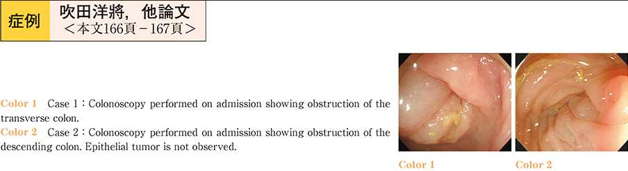
    2013 年 83 巻 1 号 p. 166-167
    発行日: 2013/12/14
    公開日: 2013/12/21
    ジャーナル フリー
    We report two cases of malignant colonic obstruction that were improved using a trans-anal decompression tube. The first case was that of an 86-year-old woman referred to our department for further investigation of advanced gastric cancer. During an outpatient visit, she was admitted as an emergency because of sudden right flank pain. Computed tomography (CT) showed that the transverse colon had been invaded by the gastric cancer, and the acute symptoms were due to obstruction of the transverse colon. A trans-anal decompression tube was inserted through the obstructed region of the transverse colon, and subsequently symptoms of intestinal obstruction improved. The second case was that of an 84-year-old woman who had been diagnosed with a left renal pelvic tumor. Due to the advanced stage of disease, this patient was followed up at the urology department without curative treatment. Four months after diagnosis, the woman developed abdominal distension and vomiting. CT revealed a left renal mass extending into the descending colon and resulting in colonic obstruction. Symptoms of intestinal obstruction were improved by insertion of the trans-anal decompression tube.
    Although these patients were at an advanced stage of disease on presentation, decompression using the trans-anal tube was useful in relieving symptoms of malignant colonic obstruction.
  • 伊藤 亜由美, 大森 鉄平, 高橋 麻衣, 合阪 暁, 米沢 麻利亜, 高山 敬子, 飯塚 文瑛, 白鳥 敬子, 番場 嘉子, 橋本 拓造, ...
    2013 年 83 巻 1 号 p. 168-169
    発行日: 2013/12/14
    公開日: 2013/12/21
    ジャーナル フリー
    A 42-year-old man visited our department chiefly complaining of abdominal pain, diarrhea and hematochezia. Colonoscopy (CS) led to a diagnosis of ischemic colitis (IC) . Although the patient was monitored, he showed recurrence of symptoms three months later. The man underwent colonoscopy again at a local hospital, which revealed longitudinal ulcers and raised a suspicion of Crohn’s disease. He was started on PSL at a dose of 30 mg/day, but as the symptoms did not improve, this was increased to 60 mg/day. The man was then admitted to our department for treatment. After admission, he was fasted and received antibiotics─achieving symptomatic relief. CS and abdominal CT were performed to determine the effect of PSL therapy. Mucosal thickening had worsened, the sigmoid mucosa appeared yellow and white, and the intestinal lumen had narrowed. CS findings were again suggestive of IC and subsequently symptoms improved. However, the pa-tient re-presented with signs of disseminated intravascular coagulation (DIC) and underwent Hartmann’s procedure in the Department of Surgery, which rapidly improved the signs of DIC. It was difficult to diagnose IC in this relatively young man, who developed recurrence in a short period of time, had the above-described CS findings, and experienced improvements in abdominal symptoms after increasing PSL dose. However, his physical state was associated with conditions of chronic circulatory insufficiency, with vascular factors such as hypertension and dialysis. Pathologically, marked microvascular blood flow disturbances were identified, suggesting that local intestinal necrosis due to microcircular vascular insufficiency occurred. It is possible that the patient had microvascular blood flow disturbance, and required close monitoring.
  • 成木良 瑛子, 鈴木 武志, 佐藤 浩一郎, 富永 健司, 伊藤 紗代, 加藤 充, 北川 智之, 平山 圭穂, 森 麻紀子, 吉田 有輝, ...
    2013 年 83 巻 1 号 p. 170-171
    発行日: 2013/12/14
    公開日: 2013/12/21
    ジャーナル フリー
    A 40-year-old man─who had been diagnosed ulcerative colitis on March 2011─presented at our emergency room with fever, frequent diarrhea and dizziness. At the same time, there was painful flat flare on his knee and toe. Laboratory data showed elevated white blood cell count and serum CRP. Lower GI endoscopy revealed ulcers extending from the rectum to the ascending colon. A few days later, another skin lesion suddenly appeared. The new cutaneous eruptions were severely painful pustules of a few centimeters in diameter. Both skin lesions were biopsied. The first lesion showed patchy adipose tissue inflammation, mainly comprising lymphocytes. The second lesion showed dense perivascular neutrophil infiltrates without vasculitis. The first lesion was suspected to be erythema nodule and the second, Sweet’s disease. The patient was treated with high dose prednisolone (60 mg/day) , and soon after the painful pustules disappeared. Abdominal symptoms and endoscopic findings also resolved. Searching on the Japana Centra Revuo Medicina database, we found only six reported cases of ulcerative colitis complicated by Sweet’s disease. Here we report this rare case with a combination of ulcerative colitis and Sweet’s.
  • 河上 唯史, 諸星 雄一, 伊藤 剛, 角田 裕也, 今村 諭, 田村 寿英, 長久保 秀一, 小池 祐司, 藤田 由里子, 小松 弘一
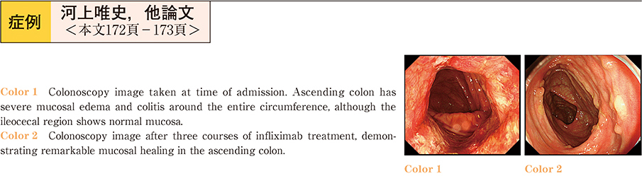
    2013 年 83 巻 1 号 p. 172-173
    発行日: 2013/12/14
    公開日: 2013/12/21
    ジャーナル フリー
    A 29-year-old woman was first diagnosed with ulcerative colitis (proctitis) six months previously. Remission was successfully induced with mesalazine, but treatment was interrupted. In spite of repeated treatment with mesalazine, symptoms exacerbated and the patient presented at our hospital. Laboratory analysis revealed leukocytosis and elevated serum CRP level. Enteropathogenic Escherichia coli and CMV antigenemia assays were negative. Colonoscopy revealed severe mucosal edema, colitis involving the entire circumference, and ulcerative lesions with a punched out appearance in the ascending colon. Examination of the transverse colon through to the sigmoid colon showed almost normal mucosa, and the rectum showed granularity of the mucosa─typical of ulcerative colitis (UC) . Differential diagnoses such as Crohn’s disease or drug-induced colitis were possible, but it was difficult to make a definitive diagnosis. The patient was admitted to our hospital, and prednisolone therapy was initiated at a dose of 60 mg/day, according to the treatment regime for severe UC. Endoscopic findings showed improvements in the mucosal edema. Conversely, pseudopolyposis of the intestinal wall was seen. On the 32nd day, the patient suddenly developed severe melena and shock. Although surgery was advised, the patient declined. Infliximab was started at a dose of 5 mg/kg, and symptoms rapidly improved. Endoscopic findings after three courses of treatment showed remarkable mucosal healing.
  • 草野 昌男, 駒沢 大輔, 渡部 敬之, 伊藤 広通, 土佐 正規, 大楽 尚弘, 池田 智之, 上野 孝治, 池谷 伸一, 中山 晴夫, ...
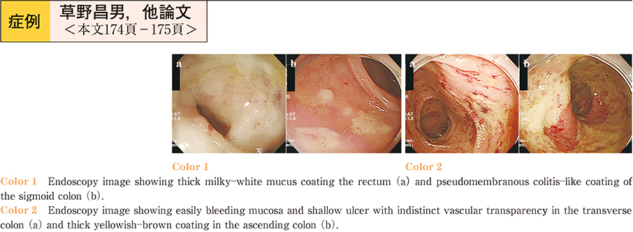
    2013 年 83 巻 1 号 p. 174-175
    発行日: 2013/12/14
    公開日: 2013/12/21
    ジャーナル フリー
    The patient was a 75-year-old woman receiving several medications─aspirin, amlodipine, candesartan, furosemide, spironolactone, rosuvastatin and triazolam─for cerebral infarction, hyperlipidemia, hypertension and sleeplessness. Furthermore the woman was being medicated with lansoprazole for reflux esophagitis. She developed watery diarrhea after taking lansoprazole for approximately four months. The woman presented at our hospital after suffering watery diarrhea for five months. Colonoscopic examination showed thick mucus coating the rectum and a pseudomembranous coating in the sigmoid colon. Colonic mucosal biopsy showed marked inflammatory cells, and a diagnosis of erosive colitis and proctitis was made. However, the diarrhea continued and a second colonoscopy was performed. This showed easily blee-ding mucosa and a shallow ulcer with indistinct vascular transparency in the transverse colon and thick yellowish-brown coating in the ascending colon. A colonic mucosal biopsy showed a thick collagen band in the subepithelial region, and collagenous colitis was diagnosed. The patient’s watery diarrhea was relieved one week after changing lansoprazole to omeprazole. Colonoscopic and pathological findings one year later showed visibly normal mucosa. In this case colonoscopic findings were atypical for collagenous colitis.
  • 金子 桂士, 西山 竜, 増田 あい, 龍 芯衣, 神津 馨里奈, 中川 太一, 大城 周, 中河原 浩史, 赤井 祐一, 小川 眞広, 中 ...
    2013 年 83 巻 1 号 p. 176-177
    発行日: 2013/12/14
    公開日: 2013/12/21
    ジャーナル フリー
    An 83-year-old man presented with right lower abdominal pain and a fever of 37.6°C. Initial blood analysis showed a WBC 10,200 /µl, CRP 1.71 mg/dl, and a slight inflammatory reaction.
    Diverticulitis of the ascending colon was diagnosed by abdominal CT. The patient was fasted, given supplementary liquid and antibiotics, and the diverticulitis healed. On the 22nd day of illness, lower gastrointestinal (GI) endoscopy was performed, confirming the diverticulum which occurred frequently in the ascending colon and sigmoid colon. Furthermore, it extended to the descending colon forming a granular mucous membrane, overrunning to an ascending colon under a coating of epithelium. Biopsy specimens from this latter part enabled a diagnosis of collagenous colitis (CC) . Lower GI endoscopy was again performed after cessation of drugs suspected to be the cause, confirming improvement of the mucus colon. This procedure also identified the mucous membrane which was a rough work with a flare, a layer of mucous membrane edema. Collagen fibers with a slightly thickened membrane were seen. If a slight mucous membrane change is expected with administration of a drug, reports of clinical signs and abnormal results from endoscopy should be followed up with a biopsy for diagnostic purposes to assess for the existence of CC.
  • 木村 典夫, 志田 勝義, 前村 誠
    2013 年 83 巻 1 号 p. 178-179
    発行日: 2013/12/14
    公開日: 2013/12/21
    ジャーナル フリー
    A 70-year-old male visited our hospital complaining of diarrhea and hematochezia.
    Abdominal CT demonstrated multiple sigmoid colonic diverticula and inflammatory changes suggestive of diverticulitis. Colonoscopy revealed diverticula scattered in the sigmoid colon with localized mucosal edema and erosion. A colonoscope could not be passed through the sigmoid colon due to the presence of a stricture. Histological analysis of biopsy specimens showed areas of inflammatory cell infiltration, crypt abscesses and granuloma formation in the sigmoid colon, and normal rectal mucosa. We diagnosed the patient with diverticular colitis, and administered steroids and 5-aminosalicylic acid. Two months later, the inflammation and stricture were shown endoscopically to have improved.
  • 天田 塩, 内 雄介, 一坂 俊介, 森 克昭, 石川 啓一, 堂脇 昌一, 藤田 晃司, 菊永 裕行, 熊井 浩一郎, 片桐 真理, 三浦 ...
    2013 年 83 巻 1 号 p. 180-181
    発行日: 2013/12/14
    公開日: 2013/12/21
    ジャーナル フリー
    A man in his seventies with diabetes mellitus was admitted to our hospital with a high fever. The patient had undergone steroid pulse therapy against sudden hearing loss five days prior to admission. A blood test showed elevations in inflammatory and hepatic parameters. Computed tomography (CT) showed swelling of the gallbladder with intracholecystic gas. The CT also identified a hepatic abscess at the perigallbladder. Emphysematous cholecystitis with peri-cholecysto-liver abscess was suspected. Percutaneous transhepatic drainage of the gallbladder and abscess was performed under ultrasonographic guidance. Preoperative colonoscopy showed a small orifice in the transverse colon near the hepatic flexure. Fistulography was performed, visualizing the fistula and gallbladder. Chinese ink was injected endoscopically near the orifice of the fistula. Choleystectomy was performed on Day 27 after admission. Ink-injection was extremely useful in locating the cholecystocolonic fistula during the procedure. The patient was discharged from the hospital on Day 101.
  • 工藤 貴章, 小澤 俊一郎, 川島 亜貴世, 鈴木 碧, 佐藤 義典, 中津 智子, 池田 佳子, 石郷岡 晋也, 細谷 浩介, 前畑 忠輝 ...
    2013 年 83 巻 1 号 p. 182-183
    発行日: 2013/12/14
    公開日: 2013/12/21
    ジャーナル フリー
    Giardia lamblia is one cause of prolonged diarrhea in overseas travelers. Demonstration of trophozoites in stool is the traditional method of diagnosis. Here we present a case of giardiasis diagnosed by lavage fluid collected during colonoscopy (CS) .A 60-year-old woman presented at our hospital with prolonged watery diarrhea since travel in Southeast Asia. She had been treated empirically due to negative results from EGD, CS and stool culture. A second CS was performed one year and nine months after the first CS, because the diarrhea persisted. Trophozoites of Giardia lamblia were detected microscopically in lavage fluid sediment collected during this CS. Oral administration of metronidazole (750 mg/day) for one week dramatically improved the patient’s symptoms. This case suggests that CS lavage fluid assessment is a useful method to detect Giardia lamblia in patients with chronic diarrhea.
  • 砂山 健一, 小里 俊幸, 瀬戸口 智彦, 清水 紀香
    2013 年 83 巻 1 号 p. 184-185
    発行日: 2013/12/14
    公開日: 2013/12/21
    ジャーナル フリー
    Aluminum potassium sulfate and tannic acid (ALTA) is a sclerosing agent for hemorrhoids. Here we report two cases in which this agent was injected via colonscopy in place of hypertonic saline epinephrine to provide hemostasis in the lower rectum.
    Case 1 : An 84-year-old female inpatient with chronic heart failure had hematochezia and acutely developed shock. Emergency colonoscopy was performed, and identified a Dieulafoy’s ulcer with active bleeding above the dentate line. ALTA (1 ml) was injected into the exposed vessel, instantly stopping the bleeding. The bleeding area was also sealed using argon plasma coagulation (APC) . Three days after treatment, swollen whitish mucosa without ulceration was observed at the treated site.
    Case 2 : A 98-year-old man was admitted with a two-day history of hematochezia after undergoing a glycerol enema and rectal mucosal scraping. Colonoscopy identified a mucosal injury with heavy bleeding above the dentate line. ALTA was injected into the point of the gush and around the ulcerated region, and almost completely suppressed the bleeding. Colonoscopy three days later showed reduced ulcer size with surrounding swollen whitened mucosa, and one week after complete healing of the ulcer.
    ALTA has a rapidly-acting astringent hemostatic effect on mucosa and a sclerotic effect due to non-bacterial inflammation with fibrosis. In addition, it has been reported that the risk of secondary ulcer by ALTA injection is between 0.45 and 4.6% in hemorrhoid treatment. Therefore, we considered that ALTA would be a suitable hemostatic agent to administer by colonoscopic injection in the lower rectum.
  • 佐藤 祥之, 加藤 慶三, 米沢 健, 立花 浩幸, 井家 麻紀子, 島田 紀朋
    2013 年 83 巻 1 号 p. 186-187
    発行日: 2013/12/14
    公開日: 2013/12/21
    ジャーナル フリー
    A 51-year-old man with alcoholic liver cirrhosis was hospitalized for treatment of rectal varices. 3D-CT revealed that the rectal varices’ inflow vessel was the superior rectal vein─branching from a mesenteric vein─and outflow vessels were the middle and inferior rectal vein draining into the internal iliac vein. The diameter of the inferior mesenteric vein was thinner than that of the left gastric vein. Therefore, it was speculated that the variceal blood flow rate would be low, and endoscopic injection sclerotherapy (EIS) was selected as the treatment method. EIS was performed using 5% ethanolamine oleate with iopamidol. Endoscopic varicealography during injection sclerotherapy (EVIS) clearly showed the rectal varices, superior rectal vein, and inferior mesenteric vein. After treatment, complete embolization of the rectal varices and inflow vessels was confirmed by contrast enhanced CT, and endoscopy revealed disappearance of the rectal varices. This case of rectal varices was successfully treated using EIS.
  • 富澤 稔, 篠崎 文信, 長谷川 留魅子, 白井 芳則, 一木 昇, 富居 一範, 杉山 隆夫, 山本 重則, 末石 眞, 吉田 孝宣
    2013 年 83 巻 1 号 p. 188-189
    発行日: 2013/12/14
    公開日: 2013/12/21
    ジャーナル フリー
    A 63-year-old man was admitted to our hospital with epigastralgia. Ultrasonography showed thickening of the wall of the gall bladder fundus. Magnetic resonance cholangiopancreatography revealed a cystic lesion on the fundus suggesting Rokitansky-Aschoff sinus (RAS) . Endoscopic retrograde cholangiopancreatography illustrated an elevated lesion on the fundus. No abnormal signal was detected by positron emission tomography. These diagnostic imaging results suggested adenomyomatosis, but the differential diagnosis of cancer remained. The patient opted for surgery instead of strict monitoring as his brother had died of gallbladder cancer. The patient underwent laparoscopic cholecystectomy. The surgically-obtained specimen revealed RAS in the thickened wall of the gall bladder fundus. Around the RAS, small muscle proliferation and lymphocyte infiltration were observed. The pathological diagnosis was adenomyomatosis of the gall bladder with chronic cholecystitis. No evidence of malignancy was found.
  • 二宮 大和, 西澤 伸恭, 木村 有希, 清水 理葉, 藤田 優裕, 高田 智司, 星野 好則, 伊藤 誉, 寺内 寿彰, 木全 大, 篠﨑 ...
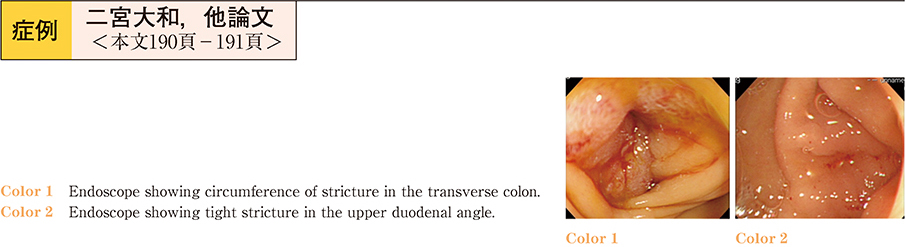
    2013 年 83 巻 1 号 p. 190-191
    発行日: 2013/12/14
    公開日: 2013/12/21
    ジャーナル フリー
    A patient in his 80ʼs was referred to our hospital with a complaint of anorexia─of one month duration─and obstructive jaundice. Abdominal CT scan and GI tract survey revealed an advanced gallbladder carcinoma causing luminal strictures in the bile duct, the duodenum and the transverse colon with extensive peritoneal dissemination. Endoscopic stenting using covered self─expandable metallic stents (SEMS) was elected for treatment. First of all, on the fifth hospital day, a Flexxus Endoscopic Biliary Stent 10×80 mm was inserted into the bile duct. Secondly, on the 12th hospital day, a Wallflex Colonic Stent 22×90 mm was inserted into the transverse colon. A third procedure was performed on the 16th hospital day, when a Wallflex Duodenal Stent 22×90 mm was inserted into the duodenum. These procedures enabled the patient to food orally three days later. He maintained a good performance status (PS 0) for the following 4 months. The sequential triple endoscopic stentings were successfully preformed. This procedure is a useful method of palliative therapy for patients with various luminal strictures derived from malignant diseases in an advanced stage, in order to maintain quality of life and permit activities of daily living.
  • 佐藤 淑, 森山 友章, 長主 直子, 佐々木 亜希子, 出張 玲子
    2013 年 83 巻 1 号 p. 192-193
    発行日: 2013/12/14
    公開日: 2013/12/21
    ジャーナル フリー
    A 64-year-old man was admitted to the hospital because of jaundice. Laboratory findings showed T-Bil had increased to 10.2 mg/dl and CA19-9 level was 62 U/ml. Abdominal CT identified an enhanced tumor at the middle to lower extrahepatic bile duct, MRCP showed disruption of the middle bile duct, and ERC revealed the tumor located at the middle and lower bile duct. Positron emission tomography showed 2-[18F]-fluoro-2-deoxy-D-glucose uptake localized to the bile duct. Biopsy specimens taken from the tumor suggested a carcinoma. The patient underwent pylorus-preserving pancreaticoduodenectomy. Histopathology showed that the tumor consisted of intermixed, adenocarcinoma and sarcomatoid spindle cells-so-called carcinosarcoma (undifferentiated carcinoma according to World Health Organization classification) . The tumor was Bmi, fm, pGinf0, pPanc0, pDuo0, int, INF-β, ly0, v0, pn1a, pHM2, pEM0, fStageI.
    Carcinosarcoma of the bile duct tends to show a polypoid lesion, and biopsy of the tumor is necessary to differentiate from adenoma, papilloma, inflammatory polyp, calculus and carcinoma.
  • 芳賀 祐規, 露口 利夫, 酒井 裕司, 坂本 大, 西川 貴雄, 杉山 晴俊, 中村 昌人, 大塚 将之, 宮崎 勝, 横須賀 收
    2013 年 83 巻 1 号 p. 194-195
    発行日: 2013/12/14
    公開日: 2013/12/21
    ジャーナル フリー
    A 54-year-old woman who had porcelain gallbladder with jaundice was referred to our hospital on suspicion of gallbladder cancer. Contrast-enhanced CT showed a mass lesion with poor vascularity between the hepatic hilum at the neck of the gallbladder. MRCP showed a stricture in the common hepatic duct. FDG/PET-CT showed strong accumulation in the mass lesion. Endoscopic retrograde cholangiography was performed with bile and tissue sampling and endoscopic ultrasound-guided fine-needle aspiration, but there was no evidence of malignancy. Peroral cholangioscopy showed inflammatory changes but no malignant findings, and pathologic evaluation of the cholangioscopy-guided targeted biopsy specimen also showed no evidence of malignancy. Inflammatory changes due to porcelain gallbladder were clinically diagnosed, and cholecystectomy was performed. The histopathological diagnosis was xanthogranulomatous cholecystitis with porcelain gallbladder. It is essensial to perform detailed investigations including histopathological examination of patients with gallbladder disease in order to distinguish non-cancerous disease from malignancy.
  • 宮澤 巧, 畑中 恒, 沼尾 規且, 矢野 智則, 牛尾 純, 冨山 剛, 玉田 喜一, 山本 博徳, 菅野 健太郎
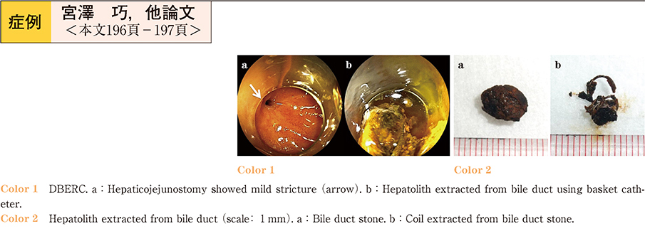
    2013 年 83 巻 1 号 p. 196-197
    発行日: 2013/12/14
    公開日: 2013/12/21
    ジャーナル フリー
    A 59-year-old woman was referred for endoscopic treatment of hepatolithiasis after living donor liver transplantation (LDLT) with Roux-en-Y hepaticojejunostomy. She underwent LDLT for liver cirrhosis B at 46 years of age, and liver biopsy 6 months later. Unfortunately, the liver biopsy resulted in biliary hemorrhage with formation of hepatic pseudoaneurysm. Treatment using transcatheter arterial embolization (TAE) with multiple coils was successful. However, the patient suffered from recurrent cholangitis with hepatolithiasis 12 years after LDLT. Double-balloon endoscopic retrograde cholangiography (DBERC) was performed to treat the hepatolithiasis at our hospital. Cholangiography revealed movable hepatolithiasis with the coils. We successfully retrieved the stones without complication using a basket catheter. The stones contained the coils used for TAE. This case is the first report of successful DBERC treatment for hepatolithiasis formed on migrated TAE coils.
  • 富澤 稔, 篠崎 文信, 長谷川 留魅子, 杉山 隆夫, 山本 重則, 末石 眞, 吉田 孝宣
    2013 年 83 巻 1 号 p. 198-199
    発行日: 2013/12/14
    公開日: 2013/12/21
    ジャーナル フリー
    A 64-year old man was admitted to our hospital with jaundice. Ultrasonography and computed tomography indicated the presence of common bile duct stones. The presence of stones was confirmed by endoscopic retrograde cholangiopancreato-graphy (ERCP) , and papillotomy was performed. Endoscopic retrograde nasobiliary drainage was initiated, and after one week an additional ERCP was performed. On this ERCP, coagula were found attached to the stomach wall. Lithotripsy was performed using basket forceps and all the stones were removed. The patient was also treated with a proton-pump inhibitor. However, four days after the second ERCP was performed, the patient’s blood pressure markedly decreased and his hemoglobin concentration drropped to 4.8 mg/dl. During an emergency endoscopy, spurting-type bleeding from a gastric ulcer was noted, which was controlled by clipping. Subsequently, the patient recovered well and was discharged; thereafter, he visited our hospital regularly for follow-up examinations.
  • 佐藤 悦基, 北村 勝哉, 山宮 知, 石井 優, 岩田 朋之, 野本 朋宏, 吉田 仁
    2013 年 83 巻 1 号 p. 200-201
    発行日: 2013/12/14
    公開日: 2013/12/21
    ジャーナル フリー
    Many publications report iatrogenic hemobilia─such as complications of percutaneous transhepatic procedures─but there are few reports of hemobilia induced by endoscopic biliary metallic stenting. Here we describe a case of hemobilia with hepatic pseudoaneurysm after placement of a self-expandable metallic stent (SEMS) for malignant biliary obstruction, treated by transcatheter arterial embolization (TAE) .
    An 84-year-old male with unresectable hilar cholangiocarcinoma received an uncovered biliary SEMS and placement of two percutaneous biliary drainage tubes. Fifty-seven days after SEMS placement, the patient developed severe melena and shock. Abdominal CT scan identified a hepatic pseudoaneurysm into the SEMS, which was treated by TAE.
    Although it is thought that hemobilia usually improves with conservative treatment, it may be better to perform hemostatic treatments such as TAE in cases complicated by pseudoaneurysm associated with SEMS.
  • 田川 徹平, 馬場 俊之, 中崎 奈都子, 大石 千歳, 金子 麗奈, 原 浩二, 金 民日, 草柳 聡, 小川 正純, 佐藤 譲
    2013 年 83 巻 1 号 p. 202-203
    発行日: 2013/12/14
    公開日: 2013/12/21
    ジャーナル フリー
    An 81-year-old man was admitted to our hospital complaining of abdominal pain. Upon admission, blood analysis showed elevated levels of serum bilirubin and hepatobiliary enzymes, together with an elevated inflammatory reaction. Acute cholangitis due to a common bile duct stone was diagnosed on the basis of imaging results. Endoscopic retrograde cholangiopancreatography was performed to drain the bile duct. Cannulation of the common bile duct was attempted, but insertion of the guide wire proved difficult. An abnormality of the cystic duct junction─where the cystic duct joined the lower bile duct─was suspected on the basis of abdominal computed tomography findings. Cannulation was eventually successful, and an endoscopic nasobiliary drainage (ENBD) tube was placed. The common bile duct stone was removed the following day. Cholangiography through the ENBD tube and magnetic resonance cholangiopancreatography revealed that the cystic duct revolved around the posterior wall of the common bile duct, and that an abnormally low junction existed between the cystic duct and the left-hand side of the common bile duct at the head of the pancreas. The patient elected not to undergo cholecystectomy due to his advanced age, and he was discharged from hospital on day ten. This case demonstrated that it is important to assess the bile duct’s course using imaging studies before initiating treatment.
  • 井上 健太郎, 相浦 浩一, 星本 相淳, 市東 昌也, 壁島 康郎, 小柳 和夫, 玉井 博修, 杉浦 仁
    2013 年 83 巻 1 号 p. 204-205
    発行日: 2013/12/14
    公開日: 2013/12/21
    ジャーナル フリー
    A 58-year-old man presented with a one-year history of remittent abdominal pain in the left upper quadrant due to acute pancreatitis. MRCP showed a significantly dilated and tortuous main pancreatic duct, which was occluded by a 10 mm-sized hypointense oval mass at the pancreas head. Computed tomography revealed the mass to be hypodense in the pre-contrast phase and slightly enhanced in the delayed phase. The most likely diagnosis was a pancreatic protein plug. However, intraductal tubulopapillary neoplasm (ITPN) could not be ruled out because of its enhancement on CT, and further diagnostic tests were required. EUS showed the pancreatic duct was incompletely filled with a 10 mm-sized homogeneously hypoechoic round mass. On ERP, the mass was slightly mobile but could not be removed using biopsy forceps. Peroral pancreatoscopy (POPS) revealed that the mass comprised opaque white anhistous cotton-like matter. Based on these findings, we concluded that the mass was more likely to be a protein plug than ITPN, and removed it completely using basket forceps. On histopathology, the mass consisted of eosinophilic mucoid matter mixed with a few non-typical columnar epithelia. The patient’s abdominal pain was relieved after the procedure. Protein plug and ITPN can have similar characteristics on CT, MRCP, EUS and ERP. POPS is useful to differentiate between a protein plug and ITPN.
  • 池内 信人, 糸井 隆夫, 糸川 文英, 祖父尼 淳, 土屋 貴愛, 栗原 俊夫, 石井 健太郎, 辻 修二郎, 梅田 純子, 田中 麗奈, ...
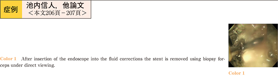
    2013 年 83 巻 1 号 p. 206-207
    発行日: 2013/12/14
    公開日: 2013/12/21
    ジャーナル フリー
    A 70-year-old man with pancreatic carcinoma underwent resection of the pancreatic body and tail with splenectomy. Postoperatively, a pancreatic fistula was identified on abdominal computed tomography (CT) . As he was asymptomatic, the patient was placed under observation. One month after surgery, the size of the pancreatic fistula─as shown on abdominal CT─had increased, and was accompanied by fever and abdominal pain. The patient was diagnosed with infected walled-off necrosis (WON) , and EUS-guided pseudocyst drainage (EUS-PCD) was performed immediately. Placement of a 4-cm plastic stent and a nasal-drainage tube were attempted using EUS-PCD. However, proximal migration of the stent into the WON occurred during the procedure. The 4-cm stent was left in situ, and a 9-cm plastic stent and a nasal-drainage tube were additionally placed. Endoscopic necrosectomy was performed five days later, and the migrated stent was retrieved at the same time using a front-view type endoscope.
  • 渡邊 薫, 杉森 一哉, 三箇 克幸, 清水 悠郎, 亀田 英里, 三輪 治生, 金子 卓, 粉川 敦史, 沼田 和司, 田中 克明, 前田 ...
    2013 年 83 巻 1 号 p. 208-209
    発行日: 2013/12/14
    公開日: 2013/12/21
    ジャーナル フリー
    Intrapancreatic accessory spleen (IPAS) is a rare benign lesion within the pancreas that frequently clinically and radiographically mimics a solid neoplasm. Distinguishing IPAS from pancreatic neoplasm/malignancy is extremely important from a treatment perspective. We report the case of a 77-year-old asymptomatic man with an incidentally detected 10 mm pancreatic tail mass. Dynamic computed tomography and contrast-enhanced harmonic endoscopic ultrasound revealed a round and well-circumscribed 10 mm hypervascular mass within the tail of the pancreas─the primary diagnostic consideration being a pancreatic neuroendocrine tumor. Superparamagnetic iron oxide-enhanced magnetic resonance imaging revealed an isointense mass relative to the spleen, consistent with IPAS. Subsequently, an endoscopic ultrasound-guided fine-needle aspiration biopsy was performed. Hematoxylin-eosin stained sections showed conspicuous thin-walled blood vessels in addition to inflammatory cells. Immunostaining for CD8 demonstrated strong positivity in endothelial cells of the thin-walled vessels. By correlating the pathological findings with the immunostaining result, we rendered the diagnosis of IPAS. Our experience in the present case supports the view that endoscopic ultrasound-guided fine-needle aspiration biopsy may enable a reliable preoperative diagnosis of IPAS and thus prevent unnecessary surgery.
  • 関根 章成, 今村 綱男, 田村 哲男, 小泉 優子, 小山 里香子, 松田 正道, 竹内 和男
    2013 年 83 巻 1 号 p. 210-211
    発行日: 2013/12/14
    公開日: 2013/12/21
    ジャーナル フリー
    A 71-year-old man had been pointed out mild dilation of pancreatic duct at regular health check in 2006. The lesion did not show remarkable change in 2009. However, significant dilation of pancreatic duct was observed, and he developed diabetes mellitus in October 2012. Laboratory data showed elevated serum pancreatic enzymes. Abdominal CT revealed pancreatic lithiasis in the tail. At that time, chronic pancreatitis was anticipated. Abdominal MRI revealed dilatation of the main pancreatic duct and its branches with cystic lesions of the pancreatic head, body, and tail. These results suggested that a cystic lesion of the pancreatic tail grew rapidly and a cystic lesion of the head was malignancy. Total pancreatectomy was performed. The histopathological examination showed IPMC of the pancreatic head and IPMN of the body. There was no neoplastic lesion other than pancreatic lithiasis in the tail. This case report shows that IPMN should be considered as a differential diagnosis when pancreatic lithiasis is observed.
  • 小嶋 啓之, 小黒 雅子, 大池 翼, 神野 彰, 菅野 真理子, 金野 朗, 市川 欧子, 櫻井 則男, 山田 俊夫
    2013 年 83 巻 1 号 p. 212-213
    発行日: 2013/12/14
    公開日: 2013/12/21
    ジャーナル フリー
    A 35-year-old man presented with high fever and abdominal pain. The patient was an alcoholic and acute pancreatitis had occurred repeatedly before this admission. Laboratory tests showed severe inflammation. CT imaging identified an enhanced cavity with a pseudocyst in the middle of the pancreatic body. The cavity contained fluid and air, and was thought to be an abscess penetrating to the stomach. EGD was performed, and detected a pocket filled with milky-white liquid in the posterior wall of the gastric upper body. Gastrography showed a fistula between the abscess and stomach. The fistula was expected to permit smooth drainage from the abscess. Intravenous antibiotics were administered for two weeks to control inflammation and diminish the abscess. Surgery was avoided in this patient and he was discharged 45 days later. Pancreatic pseudocyst is a common complication of pancreatitis. Small pseudocysts often resolve naturally. Large pseudocysts tend to cause infection, bleeding or rupture─requiring surgical treatment. In most cases reported in Japan, patients with pancreatitis and a large pseudocyst tend to be male alcoholics with hematemesis, requiring surgical treatment. Some publications report that percutaneous abscess drainage and somatostatin analogue use are effective in cases where bleeding is not present. Conservative treatment─using antibiotics and antacid─rarely cure the inflammation present with large pseudocysts.
  • 室井 歩, 港 洋平, 石橋 史明, 上山 俊介, 外山 雄三, 村松 雄輔, 宮本 勇治, 間野 真也, 古本 洋平, 村山 巌一, 堀内 ...
    2013 年 83 巻 1 号 p. 214-215
    発行日: 2013/12/14
    公開日: 2013/12/21
    ジャーナル フリー
    Pancreatic pseudocyst hemorrhage─one of the complications of pancreatitis─is a clinically severe condition, and early diagnosis and appropriate treatment are necessary.
    A 62-year-old man with heavy alcohol intake was examined by esophagogastroduodenoscopy (EGD) but no obvious bleeding site was detected. Anemia progressed during follow up and an abdominal CT-scan was performed. Two pancreatic pseudocysts were detected─the one, located at the head of pancreas, showed homogeneous high density, indicating hemorrhage pseudocyst. The patient was again monitored as no active bleeding was suspected; however the anemia worsened. A second EGD was performed and on this occasion red-brown discharge was detected emerging from the orifice of major duodenal papilla. At this point, a diagnosis of hemosuccus pancreaticus caused by pancreatic pseudocyst hemorrhage was made. The gastroduodenal artery was determined by CT angiography to be responsible for the bleeding, and arterial coil embolization was performed. The patient recovered well and no progressive anemia was seen after treatment.
お詫びと訂正
feedback
Top