64 巻, 2 号
選択された号の論文の55件中1~50を表示しています
掲載論文カラー写真集
-
2004 年 64 巻 2 号 p. 1-10
発行日: 2004年
公開日: 2014/03/28
PDF形式でダウンロード (12744K)
臨床研究
-
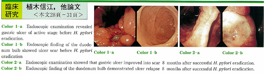
2004 年 64 巻 2 号 p. 28-31
発行日: 2004/06/10
公開日: 2014/03/28
 PDF形式でダウンロード (1077K)
 -
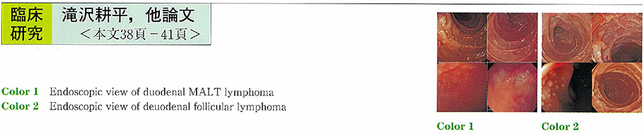
2004 年 64 巻 2 号 p. 32-37
発行日: 2004/06/10
公開日: 2014/03/28
PDF形式でダウンロード (715K) -
2004 年 64 巻 2 号 p. 38-41
発行日: 2004/06/10
公開日: 2014/03/28
 PDF形式でダウンロード (429K)
 -
2004 年 64 巻 2 号 p. 42-45
発行日: 2004/06/10
公開日: 2014/03/28
PDF形式でダウンロード (434K)
内視鏡の器械と技術
-
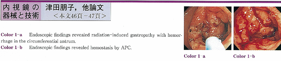
2004 年 64 巻 2 号 p. 46-47
発行日: 2004/06/10
公開日: 2014/03/28
 PDF形式でダウンロード (562K)
症例
-
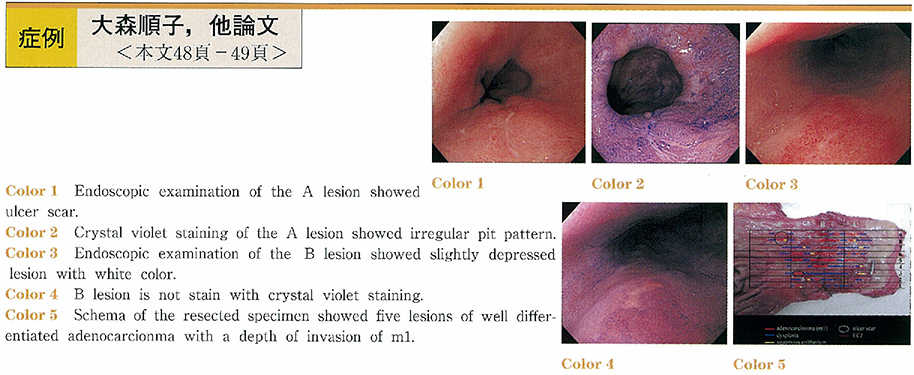
2004 年 64 巻 2 号 p. 48-49
発行日: 2004/06/10
公開日: 2014/03/28
 PDF形式でダウンロード (799K)
 -
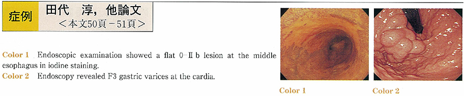
2004 年 64 巻 2 号 p. 50-51
発行日: 2004/06/10
公開日: 2014/03/28
 PDF形式でダウンロード (862K)
 -
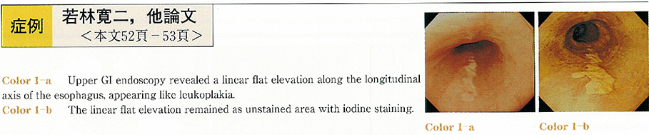
2004 年 64 巻 2 号 p. 52-53
発行日: 2004/06/10
公開日: 2014/03/28
 PDF形式でダウンロード (702K)
 -
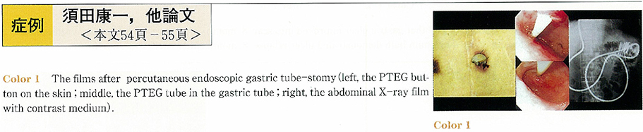
2004 年 64 巻 2 号 p. 54-55
発行日: 2004/06/10
公開日: 2014/03/28
 PDF形式でダウンロード (469K)
 -
2004 年 64 巻 2 号 p. 56-57
発行日: 2004/06/10
公開日: 2014/03/28
 PDF形式でダウンロード (587K)
 -
2004 年 64 巻 2 号 p. 58-59
発行日: 2004/06/10
公開日: 2014/03/28
 PDF形式でダウンロード (559K)
 -
2004 年 64 巻 2 号 p. 60-61
発行日: 2004/06/10
公開日: 2014/03/28
 PDF形式でダウンロード (1129K)
 -
2004 年 64 巻 2 号 p. 62-63
発行日: 2004/06/10
公開日: 2014/03/28
 PDF形式でダウンロード (258K)
 -
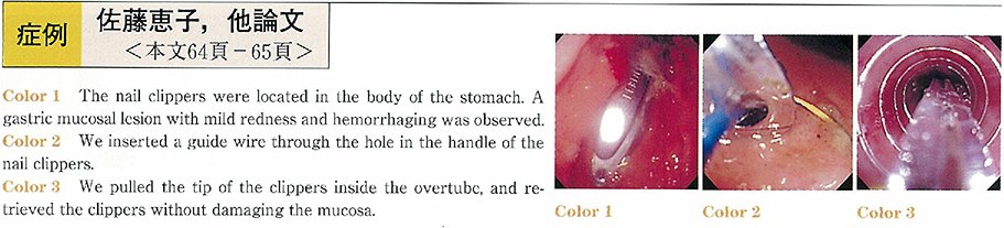
2004 年 64 巻 2 号 p. 64-65
発行日: 2004/06/10
公開日: 2014/03/28
 PDF形式でダウンロード (870K)
 -
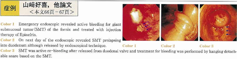
2004 年 64 巻 2 号 p. 66-67
発行日: 2004/06/10
公開日: 2014/03/28
 PDF形式でダウンロード (657K)
 -
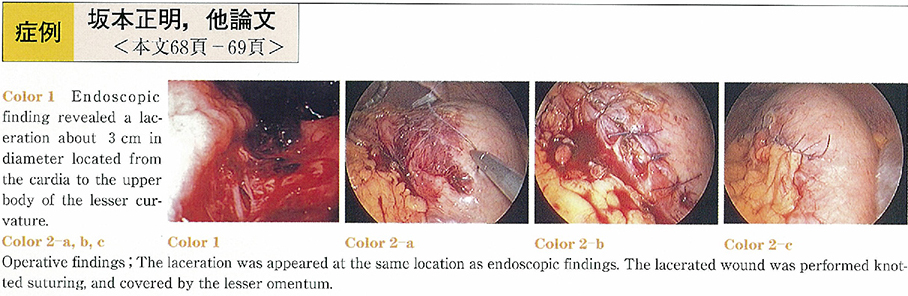
2004 年 64 巻 2 号 p. 68-69
発行日: 2004/06/10
公開日: 2014/03/28
 PDF形式でダウンロード (552K)
 -
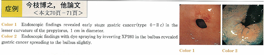
2004 年 64 巻 2 号 p. 70-71
発行日: 2004/06/10
公開日: 2014/03/28
 PDF形式でダウンロード (683K)
 -
2004 年 64 巻 2 号 p. 72-73
発行日: 2004/06/10
公開日: 2014/03/28
 PDF形式でダウンロード (916K)
 -
2004 年 64 巻 2 号 p. 74-75
発行日: 2004/06/10
公開日: 2014/03/28
 PDF形式でダウンロード (906K)
 -
2004 年 64 巻 2 号 p. 76-77
発行日: 2004/06/10
公開日: 2014/03/28
 PDF形式でダウンロード (817K)
 -
2004 年 64 巻 2 号 p. 78-79
発行日: 2004/06/10
公開日: 2014/03/28
 PDF形式でダウンロード (216K)
 -
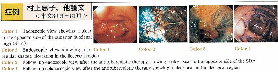
2004 年 64 巻 2 号 p. 80-81
発行日: 2004/06/10
公開日: 2014/03/28
 PDF形式でダウンロード (921K)
 -
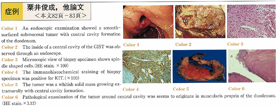
2004 年 64 巻 2 号 p. 82-83
発行日: 2004/06/10
公開日: 2014/03/28
 PDF形式でダウンロード (377K)
 -
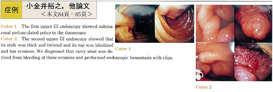
2004 年 64 巻 2 号 p. 84-85
発行日: 2004/06/10
公開日: 2014/03/28
 PDF形式でダウンロード (976K)
 -
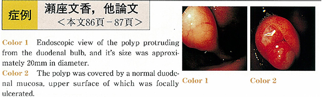
2004 年 64 巻 2 号 p. 86-87
発行日: 2004/06/10
公開日: 2014/03/28
 PDF形式でダウンロード (737K)
 -
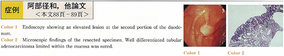
2004 年 64 巻 2 号 p. 88-89
発行日: 2004/06/10
公開日: 2014/03/28
 PDF形式でダウンロード (944K)
 -
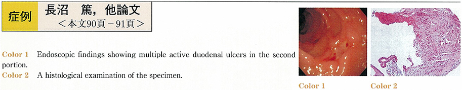
2004 年 64 巻 2 号 p. 90-91
発行日: 2004/06/10
公開日: 2014/03/28
 PDF形式でダウンロード (212K)
 -
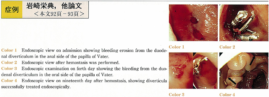
2004 年 64 巻 2 号 p. 92-93
発行日: 2004/06/10
公開日: 2014/03/28
 PDF形式でダウンロード (215K)
 -
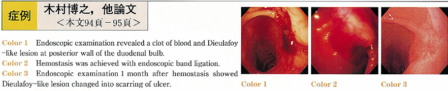
2004 年 64 巻 2 号 p. 94-95
発行日: 2004/06/10
公開日: 2014/03/28
 PDF形式でダウンロード (243K)
 -
2004 年 64 巻 2 号 p. 96-97
発行日: 2004/06/10
公開日: 2014/03/28
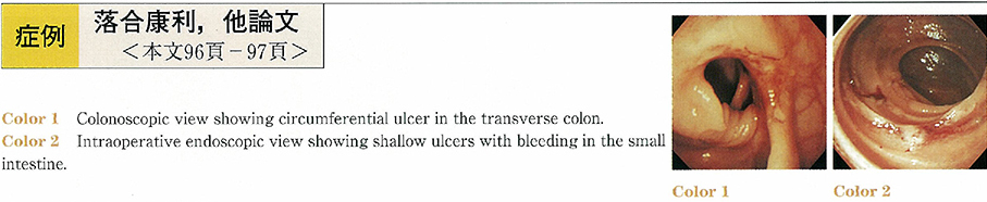 PDF形式でダウンロード (495K)
 -
2004 年 64 巻 2 号 p. 98-99
発行日: 2004/06/10
公開日: 2014/03/28
PDF形式でダウンロード (734K) -
2004 年 64 巻 2 号 p. 100-101
発行日: 2004/06/10
公開日: 2014/03/28
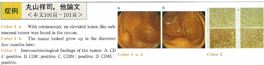 PDF形式でダウンロード (617K)
 -
2004 年 64 巻 2 号 p. 102-103
発行日: 2004/06/10
公開日: 2014/03/28
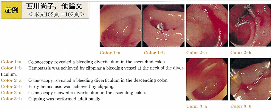 PDF形式でダウンロード (872K)
 -
2004 年 64 巻 2 号 p. 104-105
発行日: 2004/06/10
公開日: 2014/03/28
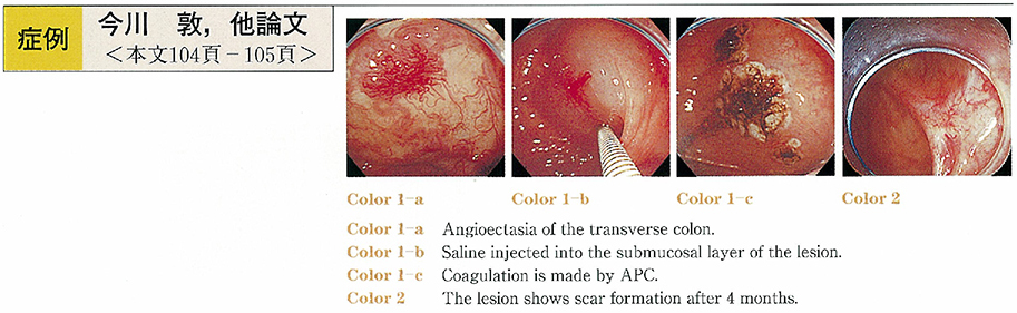 PDF形式でダウンロード (190K)
 -
2004 年 64 巻 2 号 p. 106-107
発行日: 2004/06/10
公開日: 2014/03/28
 PDF形式でダウンロード (542K)
 -
2004 年 64 巻 2 号 p. 108-109
発行日: 2004/06/10
公開日: 2014/03/28
 PDF形式でダウンロード (538K)
 -
2004 年 64 巻 2 号 p. 110-111
発行日: 2004/06/10
公開日: 2014/03/28
 PDF形式でダウンロード (528K)
 -
2004 年 64 巻 2 号 p. 112-113
発行日: 2004/06/10
公開日: 2014/03/28
 PDF形式でダウンロード (516K)
 -
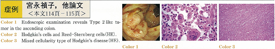
2004 年 64 巻 2 号 p. 114-115
発行日: 2004/06/10
公開日: 2014/03/28
 PDF形式でダウンロード (914K)
 -
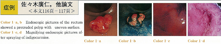
2004 年 64 巻 2 号 p. 116-117
発行日: 2004/06/10
公開日: 2014/03/28
 PDF形式でダウンロード (955K)
 -
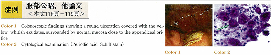
2004 年 64 巻 2 号 p. 118-119
発行日: 2004/06/10
公開日: 2014/03/28
 PDF形式でダウンロード (186K)
 -
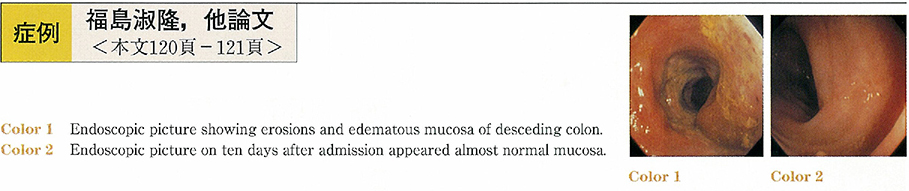
2004 年 64 巻 2 号 p. 120-121
発行日: 2004/06/10
公開日: 2014/03/28
 PDF形式でダウンロード (503K)
 -
2004 年 64 巻 2 号 p. 122-123
発行日: 2004/06/10
公開日: 2014/03/28
 PDF形式でダウンロード (224K)
 -
2004 年 64 巻 2 号 p. 124-125
発行日: 2004/06/10
公開日: 2014/03/28
 PDF形式でダウンロード (888K)
 -
2004 年 64 巻 2 号 p. 126-127
発行日: 2004/06/10
公開日: 2014/03/28
 PDF形式でダウンロード (394K)
 -
2004 年 64 巻 2 号 p. 128-129
発行日: 2004/06/10
公開日: 2014/03/28
 PDF形式でダウンロード (480K)
 -
2004 年 64 巻 2 号 p. 130-131
発行日: 2004/06/10
公開日: 2014/03/28
 PDF形式でダウンロード (304K)
 -
2004 年 64 巻 2 号 p. 132-133
発行日: 2004/06/10
公開日: 2014/03/28
 PDF形式でダウンロード (372K)
 -
2004 年 64 巻 2 号 p. 134-135
発行日: 2004/06/10
公開日: 2014/03/28
 PDF形式でダウンロード (194K)