85 巻, 1 号
選択された号の論文の50件中1~50を表示しています
- |<
- <
- 1
- >
- >|
掲載論文カラー写真集
-
2014 年 85 巻 1 号 p. 1-12
発行日: 2014年
公開日: 2014/12/17
PDF形式でダウンロード (11974K)
内視鏡の器械と技術
-
2014 年 85 巻 1 号 p. 36-39
発行日: 2014/12/06
公開日: 2014/12/17
 PDF形式でダウンロード (776K)
 -
2014 年 85 巻 1 号 p. 40-42
発行日: 2014/12/06
公開日: 2014/12/17
 PDF形式でダウンロード (364K)
臨床研究
-
2014 年 85 巻 1 号 p. 43-46
発行日: 2014/12/06
公開日: 2014/12/17
PDF形式でダウンロード (254K) -
2014 年 85 巻 1 号 p. 47-50
発行日: 2014/12/06
公開日: 2014/12/17
 PDF形式でダウンロード (283K)
 -
2014 年 85 巻 1 号 p. 51-54
発行日: 2014/12/06
公開日: 2014/12/17
 PDF形式でダウンロード (261K)
内視鏡の器械と技術
-
2014 年 85 巻 1 号 p. 56-57
発行日: 2014/12/06
公開日: 2014/12/17
 PDF形式でダウンロード (1125K)
 -
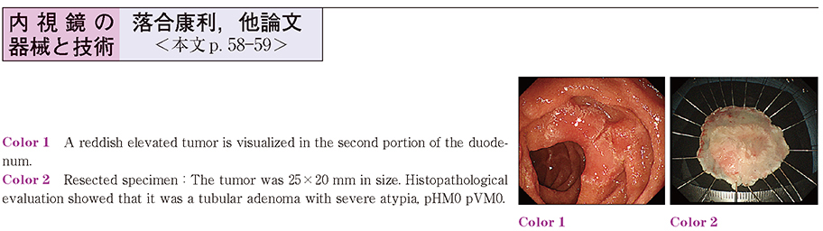
2014 年 85 巻 1 号 p. 58-59
発行日: 2014/12/06
公開日: 2014/12/17
 PDF形式でダウンロード (870K)
 -
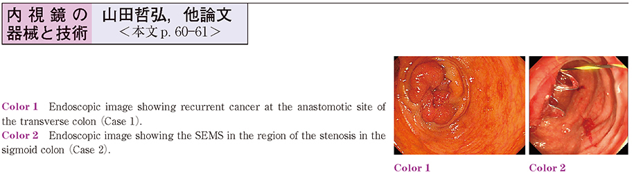
2014 年 85 巻 1 号 p. 60-61
発行日: 2014/12/06
公開日: 2014/12/17
 PDF形式でダウンロード (919K)
臨床研究
-
2014 年 85 巻 1 号 p. 62-63
発行日: 2014/12/06
公開日: 2014/12/17
 PDF形式でダウンロード (559K)
症例
-
2014 年 85 巻 1 号 p. 64-65
発行日: 2014/12/06
公開日: 2014/12/17
 PDF形式でダウンロード (487K)
 -
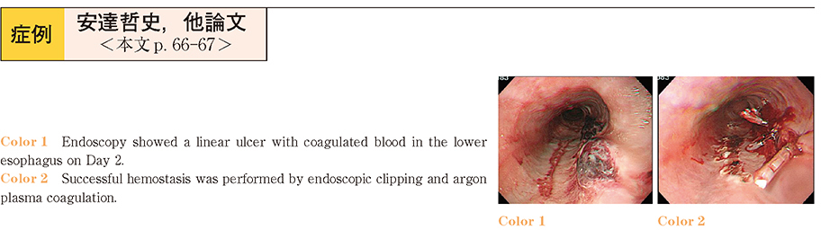
2014 年 85 巻 1 号 p. 66-67
発行日: 2014/12/06
公開日: 2014/12/17
 PDF形式でダウンロード (273K)
 -
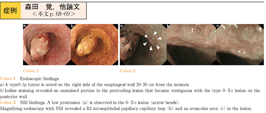
2014 年 85 巻 1 号 p. 68-69
発行日: 2014/12/06
公開日: 2014/12/17
 PDF形式でダウンロード (948K)
 -
2014 年 85 巻 1 号 p. 70-71
発行日: 2014/12/06
公開日: 2014/12/17
 PDF形式でダウンロード (633K)
 -
2014 年 85 巻 1 号 p. 72-73
発行日: 2014/12/06
公開日: 2014/12/17
 PDF形式でダウンロード (789K)
 -
2014 年 85 巻 1 号 p. 74-75
発行日: 2014/12/06
公開日: 2014/12/17
 PDF形式でダウンロード (273K)
 -
2014 年 85 巻 1 号 p. 76-77
発行日: 2014/12/06
公開日: 2014/12/17
 PDF形式でダウンロード (223K)
 -
2014 年 85 巻 1 号 p. 78-79
発行日: 2014/12/06
公開日: 2014/12/17
 PDF形式でダウンロード (894K)
 -
2014 年 85 巻 1 号 p. 80-81
発行日: 2014/12/06
公開日: 2014/12/17
 PDF形式でダウンロード (623K)
 -
2014 年 85 巻 1 号 p. 82-83
発行日: 2014/12/06
公開日: 2014/12/17
 PDF形式でダウンロード (252K)
 -
2014 年 85 巻 1 号 p. 84-85
発行日: 2014/12/06
公開日: 2014/12/17
 PDF形式でダウンロード (532K)
 -
2014 年 85 巻 1 号 p. 86-87
発行日: 2014/12/06
公開日: 2014/12/17
 PDF形式でダウンロード (747K)
 -
2014 年 85 巻 1 号 p. 88-89
発行日: 2014/12/06
公開日: 2014/12/17
 PDF形式でダウンロード (407K)
 -
2014 年 85 巻 1 号 p. 90-91
発行日: 2014/12/06
公開日: 2014/12/17
 PDF形式でダウンロード (265K)
 -
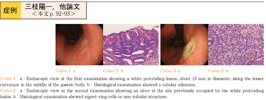
2014 年 85 巻 1 号 p. 92-93
発行日: 2014/12/06
公開日: 2014/12/17
 PDF形式でダウンロード (436K)
 -
2014 年 85 巻 1 号 p. 94-95
発行日: 2014/12/06
公開日: 2014/12/17
 PDF形式でダウンロード (413K)
 -
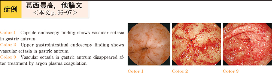
2014 年 85 巻 1 号 p. 96-97
発行日: 2014/12/06
公開日: 2014/12/17
 PDF形式でダウンロード (210K)
 -
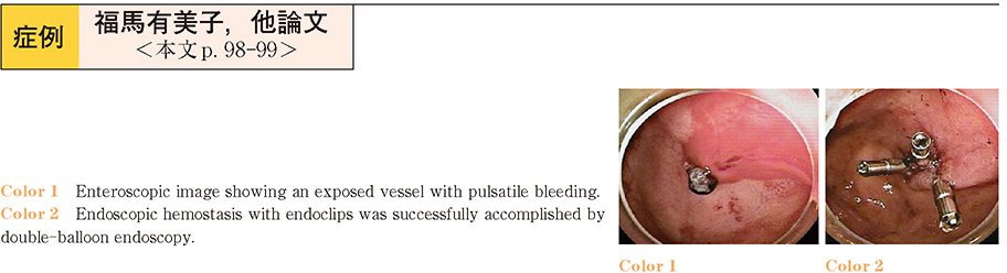
2014 年 85 巻 1 号 p. 98-99
発行日: 2014/12/06
公開日: 2014/12/17
 PDF形式でダウンロード (217K)
 -
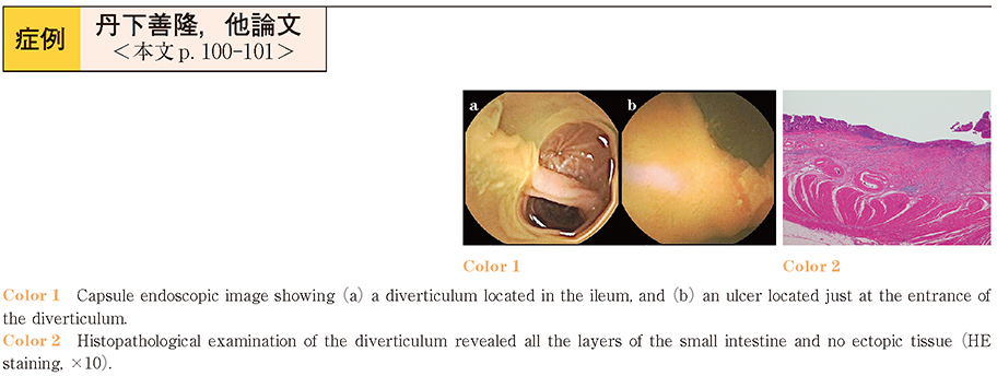
2014 年 85 巻 1 号 p. 100-101
発行日: 2014/12/06
公開日: 2014/12/17
 PDF形式でダウンロード (332K)
 -
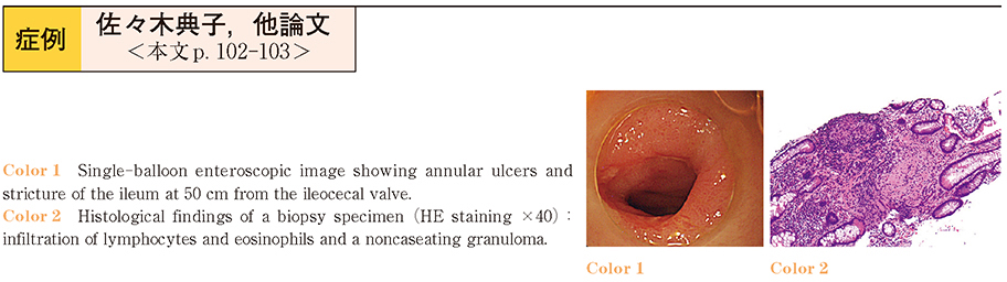
2014 年 85 巻 1 号 p. 102-103
発行日: 2014/12/06
公開日: 2014/12/17
 PDF形式でダウンロード (463K)
 -
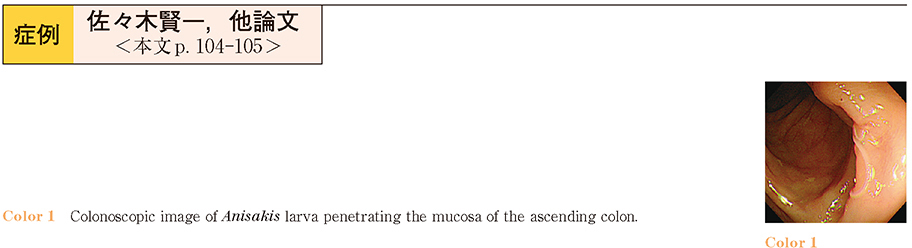
2014 年 85 巻 1 号 p. 104-105
発行日: 2014/12/06
公開日: 2014/12/17
 PDF形式でダウンロード (338K)
 -
2014 年 85 巻 1 号 p. 106-107
発行日: 2014/12/06
公開日: 2014/12/17
 PDF形式でダウンロード (358K)
 -
2014 年 85 巻 1 号 p. 108-109
発行日: 2014/12/06
公開日: 2014/12/17
 PDF形式でダウンロード (657K)
 -
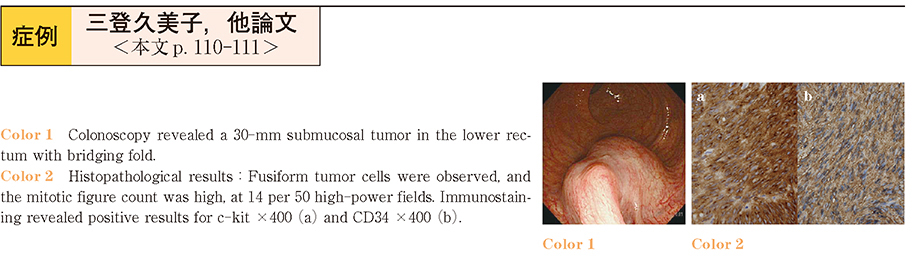
2014 年 85 巻 1 号 p. 110-111
発行日: 2014/12/06
公開日: 2014/12/17
 PDF形式でダウンロード (408K)
 -
2014 年 85 巻 1 号 p. 112-113
発行日: 2014/12/06
公開日: 2014/12/17
 PDF形式でダウンロード (564K)
 -
2014 年 85 巻 1 号 p. 114-115
発行日: 2014/12/06
公開日: 2014/12/17
PDF形式でダウンロード (493K) -
2014 年 85 巻 1 号 p. 116-117
発行日: 2014/12/06
公開日: 2014/12/17
 PDF形式でダウンロード (708K)
 -
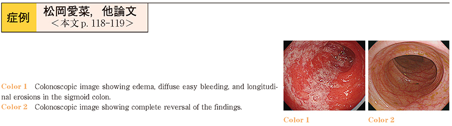
2014 年 85 巻 1 号 p. 118-119
発行日: 2014/12/06
公開日: 2014/12/17
 PDF形式でダウンロード (284K)
 -
2014 年 85 巻 1 号 p. 120-121
発行日: 2014/12/06
公開日: 2014/12/17
 PDF形式でダウンロード (552K)
 -
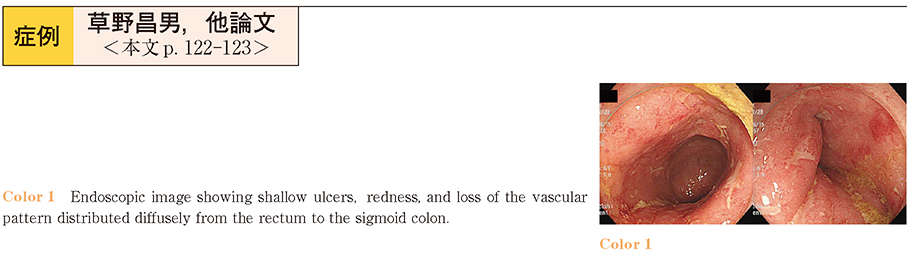
2014 年 85 巻 1 号 p. 122-123
発行日: 2014/12/06
公開日: 2014/12/17
 PDF形式でダウンロード (506K)
 -
2014 年 85 巻 1 号 p. 124-125
発行日: 2014/12/06
公開日: 2014/12/17
 PDF形式でダウンロード (773K)
 -
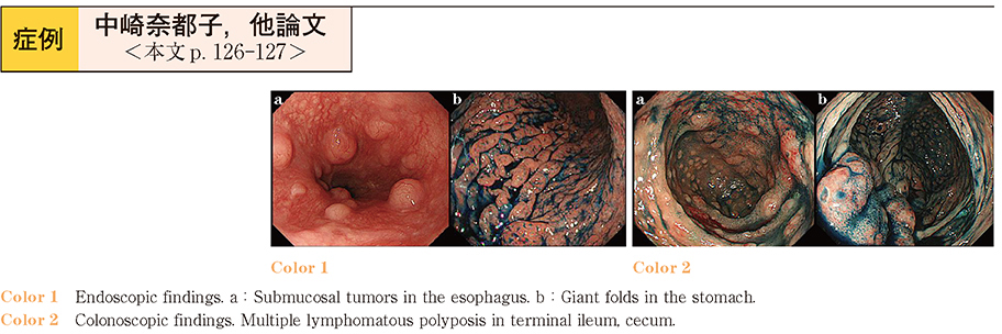
2014 年 85 巻 1 号 p. 126-127
発行日: 2014/12/06
公開日: 2014/12/17
 PDF形式でダウンロード (781K)
 -
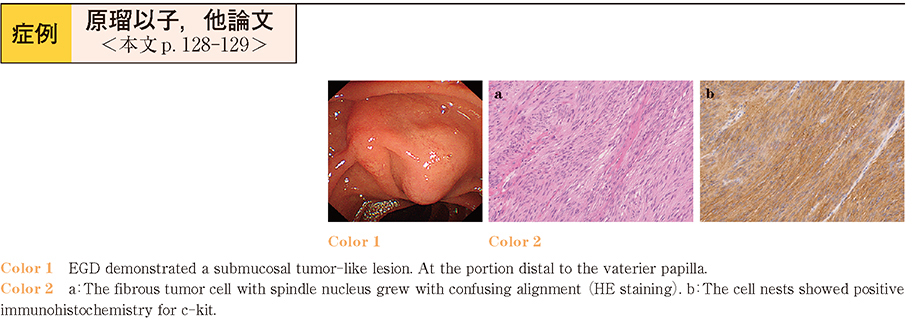
2014 年 85 巻 1 号 p. 128-129
発行日: 2014/12/06
公開日: 2014/12/17
 PDF形式でダウンロード (486K)
 -
2014 年 85 巻 1 号 p. 130-131
発行日: 2014/12/06
公開日: 2014/12/17
 PDF形式でダウンロード (584K)
 -
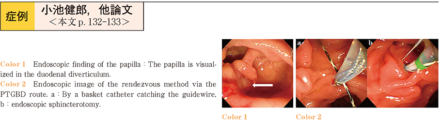
2014 年 85 巻 1 号 p. 132-133
発行日: 2014/12/06
公開日: 2014/12/17
 PDF形式でダウンロード (408K)
 -
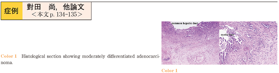
2014 年 85 巻 1 号 p. 134-135
発行日: 2014/12/06
公開日: 2014/12/17
 PDF形式でダウンロード (793K)
 -
2014 年 85 巻 1 号 p. 136-137
発行日: 2014/12/06
公開日: 2014/12/17
 PDF形式でダウンロード (540K)
 -
2014 年 85 巻 1 号 p. 138-139
発行日: 2014/12/06
公開日: 2014/12/17
PDF形式でダウンロード (429K) -
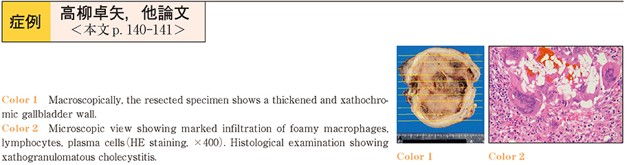
2014 年 85 巻 1 号 p. 140-141
発行日: 2014/12/06
公開日: 2014/12/17
 PDF形式でダウンロード (420K)
 -
2014 年 85 巻 1 号 p. 142-143
発行日: 2014/12/06
公開日: 2014/12/17
 PDF形式でダウンロード (467K)
- |<
- <
- 1
- >
- >|