
86 巻, 1 号
選択された号の論文の76件中1~50を表示しています
掲載論文カラー写真集
-
2015 年86 巻1 号 p. 1-16
発行日: 2015年
公開日: 2015/06/23
PDF形式でダウンロード (16805K)
内視鏡の器械と技術
-
2015 年86 巻1 号 p. 40-43
発行日: 2015/06/18
公開日: 2015/06/23
 PDF形式でダウンロード (2024K)
臨床研究
-
2015 年86 巻1 号 p. 44-48
発行日: 2015/06/18
公開日: 2015/06/23
 PDF形式でダウンロード (1294K)
 -
2015 年86 巻1 号 p. 49-52
発行日: 2015/06/18
公開日: 2015/06/23
PDF形式でダウンロード (694K) -
2015 年86 巻1 号 p. 53-57
発行日: 2015/06/18
公開日: 2015/06/23
PDF形式でダウンロード (676K) -
2015 年86 巻1 号 p. 58-62
発行日: 2015/06/18
公開日: 2015/06/23
PDF形式でダウンロード (777K) -
2015 年86 巻1 号 p. 63-65
発行日: 2015/06/18
公開日: 2015/06/23
PDF形式でダウンロード (786K) -
2015 年86 巻1 号 p. 66-69
発行日: 2015/06/18
公開日: 2015/06/23
PDF形式でダウンロード (607K) -
2015 年86 巻1 号 p. 70-73
発行日: 2015/06/18
公開日: 2015/06/23
PDF形式でダウンロード (1191K) -
2015 年86 巻1 号 p. 74-78
発行日: 2015/06/18
公開日: 2015/06/23
PDF形式でダウンロード (973K) -
2015 年86 巻1 号 p. 79-82
発行日: 2015/06/18
公開日: 2015/06/23
PDF形式でダウンロード (1016K) -
2015 年86 巻1 号 p. 83-86
発行日: 2015/06/18
公開日: 2015/06/23
 PDF形式でダウンロード (661K)
 -
2015 年86 巻1 号 p. 87-89
発行日: 2015/06/18
公開日: 2015/06/23
PDF形式でダウンロード (687K) -
2015 年86 巻1 号 p. 90-93
発行日: 2015/06/18
公開日: 2015/06/23
PDF形式でダウンロード (643K) -
2015 年86 巻1 号 p. 94-98
発行日: 2015/06/18
公開日: 2015/06/23
PDF形式でダウンロード (991K) -
2015 年86 巻1 号 p. 99-103
発行日: 2015/06/18
公開日: 2015/06/23
PDF形式でダウンロード (837K) -
2015 年86 巻1 号 p. 104-107
発行日: 2015/06/18
公開日: 2015/06/23
PDF形式でダウンロード (639K)
症例
-
2015 年86 巻1 号 p. 108-112
発行日: 2015/06/18
公開日: 2015/06/23
PDF形式でダウンロード (1726K) -
2015 年86 巻1 号 p. 114-115
発行日: 2015/06/18
公開日: 2015/06/23
 PDF形式でダウンロード (580K)
 -
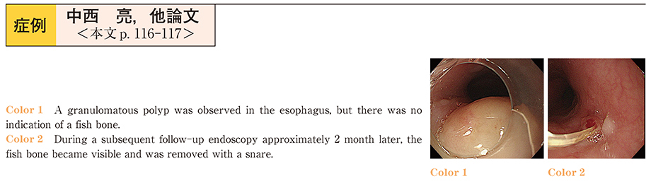
2015 年86 巻1 号 p. 116-117
発行日: 2015/06/18
公開日: 2015/06/23
 PDF形式でダウンロード (1004K)
 -
2015 年86 巻1 号 p. 118-119
発行日: 2015/06/18
公開日: 2015/06/23
 PDF形式でダウンロード (798K)
 -
2015 年86 巻1 号 p. 120-121
発行日: 2015/06/18
公開日: 2015/06/23
 PDF形式でダウンロード (582K)
 -
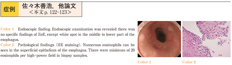
2015 年86 巻1 号 p. 122-123
発行日: 2015/06/18
公開日: 2015/06/23
 PDF形式でダウンロード (866K)
 -
2015 年86 巻1 号 p. 124-125
発行日: 2015/06/18
公開日: 2015/06/23
 PDF形式でダウンロード (1133K)
 -
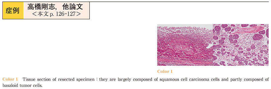
2015 年86 巻1 号 p. 126-127
発行日: 2015/06/18
公開日: 2015/06/23
 PDF形式でダウンロード (970K)
 -
2015 年86 巻1 号 p. 128-129
発行日: 2015/06/18
公開日: 2015/06/23
 PDF形式でダウンロード (625K)
 -
2015 年86 巻1 号 p. 130-131
発行日: 2015/06/18
公開日: 2015/06/23
 PDF形式でダウンロード (591K)
 -
2015 年86 巻1 号 p. 132-133
発行日: 2015/06/18
公開日: 2015/06/23
 PDF形式でダウンロード (1170K)
 -
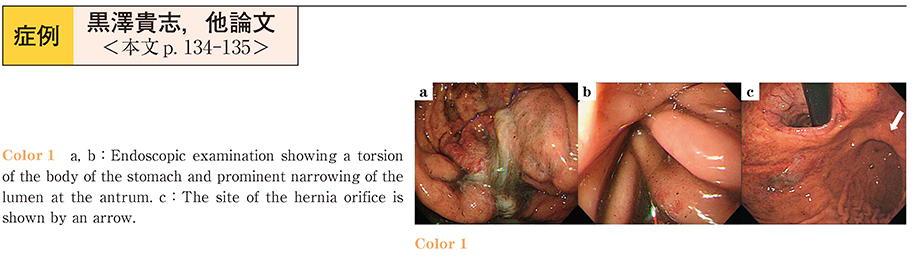
2015 年86 巻1 号 p. 134-135
発行日: 2015/06/18
公開日: 2015/06/23
 PDF形式でダウンロード (882K)
 -
2015 年86 巻1 号 p. 136-137
発行日: 2015/06/18
公開日: 2015/06/23
 PDF形式でダウンロード (749K)
 -
2015 年86 巻1 号 p. 138-139
発行日: 2015/06/18
公開日: 2015/06/23
 PDF形式でダウンロード (644K)
 -
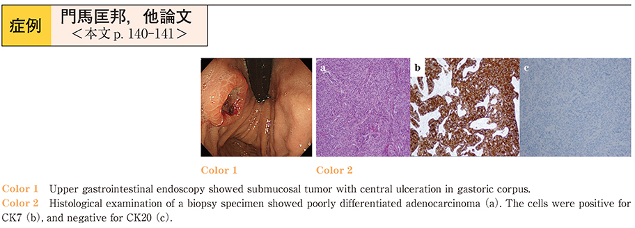
2015 年86 巻1 号 p. 140-141
発行日: 2015/06/18
公開日: 2015/06/23
 PDF形式でダウンロード (935K)
 -
2015 年86 巻1 号 p. 142-143
発行日: 2015/06/18
公開日: 2015/06/23
 PDF形式でダウンロード (1485K)
 -
2015 年86 巻1 号 p. 144-145
発行日: 2015/06/18
公開日: 2015/06/23
 PDF形式でダウンロード (792K)
 -
2015 年86 巻1 号 p. 146-147
発行日: 2015/06/18
公開日: 2015/06/23
 PDF形式でダウンロード (1180K)
 -
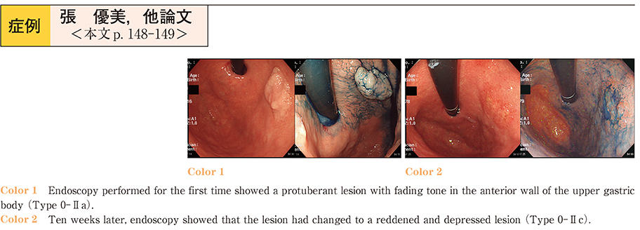
2015 年86 巻1 号 p. 148-149
発行日: 2015/06/18
公開日: 2015/06/23
 PDF形式でダウンロード (1655K)
 -
2015 年86 巻1 号 p. 150-151
発行日: 2015/06/18
公開日: 2015/06/23
 PDF形式でダウンロード (989K)
 -
2015 年86 巻1 号 p. 152-153
発行日: 2015/06/18
公開日: 2015/06/23
 PDF形式でダウンロード (771K)
 -
2015 年86 巻1 号 p. 154-155
発行日: 2015/06/18
公開日: 2015/06/23
 PDF形式でダウンロード (706K)
 -
2015 年86 巻1 号 p. 156-157
発行日: 2015/06/18
公開日: 2015/06/23
 PDF形式でダウンロード (1002K)
 -
2015 年86 巻1 号 p. 158-159
発行日: 2015/06/18
公開日: 2015/06/23
 PDF形式でダウンロード (1143K)
 -
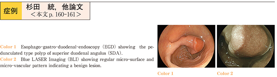
2015 年86 巻1 号 p. 160-161
発行日: 2015/06/18
公開日: 2015/06/23
 PDF形式でダウンロード (1169K)
 -
2015 年86 巻1 号 p. 162-163
発行日: 2015/06/18
公開日: 2015/06/23
 PDF形式でダウンロード (852K)
 -
2015 年86 巻1 号 p. 164-165
発行日: 2015/06/18
公開日: 2015/06/23
 PDF形式でダウンロード (896K)
 -
2015 年86 巻1 号 p. 166-167
発行日: 2015/06/18
公開日: 2015/06/23
 PDF形式でダウンロード (994K)
 -
2015 年86 巻1 号 p. 168-169
発行日: 2015/06/18
公開日: 2015/06/23
 PDF形式でダウンロード (974K)
 -
2015 年86 巻1 号 p. 170-171
発行日: 2015/06/18
公開日: 2015/06/23
 PDF形式でダウンロード (846K)
 -
2015 年86 巻1 号 p. 172-173
発行日: 2015/06/18
公開日: 2015/06/23
 PDF形式でダウンロード (934K)
 -
2015 年86 巻1 号 p. 174-175
発行日: 2015/06/18
公開日: 2015/06/23
 PDF形式でダウンロード (750K)
 -
2015 年86 巻1 号 p. 176-177
発行日: 2015/06/18
公開日: 2015/06/23
 PDF形式でダウンロード (805K)