Spine Surgery and Related Research
Online ISSN : 2432-261X
ISSN-L : 2432-261X
Volume 2, Issue 1
Displaying 1-15 of 15 articles from this issue
REVIEW ARTICLE
  • Steven Presciutti, Scott Boden
    2018 Volume 2 Issue 1 Pages 1-10
    Published: January 20, 2018
    Released on J-STAGE: January 27, 2018
    JOURNAL OPEN ACCESS

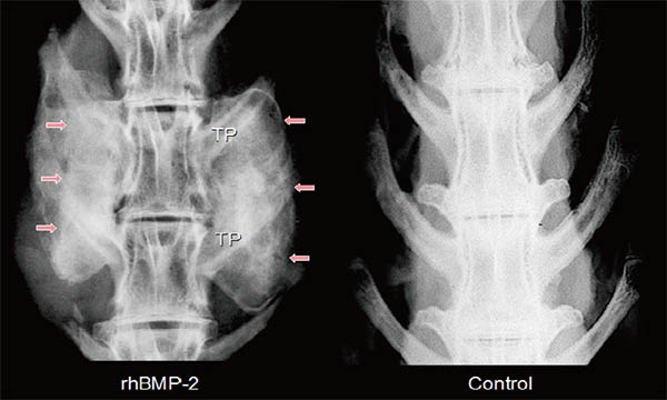
    A high rate of symptomatic spinal pseudoarthrosis and a wide range of complications associated with the use of iliac crest bone graft (the gold standard) have prompted the spine surgery community to seek alternative options to promote spinal fusion. Emory University has been one of the global leaders in this endeavor. This invited review covers the last 25 years of Emory's contributions to translational spine research, focusing specifically on our work with bone morphogenetic proteins (BMP) and the BMP signaling pathway. As a result of this work, recombinant human BMP-2 is the only Food and Drug Administration approved biologic bone graft substitute. It has been shown to significantly increase spinal fusion rates across the spinal column because of its potent ability to stimulate local bone formation through the recruitment of mesenchymal stem cells. This review covers our development of animal models of spinal fusion, our body of work regarding the translation of BMP from the benchtop to the clinic, the discovery of LMP-1 and strategies to enhance cellular responsiveness to BMPs, and the design of various small molecule drugs that can enhance local bone formation.

    Download PDF (865K)
  • Seiji Ohtori, Masayuki Miyagi, Gen Inoue
    2018 Volume 2 Issue 1 Pages 11-17
    Published: January 20, 2018
    Released on J-STAGE: January 27, 2018
    JOURNAL OPEN ACCESS

    Introduction: Many patients suffer from discogenic low back pain. However, the mechanisms, diagnosistic strategy, and treatment of discogenic low back pain all remain controversial. The purpose of this paper was to review the pathological mechanisms of discogenic low back pain.

    Methods: Many authors have investigated the pathological mechanisms of discogenic low back pain using animal models and examining human patients. Central to most investigations is understanding the innervation and instabilities of diseased intervertebral discs and the role of inflammatory mediators. We discuss three pathological mechanisms of discogenic low back pain: innervation, inflammation, and mechanical hypermobility of the intervertebral disc.

    Results: Sensory nerve fibers include C-fibers and A delta-fibers, which relay pain signals from the innervated outer layers of the intervertebral disc under normal conditions. However, ingrowth of these sensory nerve fibers into the inner layers of intervertebral disc occurs under disease conditions. Levels of neurotrophic factors and some cytokines are significantly higher in diseased discs than in normal discs. Stablization of the segmental hypermobility, which can be induced by intervertebral disc degeneration, suppresses inflammation and prevents sensitization of sensory nerve fibers innervating the disc.

    Conclusions: Pathological mechanisms of discogenic low back pain include sensory nerve ingrowth into inner layers of the intervertebral disc, upregulation of neurotrophic factors and cytokines, and instability. Inhibition of these mechanisms is important in the treatment of discogenic low back pain.

    Download PDF (246K)
  • Hiroshi Takahashi, Yasuaki Iida, Yuichirou Yokoyama, Keiji Hasegawa, S ...
    2018 Volume 2 Issue 1 Pages 18-22
    Published: January 20, 2018
    Released on J-STAGE: January 27, 2018
    JOURNAL OPEN ACCESS

    Local application of vancomycin has recently become widely used in spine surgery. However, local application is not included in the indication and has not been approved by the US Food and Drug Administration (FDA). Thus, we searched for reports with "intra wound-vancomycin" and "SSI" as keywords in the MEDLINE database, and investigated the efficacy, problems with use, and future prospects based on these reports. Intrawound vancomycin was described as effective in most of the reports, but was found to have no effect or to aggravate the condition in some reports. A toxic effect on osteoblasts due to a high local concentration was described in some reports, whereas local application was found to be safe in other studies. The amount of vancomycin used and the administration method varied among the reports. Overall, the results suggest that intrawound vancomycin is clinically effective, but this has yet to be established in a randomized controlled trial. There is a need to identify cases that should be selected for this treatment and to investigate the dose and optimum concentration of vancomycin for clinical use.

    Download PDF (68K)
  • Kazuyoshi Nakanishi, Nobuhiro Tanaka, Naosuke Kamei, Shinji Kotaka, Mi ...
    2018 Volume 2 Issue 1 Pages 23-29
    Published: January 20, 2018
    Released on J-STAGE: January 27, 2018
    JOURNAL OPEN ACCESS

    Introduction: The clinical entity of cervical spondylotic amyotrophy (CSA) is characterized by severe muscle atrophy in the upper extremities with insignificant sensory deficits in patients with cervical spondylosis. However, the pathogenesis of CSA is still unclear.

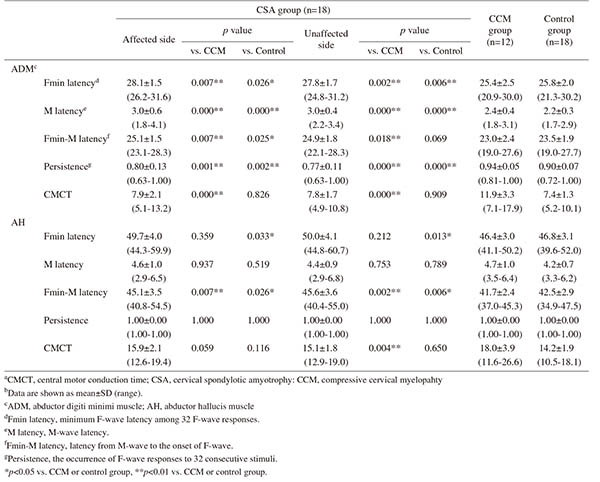
    Methods: We assessed electrophysiological motor conduction through the corticospinal tract and ulnar and tibial nerves, which do not supply the deltoid or biceps muscles, of 18 patients with CSA, 12 patients with compressive cervical myelopathy, and 18 control subjects with cervical spondylotic radiculopathy. Motor evoked potentials following transcranial magnetic stimulation and M-waves and F-waves following electrical stimulation were measured from the bilateral abductor digiti minimi muscles (ADMs) and abductor hallucis muscles (AHs). The peripheral conduction time (PCT) was calculated from the latencies of the CMAPs and F-waves as follows: (latency of CMAPs + latency of F-waves - 1) / 2. The central motor conduction time (CMCT) was calculated by subtracting the PCT from the onset latency of the MEPs.

    Results: The M-wave (M) latency and minimum F-wave (Fmin) latency from the ADM, and Fmin-M latency from the ADM/AH were significantly longer in the CSA group than in the other groups, on both the affected (p = 0.000-0.007) and unaffected sides (p = 0.000-0.033). F-wave persistence from the bilateral ADMs was significantly lower in the CSA group than in the other groups (p = 0.000-0.002). Among the CSA patients, there were no significant differences in these parameters between the affected and unaffected sides. The CMCT showed no significant differences between the CSA and control groups, but significant differences between the CSA and CCM groups (p = 0.000-0.004).

    Conclusions: CSA patients with unilateral deltoid muscle atrophy had subclinical impairments of lower motor neurons and/or peripheral axons in the ulnar nerve, and subclinical impairments of peripheral axons in the tibial nerve. These motor impairments may have originally existed in these individuals before the onset of CSA.

    Download PDF (234K)
  • Ryoma Aoyama, Tateru Shiraishi, Masahiro Kato, Junichi Yamane, Ken Nin ...
    2018 Volume 2 Issue 1 Pages 30-36
    Published: January 20, 2018
    Released on J-STAGE: January 27, 2018
    JOURNAL OPEN ACCESS

    Introduction: The characteristics of cervical spondylolisthesis are not currently fully understood, because of the shortage of reports covering the large population of patients with cervical spondylolisthesis. The purpose of this study was to elucidate the characteristics of cervical spondylolisthesis by examining a relatively large number of cases.

    Methods: We analyzed 101 cases with more than 2 mm of vertebral listhesis as determined from X-ray or computed tomography (CT) images among 731 patients who underwent surgery at a single institute. We considered the C2-7 angle, range of motion, and C2-7 sagittal vertical axis on lateral X-ray images. From sagittal CT images, classifications into five grades based on the slipped disc and adjacent caudal levels were made. We examined the orientation of facet joints at the slipped level using axial CT images.

    Results: Spondylolisthesis was recognized in 101 cases at 124 levels. Anterior and posterior spondylolisthesis were detected in 68 and 40 cases, respectively. Anterior spondylolisthesis developed predominantly at C3 or C4, usually at the level adjacent to the narrowed disc, or at C7, adjacent to the stiffened thoracic spine. The disc height was relatively preserved at the anterior slipped level. Posterior spondylolisthesis developed predominantly at the level of the significantly narrowed disc associated with advanced intervertebral osteoarthritis. At the segment with listhesis in the lower cervical spine, the direction of the facet joint in the axial plane tended to be posteromedial.

    Conclusions: Cervical degenerative spondylolisthesis was classified into two types. The first and more common listhesis occurred adjacent to stiffened levels, and anterior slippage was common in this type. The second and less common listhesis occurred within progressively degenerated segments, and posterior slippage was prominent. We have uniquely described the morphological changes in orientation of the cervical facet joints at the slipped level in the transverse plane.

    Download PDF (350K)
  • Ryoji Tauchi, Yoshitaka Suzuki, Taichi Tsuji, Tetsuya Ohara, Toshiki S ...
    2018 Volume 2 Issue 1 Pages 37-41
    Published: January 20, 2018
    Released on J-STAGE: January 27, 2018
    JOURNAL OPEN ACCESS

    Introduction: The purpose of this study is to demonstrate the clinical characteristics and thoracic factors such as sternal tilt angle and Haller index in patients with idiopathic or syndromic scoliosis associated with pectus excavatum.

    Methods: We performed a retrospective review on a cohort of 70 patients (37 males and 33 females) diagnosed with idiopathic and syndromic scoliosis associated with pectus excavatum between 1985 and 2014. We investigated age, location and Cobb angle of the main curve, and thoracic factors including sternal deviation and tilting angle and Haller index using radiographs and computed tomography of the chest.

    Results: Patients' mean age at the first visit to our hospital was 10.3 years (1-18 years old). There were 41 patients with idiopathic scoliosis and 29 with syndromic scoliosis. Main curve locations were thoracic in 52 patients, thoracolumbar in 10, and lumbar in 8. The mean Cobb angle of the main curve was 45.0 degrees (11-109 degrees). The sternum was displaced on the left side in 72% of patients, central in 23%, and right in 5%. Mean sternal tilt angle was 12.4 degrees (2.3-34 degrees), and mean Haller index score was 4.9 (2.9-9.2). There was no significant correlation between Cobb angle and sternal tilt angle/Haller index. However, a significant difference was found between sternal tilt angle and Haller index.

    Conclusion: Most patients with both scoliosis and pectus excavatum have left side deviated sternum and a higher Haller index score; therefore this can negatively impact cardiac function. Prone positioning and the corrective force applied during scoliosis surgery as well as thoracic compression during cast or brace treatment may have a negative effect on cardiac function in these patients.

    Download PDF (250K)
  • Jun Sato, Kazuhide Inage, Masayuki Miyagi, Yoshihiro Sakuma, Kazuyo Ya ...
    2018 Volume 2 Issue 1 Pages 42-47
    Published: January 20, 2018
    Released on J-STAGE: January 27, 2018
    JOURNAL OPEN ACCESS

    Introduction: Discogenic back pain remains poorly understood with respect to etiopathogenesis, despite being a considerable burden. We sought to examine the expression of vascular endothelial growth factor in injured intervertebral discs in rat caudal vertebrae.

    Methods: Forty-eight male Sprague Dawley rats were assigned to 2 groups according to disc puncture injury: puncture (n = 32) or non-puncture (n = 16). Disc puncture was performed percutaneously such that the incision would be in the primary plane of motion for the coccygeal discs 5-6, 6-7, and 7-8. A 26-gauge needle was used to puncture each disc 10 times. Punctured discs were examined histologically by hematoxylin and eosin staining at 1, 7, 14, and 28 days post-injury.

    Results: Vascular endothelial growth factor was localized immunohistochemically, and determined quantitatively using an enzyme-linked immunosorbent assay. Peak inflammation occurred on the 7th day post-injury, but tissue degeneration continued until day 28. Local expression of vascular endothelial growth factor tended to be highest in the annulus fibrosus on the 7th and 14th days after puncture injury. The level of vascular endothelial growth factor was highest 1-day post-injury, and then gradually decreased thereafter. Furthermore, vascular endothelial growth factor levels in the puncture group were significantly higher than those in the non-puncture control group (p < 0.05).

    Conclusions: We found increased expression of the inflammatory cytokine vascular endothelial growth factor in injured intervertebral discs, suggesting that vascular endothelial growth factor may be clinically important in discogenic back pain.

    Download PDF (1094K)
  • Wataru Saito, Gen Inoue, Takayuki Imura, Toshiyuki Nakazawa, Masayuki ...
    2018 Volume 2 Issue 1 Pages 48-52
    Published: January 20, 2018
    Released on J-STAGE: January 27, 2018
    JOURNAL OPEN ACCESS

    Introduction: Echocardiography is an important component of perioperative cardiac risk stratification in patients with neuromuscular scoliosis (NMS). However, there are little data regarding the relationship between preoperative echocardiographic findings and spinal deformity. We retrospectively reviewed preoperative echocardiographic data to investigate the relationship between echocardiographic evaluation and spinal deformity in NMS.

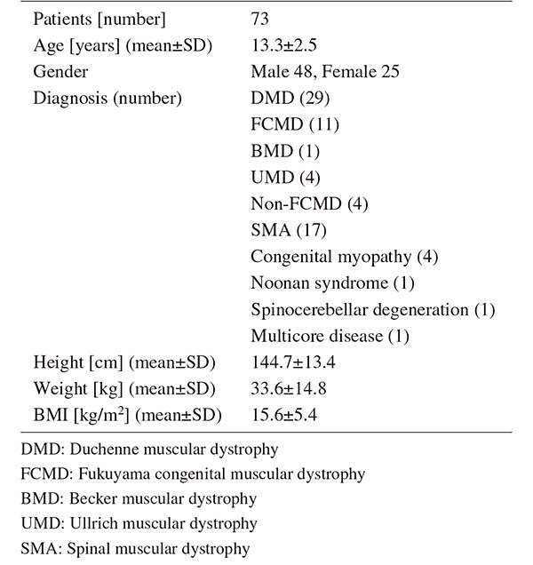
    Methods: We reviewed 73 NMS patients (mean age: 13.3 years, male 66%) who underwent spinal correction surgery between 2008 and 2016. Echocardiographic data including ejection fraction (EF), valvar disease, and inferior vena cava diameter were collected from the preoperative exam. Demographic and radiographic data were also collected.

    Results: Preoperative diagnoses included Duchenne muscular dystrophy, Fukuyama congenital muscular dystrophy, other dystrophy, spinal muscular atrophy, and congenital myopathies. Mean Body Mass Index (BMI) was 15.6 kg/m2. Mean major Cobb angle before surgery was 86.6 ± 28.2°. Because of technical difficulty, complete echocardiographic data could only be collected and evaluated in 49.3% of patients. Neither right nor left sided cardiac evaluation could be completed in 20.5%. Patients in whom complete echocardiographic data could not be collected had significantly more extensive thoracic scoliosis with a more rigid curve and hypokyphosis, and were of lower weight and BMI than patients in whom complete data could be collected. Ten cases (13.7%) were diagnosed as having minor heart-related complications immediately after surgery, and they had higher right atrial pressures preoperatively.

    Conclusions: Echocardiography can be technically difficult in NMS patients with extensive spinal deformities. We found that perioperative cardiac function could only be evaluated by echocardiogram in about half of NMS patients undergoing spinal correction surgery. The absence of an adequate preoperative cardiac evaluation could render these patients more susceptible to perioperative heart-related complications. Echocardiography may not be sufficient to evaluate cardiac conditions in children with extensive NMS.

    Download PDF (75K)
  • Shigeto Ebata, Tetsuro Ohba, Hirotaka Haro
    2018 Volume 2 Issue 1 Pages 53-59
    Published: January 20, 2018
    Released on J-STAGE: January 27, 2018
    JOURNAL OPEN ACCESS

    Introduction: Through an extreme lateral retroperitoneal and transpsoas approach to intervertebral disc and fusion surgery, a large lordosis cage can be placed for solid and stable intervertebral fusion and to provide strong anterior support, disc height restoration, favorable alignment, and indirect nerve decompression. However, appropriate placement of the interbody cage remains insufficiently researched. We sought to determine both appropriate cage placement as well as other factors affecting nerve decompression in extreme lateral interbody fusion (XLIF) surgery.

    Methods: We included 53 consecutive patients suffering from lumbar degenerative diseases with an indication for XLIF. Radiographic analysis using a sagittal computed tomography (CT) and axial magnetic resonance imaging (MRI) views was conducted to determine intervertebral disc height and angle, degree of disc bulging and thickness of the flavum, the area of the dural tube, cage height, pre- and postoperative disc bulging, change of disc bulging after surgery, cage subsidence, and cage placement at the rostral and caudal endplates.

    Results: Intervertebral disc height and angle were significantly increased at all levels (L2/3, 3/4, 4/5) (p < 0.05). The area of the dural tube was significantly increased (p < 0.05), whereas the degree of disc bulging and thickness of the flavum were significantly decreased at all disc levels (p < 0.05). The enlarged area of the dural tube showed significant correlation with increased disc height (p = 0.019), preoperative flavum thickness (p = 0.008), change of flavum thickness (p < 0.0001), and cage placement at the rostral endplate (p = 0.014).

    Conclusions: A decrease in flavum buckling is more important than disc protrusion as a consideration for obtaining indirect decompression. Central placement may be advantageous for indirect decompression.

    Download PDF (410K)
  • Atsushi Kojima, Atsushi Fujii, Shigeta Morioka, Yoshiaki Torii, Kenich ...
    2018 Volume 2 Issue 1 Pages 60-64
    Published: January 20, 2018
    Released on J-STAGE: January 27, 2018
    JOURNAL OPEN ACCESS

    Introduction: This study compared traditional manual methods and power tool use with regard to the speed and accuracy of percutaneous pedicle screw (PPS) placement and determined the advantages associated with the use of power tools.

    Although the indication of PPS placement in minimally invasive spine stabilization (MISt) procedures has been recently expanded, there are no reports on PPS insertion using a power tool.

    Methods: We evaluated 35 patients who underwent PPS insertion using a power tool during MISt procedures. On one side, PPS insertion was performed using the manual (M) method, whereas on the contralateral side, insertion was performed using the power tool (P) method. We assessed the number of implanted PPSs, time taken to implant PPSs after guidewire insertion, and accuracy of PPS placement as ranked postoperatively using computed tomography images.

    Results: A total of 294 PPSs were inserted (147 using the M method and 147 using the P method). The mean PPS insertion time was 10.5 s using the P method and 27.4 s using the M method. The time required for inserting a screw using the P method remained consistent in the range of 10-15 s, whereas the time using the M method tended to increase from the second screw onward, with a range of 25-30 s. With regard to PPS insertion accuracy, a 2 mm or more pedicle breach was noted in 2 (1.4%) case after the P method and in 2 (1.4%) case after the M method.

    Conclusions: PPS placement using power tools has the potential to save the surgical time during MISt procedures.

    Download PDF (212K)
  • Ryota Takatori, Taku Ogura, Wataru Narita, Tatsuro Hayashida, Kazuya T ...
    2018 Volume 2 Issue 1 Pages 65-71
    Published: January 20, 2018
    Released on J-STAGE: January 27, 2018
    JOURNAL OPEN ACCESS

    Introduction: Corrective surgery for adult degenerative scoliosis using lateral interbody fusion (LIF) and additional posterior fixation is an efficient procedure. However, it is unclear how this procedure affects rotational deformity correction. Therefore, the goal of the present study was to use three-dimensional (3D) images, taken during surgery, to investigate rotational deformity correction in the treatment of adult degenerative scoliosis using LIF and posterior fixation using a pedicle screw system.

    Methods: The subjects were 12 females who were treated using LIF and posterior fixation for adult degenerative scoliosis. The patients had a mean age of 72 (65-76) years. 3D images were acquired before surgery, after LIF, and after additional posterior fixation. Rotational angles of the upper vertebra with respect to the lower vertebra of each fixed segment were measured in 3 planes. Correction factors for rotational deformity were investigated after LIF and additional posterior fixation.

    Results: There were significant improvements in radiographical parameters for global spinal balance. The correction angles per segment were 4.7° for lateral bending, 6.9° for lordosis, and 4.5° for axial rotation. LIF was responsible for correction of four-fifths of lateral bending and axial rotation, and two-thirds of lordotic changes.

    Conclusions: Lateral bending, axial rotational deformities, and lordosis were primarily corrected by LIF. Further lordosis correction was achieved using additional posterior fixation. These results indicate that corrective surgery for adult degenerative scoliosis using these procedures is effective for rotational deformity correction and leads to an ideal global spinal alignment.

    Download PDF (329K)
  • Takashi Ohnishi, Akira Iwata, Masahiro Kanayama, Fumihiro Oha, Tomoyuk ...
    2018 Volume 2 Issue 1 Pages 72-76
    Published: January 20, 2018
    Released on J-STAGE: January 27, 2018
    JOURNAL OPEN ACCESS

    Introduction: Numerous studies have reported the risk factors of osteoporotic vertebral collapse. However, whether spino-pelvic and global spinal alignments are associated with the occurrence of osteoporotic vertebral collapse remains unclear. This study aimed to investigate the association between spino-pelvic and global spinal alignments and the occurrence of osteoporotic vertebral collapse.

    Methods: A total of 46 consecutive patients who underwent a nonoperative treatment for a single-level fresh osteoporotic thoracolumbar vertebral compression fracture (T10 to L3) were retrospectively reviewed. The parameters evaluated were the pelvic incidence, anterior deviation of the C7 plumb line, distance between the C7 plumb line and the center of the fractured vertebra, and kyphotic wedge angle of the fractured vertebra in a standing whole-spine radiograph at the beginning of the nonoperative treatment. As an outcome measure, the presence or absence of osteoporotic vertebral collapse was radiographically evaluated at the final follow-up. Multiple logistic regression analysis was used to determine significant risk factors of osteoporotic vertebral collapse.

    Results: The mean values for each parameter were as follows: pelvic incidence, 58.0 degrees; anterior deviation of the C7 plumb line, 3.0 cm; distance between the C7 plumb line and the center of the fractured vertebra, 5.7 cm; and kyphotic wedge angle of the fractured vertebra, 14.6 degrees. Multiple logistic regression analysis revealed that the distance between the C7 plumb line and center of the fractured vertebra was a significant risk factor of osteoporotic vertebral collapse (p = 0.012; odds ratio, 1.025). The anterior deviation of the C7 plumb line (p = 0.214), pelvic incidence (p = 0.728), and kyphotic wedge angle of the fractured vertebra (p = 0.07) did not affect the occurrence of osteoporotic vertebral collapse.

    Conclusions: A large distance between the C7 plumb line and center of the fractured vertebra was a significant risk factor of osteoporotic vertebral collapse. The distance approximately represents that of between gravity center of trunk cranial to the fractured vertebra and the fractured vertebra. Accordingly, the large distance may cause larger flexion moment to the fractured site, leading to stress concentration that results in insufficient bone healing.

    Download PDF (106K)
  • Teruaki Ono, Kiyoshi Tarukado, Osamu Tono, Katsumi Harimaya, Yuichiro ...
    2018 Volume 2 Issue 1 Pages 77-81
    Published: January 20, 2018
    Released on J-STAGE: January 27, 2018
    JOURNAL OPEN ACCESS

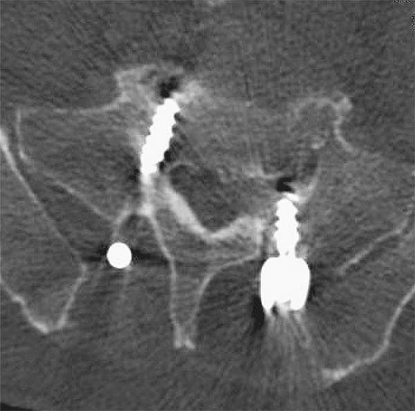
    Introduction: The clinical significance of lumbosacral transitional vertebrae (LSTV) has been reported. However, the association between LSTV and lumbosacral pedicle anatomical anomaly has not been investigated. We hypothesized that LSTV might be associated with lumbosacral anatomical anomaly. The purpose of this study was to examine the morphological association between LSTV and lumbosacral pedicle asymmetry (PA) using computed tomography (CT).

    Methods: A retrospective review of CT images of 347 lumbosacral degenerative disease patients was performed. We divided the subjects into two groups: the normal and LSTV groups. LSTV was classified based on Castellvi's classification. PA was defined as a difference of more than 20° between the right and left angles of the pedicle.

    Results: Seventy out of 347 lumbosacral degenerative disease patients (20.17%) were diagnosed with LSTV. In the normal group, only a 0.54% incidence of PA was seen; however, with respect to the LSTV group, a 9.29% incidence of PA was seen. A significant difference in PA incidence was observed between the groups (p < 0.001). Type IIIa and Type IV in the LSTV group showed a statistically significant PA incidence rate (p = 0.004 and p = 0.039, respectively).

    Conclusions: Our study demonstrated that there was a significant difference in the incidence of PA between LSTV subjects and normal subjects. Moreover, the incidence of PA was significantly higher in LSTV subjects with severe anomaly. These results suggested that lumbosacral spine anomaly might have a close relationship with the incidence of PA and lumbosacral nerve root asymmetry. Therefore, morphological evaluation of the pedicle is important for preoperative surgical management, especially in cases using pedicle screws. This information could lower the incidence of pedicle screw malposition when pedicle screws are inserted at the lumbosacral spine.

    Download PDF (637K)
  • Kenyu Ito, Shiro Imagama, Kei Ando, Kazuyoshi Kobayashi, Mikito Tsushi ...
    2018 Volume 2 Issue 1 Pages 82-85
    Published: January 20, 2018
    Released on J-STAGE: January 27, 2018
    JOURNAL OPEN ACCESS

    Introduction: An occipital-cervical surgery for children is challenging for surgeons because of the immature bone quality, extensive anatomical variability, and small osseous structures. Furthermore, occipital-C2 fusion in children results in great stress on the C2 screws. We report a technique that uses both C2 pedicle and bilateral lateral mass screws (C2 hybrid screws) in children with an upper cervical disorder to preserve motion segment and secure strength in those who require occipital-cervical fusion.

    Case Report: Case 1 was that of a 5-year-old girl with Down syndrome who had atlantoaxial dislocation and os odontoideum. Owing to the C1 hypoplasia, the posterior arch was fractured by the C1 lateral mass screw. Therefore, O-C2 fusion was performed. C2 bilateral lamina screws were added along with the C2 bilateral pedicle screws for reinforcement. Case 2 was that of an 8-year-old boy who presented with torticollis and neck pain. The patient was diagnosed as having atlantoaxial rotatory fixation. The right vertebral artery was obstructed, and the left vertebral artery was dominant. The C1 posterior arch was bifid and assimilated with the occipital bone. C2 bilateral lamina screws were added with the right C2 pedicle screw for reinforcement. Both cases attained bone union after O-C2 fusion surgery using hybrid screws.

    Conclusions: The use of C2 hybrid screws with both C2 pedicle and bilateral lateral mass screws can preserve mobile segments in the fusion area in young children who require occipital-cervical fixation.

    Download PDF (891K)
  • Sumihisa Orita, Takao Nakajima, Kenta Konno, Kazuhide Inage, Takeshi S ...
    2018 Volume 2 Issue 1 Pages 86-92
    Published: January 20, 2018
    Released on J-STAGE: January 27, 2018
    JOURNAL OPEN ACCESS

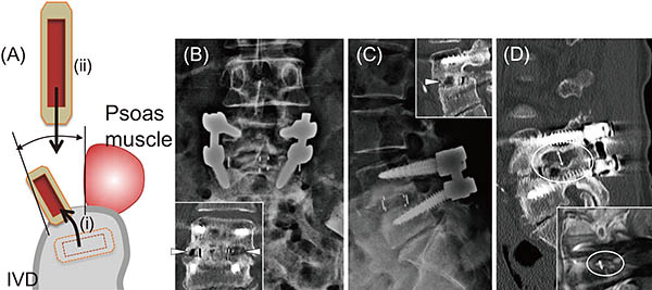
    Introduction: Failed spinal fusion surgery sometimes requires salvage surgery when symptomatic, especially with postsurgical decrease in intervertebral disc height followed by foraminal stenosis. For such cases, an anterior approach to lumbar lateral interbody fusion (LLIF) provides safe, direct access to the pathological disc space and a potential improvement in the fusion rate. One LLIF approach, oblique lateral interbody fusion (OLIF), targets the oblique lateral window of the intervertebral discs to achieve successful lateral interbody fusion. The current technical note describes spinal revision surgery using the OLIF procedure.

    Technical Note: The subjects were patients with leg pain and/or lower back pain derived from decreased intervertebral height followed by foraminal stenosis due to failed spinal fusion surgery. These patients underwent additional OLIF surgery and posterior fusion with no additional posterior direct decompression. Their outcomes were evaluated using the Japanese Orthopaedic Association (JOA) scores at baseline and final follow-up. Bony union was also evaluated using computed tomography images at final follow-up.

    Six subjects were evaluated, with two representative cases described in detail. Four patients had an adjacent segment disorder, and the other two patients had pseudarthrosis due to postoperative infection. The mean JOA score improved from 5.7 ± 5.4 to 21.2 ± 2.3, with a mean recovery rate of 65.0%. All cases showed intervertebral bony union.

    Conclusions: We introduced a salvage strategy for failed posterior spine fusion surgery cases using the OLIF procedure. Patients effectively achieved recovered intervertebral and foraminal height with no additional posterior direct decompression.

    Download PDF (1496K)
feedback
Top