Spine Surgery and Related Research
Online ISSN : 2432-261X
ISSN-L : 2432-261X
Volume 5, Issue 3
Displaying 1-17 of 17 articles from this issue
INSTRUCTIONAL LECTURE
  • Tetsuya Tamaki, Muneharu Ando, Yukihiro Nakagawa, Hiroshi Iwasaki, Shu ...
    2021 Volume 5 Issue 3 Pages 120-132
    Published: May 27, 2021
    Released on J-STAGE: May 27, 2021
    Advance online publication: March 10, 2021
    JOURNAL OPEN ACCESS

    An intraoperative functional spinal cord monitoring system is a technology used by spine and spinal cord surgeons to perform a safe surgery and to gain further surgical proficiency. However, no existing clinical neurophysiological method used in the operating room can monitor all complex spinal cord functions. Therefore, by observing the activities of certain neural action potentials transferred via limited neural tissues, surgeons need to deductively estimate the function of the whole spinal cord. Thus, as the number of spinal cord functions that need to be observed increases, spinal cord monitoring can be more reliable. However, in some situations, critical decision-making is affected by the limited capability of these methods. Nevertheless, good teamwork enables sharing of seamless information within the team composed of a surgeon, anesthesiologist, monitoring technician and nurses greatly contributes to making quick and accurate decisions. The surgeon, who is the person in charge of the team, should communicate with multidisciplinary team members using common technical terms. For this reason, spine and spinal cord surgeons must have appropriate knowledge of the methods currently used, especially of their utility and limitations. To date, at least six electrophysiological methods are available for clinical utilization: three are used to monitor sensory-related tracts, and three are used to monitor motor-related spinal cord functions. If surgeons perform electrode setting, utilizing their expertise, then the range of available methods is broadened, and more meticulous intraoperative functional spinal cord monitoring can be carried out. Furthermore, if the team members share information effectively by utilizing a clinically feasible judicious checklist or tools, then spinal cord monitoring will be more reliable.

    Download PDF (463K)
REVIEW ARTICLE
  • Kumiko Yotsuya, Tomohiko Hasegawa, Yu Yamato, Go Yoshida, Sho Kobayash ...
    2021 Volume 5 Issue 3 Pages 133-143
    Published: May 27, 2021
    Released on J-STAGE: May 27, 2021
    Advance online publication: September 23, 2020
    JOURNAL OPEN ACCESS

    Background: Since 1929, only 171 epidural hemangioma cases have been reported. We report five epidural hemangiomas and review cases reported over the past decade in terms of radiological features and clinical findings.

    Methods: Among patients operated on for spinal tumors at our hospital between 2009 and 2020, five had epidural hemangiomas. We retrospectively examined patient records and images and reviewed relevant English literature in PubMed from 2009 to 2019. Eighty-seven epidural hemangioma cases were reported in the last 10 years.

    Results: Among 87 cases, the average age was 49.58 years; 43 and 44 cases were male and female, respectively. The most common lesion level was thoracic (59.8%), while common symptoms were back pain (42.5%); numbness, hypoesthesia, or anesthesia (37.9%); paraparesis (34.5%); and radicular pain (20.7%). On magnetic resonance imaging (MRI), 77.1% showed hypo-isointensity on T1-weighted image (WI), hyperintensity on T2WI, and homogenous enhanced patterns with contrast. Total resection was performed in most cases, with good clinical outcomes. Preoperative embolization was performed in four cases, with good surgical outcomes. The median follow-up duration of postoperative MRI was 16 months, and no case had recurrence.

    Conclusions: Epidural hemangiomas are extremely rare and may be difficult to diagnose preoperatively. They should be considered if a dumbbell-shaped or epidural hypervascular lesion is noted. The treatment of choice for epidural hemangioma is total resection, with most cases having good clinical outcomes. Preoperative embolization may be useful for reducing bleeding. Although there were almost no recurrences after resection, careful follow-up for longer periods is required.

    Download PDF (3185K)
ORIGINAL ARTICLE
  • Motoya Kobayashi, Junichi Ohya, Yuki Onishi, Junichi Kunogi, Naohiro K ...
    2021 Volume 5 Issue 3 Pages 144-148
    Published: May 27, 2021
    Released on J-STAGE: May 27, 2021
    Advance online publication: November 20, 2020
    JOURNAL OPEN ACCESS

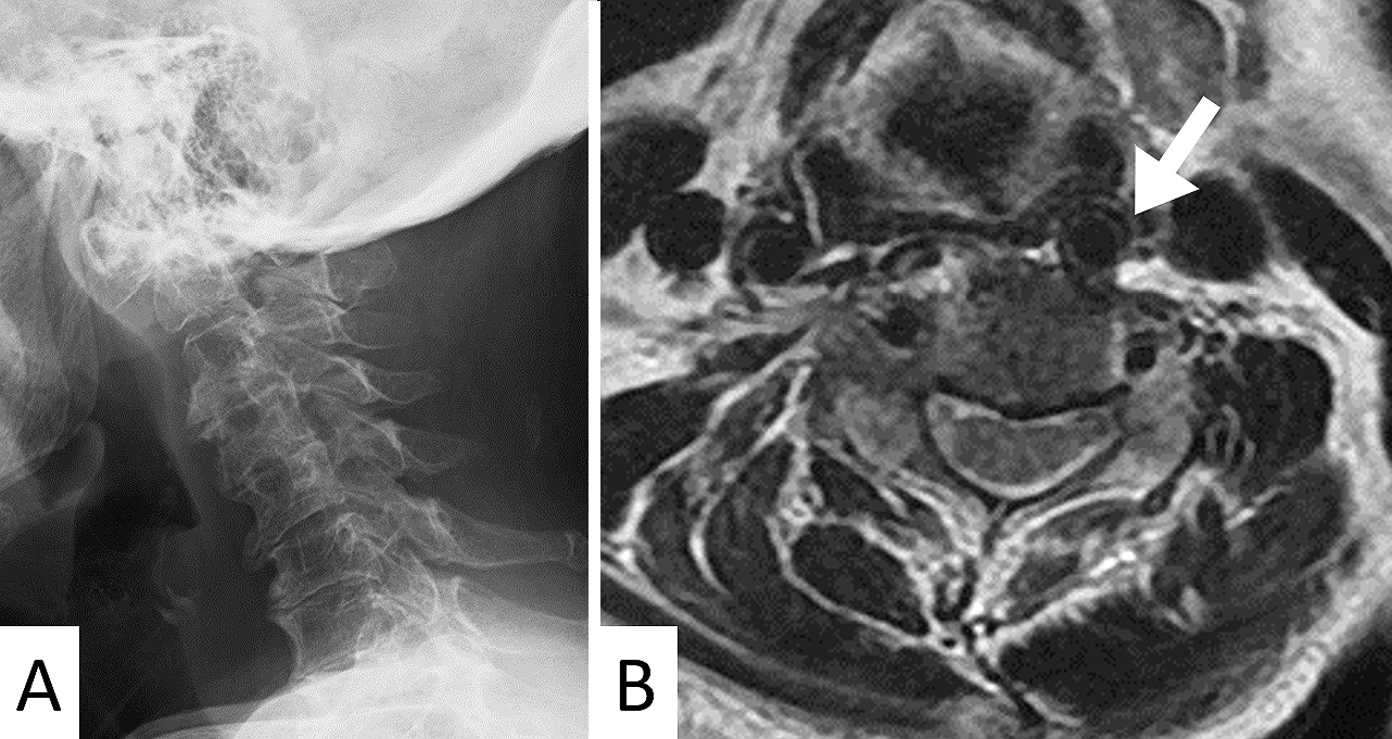
    Introduction: Reportedly, the medialization of the common carotid artery (MCCA) to be a vascular anomaly with a potential risk of intraoperative carotid artery injury. Nevertheless, among spine surgeons, the presence of MCCA has not been well recognized.

    Methods: We retrospectively reviewed consecutive patients who underwent cervical radiographs and magnetic resonance imaging (MRI) examinations in a single spine center. Using MRI, the MCCA grade was classified into grades 1 to 3 in order of severity. Radiographic measurement included C2-C7 angles as cervical lordosis, cervical sagittal vertical axis (C-SVA), T1 slope (T1S), and T1S-cervical lordosis mismatch. We compared each patient's background and radiographic parameters between patients with each of the three MCCA grades. The continuous variables were compared using the Jonckheere-Terpstra trend test and the proportions were compared using the Cochran-Armitage trend test to investigate the trend of variables in three grades.

    Results: The present study included data from 133 eligible patients (65 males and 68 females) with a mean age of 63.7 (±14.2) years. The details of MCCA grading were as follows: grade 1, n=101; grade 2, n=27; and grade 3, n=5. With an increasing MCCA grade, age (61.9±14.0, 68.2±13.8, and 76.4±9.4 years for grades 1, 2, and 3, respectively, p=0.005) and proportion of female (p<0.001) had an increasing trend, whereas cervical lordosis had a decreasing trend (11.7±13.5°, 7.0±14.5°, and −10.0±19.2° for grades 1, 2, and 3, respectively, p=0.011).

    Conclusions: Several patient backgrounds including the female gender, older age, and kyphotic alignment were determined as MCCA risk factors. Careful preoperative neck vasculature assessment would avoid a catastrophic complication during anterior cervical surgery.

    Download PDF (429K)
  • Shereen I. Fawaz, Mohamed A. Elgebeily, Heba G. Saber, Hany El Zahlawy
    2021 Volume 5 Issue 3 Pages 149-153
    Published: May 27, 2021
    Released on J-STAGE: May 27, 2021
    Advance online publication: December 05, 2020
    JOURNAL OPEN ACCESS

    Introduction: The modified Japanese Orthopaedic Association (mJOA) score is considered to be among the most comprehensive scores in the assessment of patients with cervical myelopathy. Hence, providing reliable, translated, and cross-culturally adapted versions in different languages is required to standardize the evaluation of patients. This study aimed to translate a reliable Arabic version of the mJOA score.

    Methods: A total of 65 patients of variable age and with etiologies for compressive cervical myelopathy were recruited. Both forward and backward translations were performed. Then, intraobserver and interobserver reliabilities were measured using the intraclass correlation coefficient and Cronbach's alpha coefficient.

    Results: The mean age of the patients was 58.08 years, and most of them were male (69.2%). The intraobserver and interobserver reliabilities were almost in perfect agreement for the different sections and the total score, which were 96.8% and 97.4%, respectively.

    Conclusions: In this study, a reliable, cross-culturally adapted Arabic version of the mJOA score for patients with cervical myelopathy is provided. Although the study was conducted on Egyptian patients, we believe that it could be implemented in majority of the Arabic-speaking population.

    Download PDF (254K)
  • Maho Okamura, Wataru Saito, Masayuki Miyagi, Eiki Shirasawa, Takayuki ...
    2021 Volume 5 Issue 3 Pages 154-159
    Published: May 27, 2021
    Released on J-STAGE: May 27, 2021
    Advance online publication: November 20, 2020
    JOURNAL OPEN ACCESS

    Introduction: Intraoperative hypothermia is associated with perioperative complications such as blood loss and wound infection. Thus, perioperative heat retention methods to prevent perioperative hypothermia such as providing a warmed blanket and active patients' warming are important. Although major surgery and pediatric patient age are noted as risk factors, only a few studies focus on hypothermia as an intraoperative complication in pediatric scoliosis surgery. The aim of this study is to investigate the incidence of intraoperative hypothermia in pediatric scoliosis surgery and the associated preoperative risk factors.

    Methods: We retrospectively reviewed the records of pediatric patients who underwent posterior spinal fusion at a single institution between 2015 and 2019. We recorded the background data, perioperative data, lowest recorded core temperature, and perioperative complications. Patients were divided into those whose temperature decreased below 36°C (Group H) and those who maintained a temperature of 36°C or greater (Group N) during surgery. We compared the two groups and performed multivariate analysis to identify preoperative risk factors for intraoperative hypothermia.

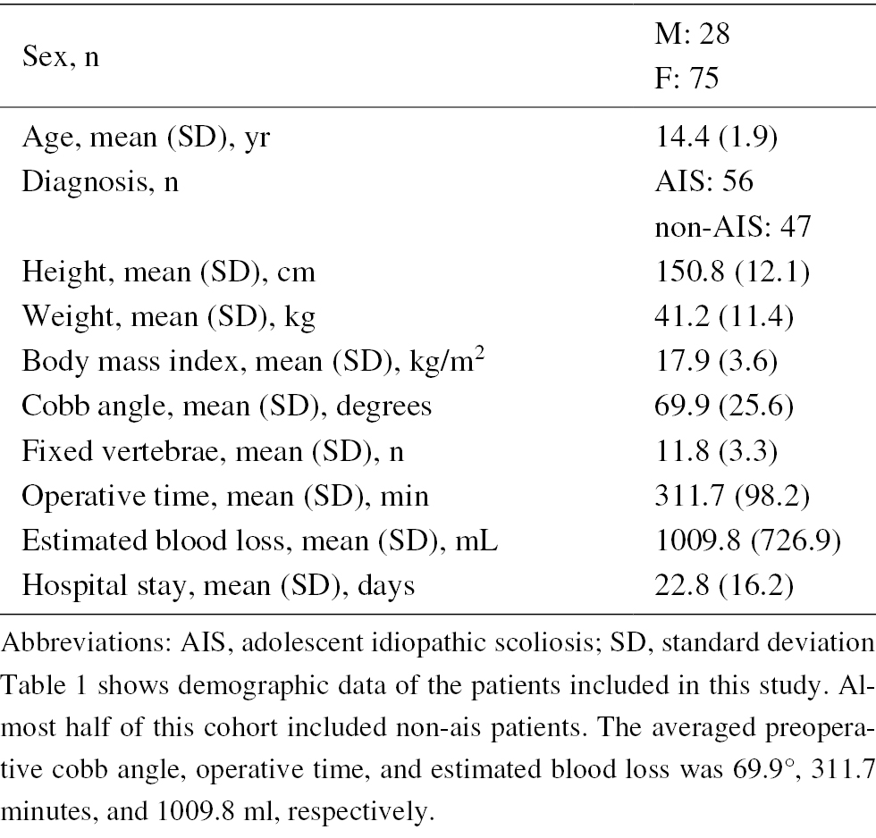
    Results: A total of 103 patients underwent posterior spinal fusion; 56 for adolescent idiopathic scoliosis and 47 for neuromuscular scoliosis. Hypothermia was observed in 40 patients (38.8%). Group H had more non-adolescent idiopathic scoliosis (AIS) patients, lower mean body mass index, greater mean blood loss, greater number of fused vertebrae, larger preoperative Cobb angle, and lower initial core body temperature (immediately after induction of anesthesia). On multivariate analysis, a diagnosis of neuromuscular scoliosis, a lower body mass index, and a lower initial core body temperature were identified as independent risk factors for intraoperative hypothermia.

    Conclusions: The incidence of hypothermia in pediatric posterior scoliosis surgery is 38.8%. Diagnosis of non-AIS, lower body mass index, and lower core body temperature at the time of anesthesia induction are preoperative risk factors for intraoperative hypothermia.

    Download PDF (85K)
  • Kenyu Ito, Kei Ando, Kazuyoshi Kobayashi, Hiroaki Nakashima, Yukihiro ...
    2021 Volume 5 Issue 3 Pages 160-164
    Published: May 27, 2021
    Released on J-STAGE: May 27, 2021
    Advance online publication: September 23, 2020
    JOURNAL OPEN ACCESS

    Introduction: Recent research has shown that spinal sagittal alignment plays a critical role in health-related quality of life. However, most of these studies were cross-sectional in nature, and longitudinal studies of lumbar lordosis (LL) in healthy subjects were few. This study aims to evaluate the change in lumbar sagittal parameters during a 10-year period.

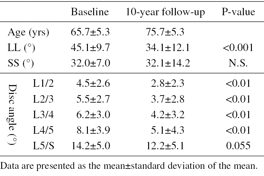
    Methods: The study population included 45 individuals (mean age, 65.7 years; male, n=20; female, n=25) who underwent sagittal lumbar radiography and a basic health checkup during a 10-year period. The radiologic parameters were LL, disc angle, sacral slope angle (SS), and pelvic incidence (PI). The change of LL during the 10-year period was defined as ΔLL. The subjects were divided into the LL maintenance group (n=33) and the LL non-maintenance group (n=12) based on their LL values.

    Results: The radiologic baseline/final parameters were as follows: LL, 45/34 degrees (P<0.001); L1/L2 disc angle, 4.5/2.5 degrees; L2/L3 disc angle, 5.5/2.7 degrees; L3/L4 disc angle, 6.2/4.2 degrees; L4/L5 disc angle, 8.1/5.1 degrees; L5/S disc angle, 14.2/12.2 degrees; and SS, 32.0/32.1 degrees.

    The mean PI (50.5 degrees) was tended to be associated with the final LL (R=0.31, P=0.044) and was correlated with the ΔLL (R=0.43, P<0.01).

    The data of the LL maintenance/non-maintenance groups were as follows: age, 65.0/67.0; primary LL, 43.2/50.2 degrees (P<0.05); final LL, 36.2/27.8 degrees (P<0.05); and PI, 52.8/43.8 degrees (P<0.01).

    Conclusions: During the 10-year study period, the LL in middle-aged and elderly volunteers decreased by 11 degrees. The factor of maintenance of LL was PI.

    Download PDF (137K)
  • Keisuke Masuda, Hideki Shigematsu, Masato Tanaka, Sachiko Kawasaki, Yu ...
    2021 Volume 5 Issue 3 Pages 165-170
    Published: May 27, 2021
    Released on J-STAGE: May 27, 2021
    Advance online publication: October 22, 2020
    JOURNAL OPEN ACCESS

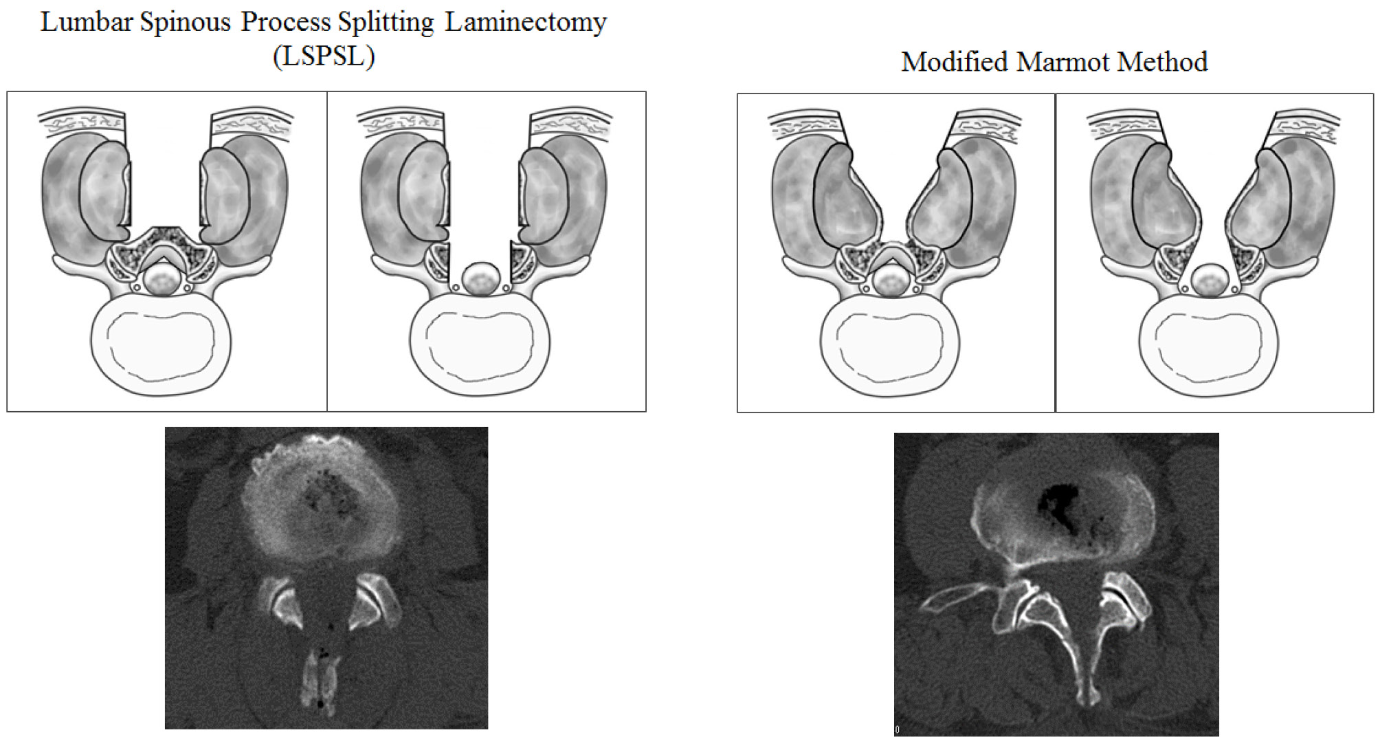
    Introduction: Compared with the conventional posterior lumbar decompression surgery, the spinous process splitting approach for lumbar spinal stenosis is less invasive. There are currently two types of the spinous process splitting approach that are performed. First is the lumbar spinous process splitting laminectomy (LSPSL), which involves the detachment of the spinous process from the lamina. Second is the modified Marmot method, which involves leaning of the spinous process without detachment from the lamina. To the best of our knowledge, this is the first study comparing the 2-year surgical outcomes of the modified Marmot method and LSPSL in cases of lumbar spinal canal stenosis.

    Methods: We recruited 69 patients who underwent decompression surgery. A total of 32 patients underwent the modified Marmot method (M group), and 37 patients underwent LSPSL (S group). We compared the clinical results, laboratory data of surgical invasion, wound pain, and safety.

    Results: No significant difference was observed in terms of the demographic data and operative time between the two groups. The number of decompressed segments and intraoperative and postoperative blood loss volume in the M group were greater than that in the S group. In the S group, the postoperative Japanese Orthopedic Association scores and recovery rates were significantly greater compared with those in the M group. Perioperative complications did not significantly differ between the two groups. On postoperative day 1, the Postoperative Visual Analog Scale scores at rest in the M group were lower than those in the S group.

    Conclusions: In clinical practice, we believe that posterior lumbar decompression surgery is safe, effective, and minimally invasive. Although the modified Marmot method may be less invasive and result in the reduction of wound pain during early postoperative periods, the clinical results did not exhibit greater long-term improvements with regard to surgical complications and neurological improvement, when compared with LSPSL.

    Download PDF (280K)
  • Takeshi Sasagawa, Yosuke Takeuchi, Ikuo Aita
    2021 Volume 5 Issue 3 Pages 171-175
    Published: May 27, 2021
    Released on J-STAGE: May 27, 2021
    Advance online publication: October 22, 2020
    JOURNAL OPEN ACCESS

    Introduction: Some reports revealed that hidden blood loss (HBL) during surgery for traumatic thoracolumbar fracture cannot be ignored, even when using a percutaneous approach. Using percutaneous pedicle screws (PPS) for traumatic thoracolumbar fracture, this study aimed to compare estimate blood loss (EBL), including HBL, between early and late fixation.

    Methods: This investigation was a retrospective study. In the present study, data from 39 patients who underwent posterior spinal stabilization using PPS for single-level thoracolumbar fracture have been included. We divided the patients into an early group (group E) (n=20) in whom surgery was conducted within 3 days of fracture and a late group (group L) (n=19) in whom surgery was conducted more than 3 days after fracture. We evaluated hemoglobin (Hb) on the day of injury, and 1, 3 or 4, and 7 days after surgery, EBL, HBL, and transfusion requirement.

    Results: Hb on day 1 (group E: 12.2±1.7 g/dL, group L: 12.3±1.6 g/dL) was significantly less than that on the injured day (group E: 14.2±1.7 g/dL, group L: 13.9±1.7 g/dL) in both groups. The values of Hb and EBL were not significantly different at any time between the two groups. HBL (group E: 487±266 mL, group L: 386±305 mL) was not significantly different between the two groups. No patients required transfusion in either group.

    Conclusions: EBL in early fixation using PPS for traumatic thoracolumbar fracture is not significantly different compared with that in late surgery from days 1 to 7 postoperatively. Early fixation using PPS for traumatic thoracolumbar fracture does not result in negative outcomes any more than those in late surgery in terms of blood loss.

    Download PDF (228K)
  • Tsunehiko Konomi, Shinjiro Kaneko, Amir Fariz Zakaria, Kanehiro Fujiyo ...
    2021 Volume 5 Issue 3 Pages 176-181
    Published: May 27, 2021
    Released on J-STAGE: May 27, 2021
    Advance online publication: October 22, 2020
    JOURNAL OPEN ACCESS

    Introduction: An anterior surgical approach for severe infectious spondylodiscitis in the lumbar region is optimal but not always atraumatic. The aim of this study was to evaluate the efficacy and safety of a minimal anterior-lateral retroperitoneal approach, also known as a surgical approach for oblique lumbar interbody fusion, for cases with severe infectious spondylodiscitis with osseous defects.

    Methods: Twenty-four consecutive patients who underwent anterior debridement and spinal fusion with an autologous strut bone graft for infectious spondylodiscitis with osseous defects were reviewed retrospectively. Eleven patients underwent the minimal retroperitoneal approach (Group M), and 13 underwent the conventional open approach (Group C). Peri- and postoperative clinical outcomes, that is, estimated blood loss (EBL), operative time (OT), creatine kinase (CK) level, visual analog scale (VAS), and rates of bone union and additional posterior instrumentation, were evaluated, and the differences between both groups were assessed statistically.

    Results: Mean EBL, serum CK on the 1st postoperative day, and VAS on the 14th postoperative day were 202.1 mL, 390.9 IU/L, and 9.5 mm in Group M and 648.3 mL, 925.5 IU/L, and 22.3 mm in Group C, respectively, with statistically significant differences between the groups. There were no statistically significant intergroup differences in OT and rates of bone union and additional posterior instrumentation.

    Conclusions: Anterior debridement and spinal fusion using the minimal retroperitoneal approach is a useful and safe surgical technique. Although a preponderance of the minimal approach regarding early bone union is not validated, this technique has the advantages of conventional open surgery, but reduces blood loss, muscle injury, and pain postoperatively.

    Download PDF (186K)
  • Chad E Cook, Alessandra N Garcia, Christopher Shaffrey, Oren Gottfried
    2021 Volume 5 Issue 3 Pages 182-188
    Published: May 27, 2021
    Released on J-STAGE: May 27, 2021
    Advance online publication: November 20, 2020
    JOURNAL OPEN ACCESS

    Introduction: Employment status plays an essential role as a social determinant of health.

    Unemployed are more likely to have a longer length of hospital stay and a nearly twofold greater rate of 30 day readmission than those who were well employed at the time of back surgery. This study aimed to investigate whether employment status influenced post-surgery outcomes and if so, the differences were clinically meaningful among groups.

    Methods: This retrospective observational study used data from the Quality Outcomes Database Lumbar Registry. Data refinement was used to isolate individuals 18 to 64 who received primary spine surgeries and had a designation of employed, unemployed, or disabled. Outcomes included 12 and 24 month back and leg pain, disability, patient satisfaction, and quality of life. Differences in descriptive variables, comorbidities, and outcomes measures (at 12 and 24 months) were analyzed using chi-square and linear mixed-effects modeling. When differences were present among groups, we evaluated whether they were clinically significant or not.

    Results: Differences (between employed, unemployed, and disabled) among baseline characteristics and comorbidities were present in nearly every category (p<0.01). In all cases, those who were disabled represented the least healthy, followed by unemployed, and then employed. Clinically meaningful differences for all outcomes were present at 12 and 24 months (p<0.01). In post hoc analyses, differences between each group at nearly all periods were found.

    Conclusions: The findings support that the health-related characteristics are markedly different among employment status groups. Group designation strongly differentiated outcomes. These findings suggest that disability and unemployment should be considered when determining prognosis of the individual.

    Download PDF (97K)
  • Daisuke Kurosawa, Eiichi Murakami, Toshimi Aizawa, Takashi Watanabe
    2021 Volume 5 Issue 3 Pages 189-195
    Published: May 27, 2021
    Released on J-STAGE: May 27, 2021
    Advance online publication: January 21, 2021
    JOURNAL OPEN ACCESS

    Introduction: Sacroiliac joint (SIJ) arthrodesis is the last resort for patients with severe SIJ pain. However, this technique does not always provide good outcomes regarding activities of daily living (ADL). This study aims to reveal the preoperative clinical features associated with poor outcomes of SIJ arthrodesis.

    Methods: Twenty-six consecutive patients who underwent SIJ arthrodesis between 2009 and 2018 were evaluated. Good-outcome was defined as ≥30% improvement in ADL, quantified by the Roland-Morris Disability Questionnaire (RDQ). The good-outcome group (17 patients; 10 men and 7 women, 42.5±8.4 years old) and the poor-outcome group (9 patients; 1 man and 8 women, 47.0±17.9 years old) were compared to identify the preoperative clinical features of poor surgical outcomes.

    Results: No significant differences were observed between the two groups regarding age, preoperative RDQ score, time between onset and the confirmed diagnosis of SIJ pain, and the time between diagnosis and surgical treatment. The following preoperative clinical features associated with poor surgical outcomes were identified: female sex, pain in multiple regions, walking with a cane, and the use of a wheelchair before surgery (P<0.05).

    Conclusions: The present study demonstrated that poor postoperative outcomes in patients with severe SIJ pain were associated with the following preoperative clinical features: female sex, pain in multiple regions, walking with a cane, or use of a wheelchair. Ample attention is warranted in patients with such features who are indicated for surgical treatments.

    Download PDF (514K)
  • Masashi Sato, Yasuhiro Shiga, Naoya Takayama, Masamitsu Sone, Kentaro ...
    2021 Volume 5 Issue 3 Pages 196-204
    Published: May 27, 2021
    Released on J-STAGE: May 27, 2021
    Advance online publication: February 22, 2021
    JOURNAL OPEN ACCESS

    Introduction: Platelet-rich plasma (PRP) is drawing attention as a substance that can promote bone formation. The growth factors present in PRP are stable for a long time after freeze-drying. However, the effects of PRP are inconsistent, and its effects on bone union in spinal surgery remain controversial. The immortalized megakaryocyte cell lines (imMKCLs) derived from human-induced pluripotent stem cells (hiPSCs) have been developed to produce numerous stable and clinically functional platelets. In this study, growth factors present in freeze-dried hiPSC-derived imMKCLs and platelets (iPS-MK/Plts) were evaluated, and their ability to promote bone formation was examined using a rat lumbar artificial bone grafting model.

    Methods: We prepared freeze-dried iPS-MK/Plts and quantified their growth factors by enzyme-linked immunosorbent assays. Surgical grafting of artificial bone to the lumbar transverse processes was performed in 8-week-old female rats, which were divided into two groups: artificial bone graft (control) and artificial bone graft plus freeze-dried iPS-MK/Plts (iPS group). Transplantation was performed only on the left side. Eight weeks after the surgery, we captured computed tomography images and compared bilateral differences in the bone volume of the graft site in each rat. We also compared the left side/right side bone volume ratio between the two groups.

    Results: The freeze-dried iPS-MK/Plts contained numerous growth factors. While there was no significant increase in bone volume on the transplanted side than that on the non-grafted side in the control group, bone volume significantly increased on the transplanted side in the iPS group, as evidenced by augmented mean left/right bone volume ratio of the iPS group compared with that of the control group. But the new bone observed in the iPS group was histologically normal.

    Conclusions: Freeze-dried hiPSC-derived MKCLs and platelets contain several stable growth factors and have the potential for promoting new bone formation.

    Download PDF (2079K)
TECHNICAL NOTE
  • Shinji Takahashi, Hidetomi Terai, Hiromitsu Toyoda, Masatoshi Hoshino, ...
    2021 Volume 5 Issue 3 Pages 205-210
    Published: May 27, 2021
    Released on J-STAGE: May 27, 2021
    Advance online publication: December 05, 2020
    JOURNAL OPEN ACCESS

    Introduction: Because of adolescent idiopathic scoliosis (AIS), most surgeons use rod rotation on the concave side for Lenke types 1 and 2 curves. Nevertheless, the accurate placement of pedicle screws within dysplastic pedicles, especially on the concave side, is sometimes challenging. Conversely, there is a concern that apical rotation might be exacerbated after convex rod rotation maneuver (RRM) because the rod is rotated in the same direction as vertebral rotation. This study aims to demonstrate the surgical technique and outcomes of a convex RRM with direct vertebral rotation (DVR) for the correction of AIS.

    Technical Note: Multilevel pedicle screws were inserted into the vertebrae. The pre-bent pure titanium rod was set on the convex side and then derotated to nearly 90°. DVR was conducted for the desired vertebrae. Another pre-bent titanium alloy rod, for placement on the concave side, was contoured the same as the rod on the convex side. Using a reduction tube that allowed easier capture of the rod, the rod was connected to the concave side screws. DVR was again conducted for the desired vertebrae. Among the 59 patients, the correction rate of the main thoracic curve in Lenke types 1 and 2 AIS was 75.1% and 65.0%, respectively. The absolute value of the change in apical vertebral rotation between pre- and post-operative computed tomography (CT) scans in Lenke types 1 and 2 curves was 4.8° and 4.2°, respectively.

    Conclusions: The convex RRM improved vertebral rotation in Lenke types 1 and 2 AIS. This procedure should be regarded as one of the surgical options for AIS, especially in patients with a narrow pedicle width on the concave side.

    Download PDF (782K)
CLINICAL CORRESPONDENCE
ERRATUM
feedback
Top