Spine Surgery and Related Research
Online ISSN : 2432-261X
ISSN-L : 2432-261X
Volume 4, Issue 2
Displaying 1-16 of 16 articles from this issue
REVIEW ARTICLE
  • Garrett K Harada, Zakariah K Siyaji, Sadaf Younis, Philip K Louie, Din ...
    2020 Volume 4 Issue 2 Pages 99-110
    Published: April 27, 2020
    Released on J-STAGE: April 27, 2020
    Advance online publication: February 26, 2020
    JOURNAL OPEN ACCESS

    Objective: To review and highlight the historical and recent advances of imaging in spine surgery and to discuss current applications and future directions.

    Methods: A PubMed review of the current literature was performed on all relevant articles that examined historical and recent imaging techniques used in spine surgery. Studies were examined for their thoroughness in description of various modalities and applications in current and future management.

    Results: We reviewed 97 articles that discussed past, present, and future applications for imaging in spine surgery. Although most historical approaches relied heavily upon basic radiography, more recent advances have begun to expand upon advanced modalities, including the integration of more sophisticated equipment and artificial intelligence.

    Conclusions: Since the days of conventional radiography, various modalities have emerged and become integral components of the spinal surgeon's diagnostic armamentarium. As such, it behooves the practitioner to remain informed on the current trends and potential developments in spinal imaging, as rapid adoption and interpretation of new techniques may make significant differences in patient management and outcomes. Future directions will likely become increasingly sophisticated as the implementation of machine learning, and artificial intelligence has become more commonplace in clinical practice.

    Download PDF (1192K)
  • Aakash Agarwal, Boren Lin, Hossein Elgafy, Vijay Goel, Chris Karas, Ch ...
    2020 Volume 4 Issue 2 Pages 111-116
    Published: April 27, 2020
    Released on J-STAGE: April 27, 2020
    Advance online publication: June 21, 2019
    JOURNAL OPEN ACCESS

    The current communication seeks to provide an updated narrative review on latest methods of reducing implant contaminations used during spine surgery. Recent literature review has shown that both preoperative reprocessing and intraoperative handling of implants seem to contaminate implants. In brief, during preoperative phase, the implants undergo repeated bulk cleaning with dirty instruments from the OR, leading to residue buildup at the interfaces and possibly on the surfaces too. This, due to its concealed nature, remains unnoticed by the SPD (sterile processing department) or other hospital staff. Nevertheless, these can be avoided by using individually prepackaged presterilized implants. In the intraoperative phase, the implants (in the sterile field) are directly touched by the scrub tech with soiled (assisting the surgeon dispose the tissues from the instruments in use) gloves for loading onto an insertion device. It is then kept exposed on the working table (either separately or next to the used instruments as the pedicles hole are being prepared). Latest investigation has shown that by the time it is implanted in the patient, it can harbor up to 10e7 bacterial colony-forming units. The same implants were devoid of such colony-forming units, when sheathed by an impermeable sterile sheath around the sterile implant.

    Download PDF (1518K)
ORIGINAL ARTICLE
  • Satoshi Ujigo, Daniel Jonsson, Yalda Bogestål, Joakim Håkansson, Jenni ...
    2020 Volume 4 Issue 2 Pages 117-123
    Published: April 27, 2020
    Released on J-STAGE: April 27, 2020
    Advance online publication: July 10, 2019
    JOURNAL OPEN ACCESS

    Introduction: A disc herniation has traditionally been considered as disc tissue that has slipped out from an intervertebral disc. However, it was recently suggested that the disc herniation mass is a product of bioactive substances from the disc and that the disc hernia would more likely be scar tissue than herniated disc material. In this study, we aimed to analyze the structural components of experimentally induced disc herniations and compare with scar tissue and nucleus pulposus, in the rat.

    Methods: Twenty-eight rats had their L4-5 discs punctured. After three weeks, the nodule that had been formed over the puncture site, scar tissue from the spine musculature, and normal nucleus pulposus were harvested and processed for further analysis.

    Results: Proteomics analysis demonstrated that the formed nodule was more similar to scar tissue than to nucleus pulposus. Gene expression analysis showed that there was no resemblance between any tissues when looking at inflammatory genes but that, there was a clear resemblance between the nodule and scar tissue when analyzing extracellular matrix-related genes. Analysis of the GAG and polysaccharide size distribution revealed that only the nodule and scar tissue contained the shorter versions, potentially short chain hyaluronic acid that is known to induce inflammatory responses. The hematoxylin and eosin stained sections of the nodule, disc tissue, and scar tissue indicated that the morphology of the nodule and scar tissue was very similar.

    Conclusions: The nodule formed after experimental disc puncture, and that resembles a disc hernia, has a more structural resemblance to scar tissue than disc tissue. The nodule is, therefore, more likely to be induced by disc-derived bioactive substances than being formed by herniated disc material.

    Download PDF (615K)
  • Atsushi Nakano, Yoshiharu Nakaya, Takashi Fujishiro, Sachio Hayama, Ta ...
    2020 Volume 4 Issue 2 Pages 124-129
    Published: April 27, 2020
    Released on J-STAGE: April 27, 2020
    Advance online publication: September 04, 2019
    JOURNAL OPEN ACCESS

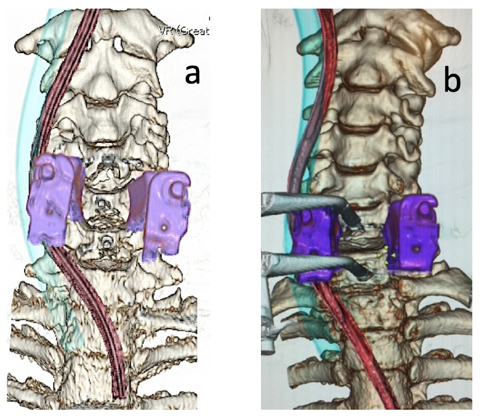
    Introduction: Using intraoperative computed tomography (iCT), we aimed to clarify the course of the esophagus and pharynx during anterior cervical spine surgery to estimate the risk of intraoperative injury.

    Methods: Sixteen patients who underwent anterior cervical spine surgery with intraoperative CT for registration of a navigation system without release of blade retraction were included. To investigate the status of the retracted esophagus and pharynx, the distance between the nasogastric tube and center of the vertebra (NVD) was measured at each disc and vertebral level (C4-7) using axial CT. The location of the cricoid cartilage, which may affect the shift of the esophagus and pharynx, was noted. Presence or absence of contact between the esophagus and the edge of the surgical blade was investigated.

    Results: The NVDs were 28.0, 28.3, 28.9, 27.2, 24.7, 19.9, and 13.8 mm at C4, C4/5, C5, C5/6, C6, C6/7, and C7, respectively; NVDs at C6/7 or more caudal levels were significantly shorter than those at C6 or more cranial levels (P < 0.001). The cricoid cartilage was observed at the C4-C5/6 level. Esophageal contact with the edge of the blade was observed in nine cases at C6 or more caudal levels.

    Conclusions: The esophagus, which was placed at C6 or more caudal levels, was directly retracted by the blade. Nevertheless, the pharynx, which was placed at C6 or more cranial levels, was mostly retracted with the cricoid cartilage. Thus, the risk of direct esophageal injury was higher at C6 or more caudal levels than at cranial levels.

    Download PDF (525K)
  • Soichiro Masuda, Shunsuke Fujibayashi, Mitsuru Takemoto, Youngwoo Kim, ...
    2020 Volume 4 Issue 2 Pages 130-134
    Published: April 27, 2020
    Released on J-STAGE: April 27, 2020
    Advance online publication: November 01, 2019
    JOURNAL OPEN ACCESS

    Introduction: Symptomatic postoperative hematoma after spine surgery is a rare but serious complication. The objective of this study was to investigate the incidence and clinical features of symptomatic postoperative hematoma after spine surgery.

    Methods: We retrospectively identified 10,680 patients who underwent spine surgery between 2002 and 2012 in nine hospitals. We reviewed the incidence of postoperative hematoma and its clinical features, including time before onset, main symptoms, and neurological outcomes.

    Results: The overall incidence of symptomatic postoperative hematoma after spine surgery was 0.4% (45/10,680). Postoperative hematoma was more frequent after thoracic spine surgery than after cervical or lumbar surgery. The onset of postoperative hematoma occurred at an average of 2.6 days (range 0-14 days) postoperatively. The chief symptoms caused by postoperative hematoma after spine surgery were tetra/paraplegia in 30 patients, hemiplegia in eight patients, intractable pain in five patients, and airway dysfunction in two patients. Surgical evacuation of the spinal epidural hematoma resulted in improvement of at least one grade in 35 patients, while four patients had complete motor paralysis even after evacuation surgery.

    Conclusions: We report the clinical details of 45 patients with postoperative hematoma after spine surgery. This information could assist surgeons to make a prompt diagnosis and perform early evacuation surgery for postoperative hematoma following spine surgery.

    Download PDF (116K)
  • Izaya Ogon, Hiroyuki Takashima, Tomonori Morita, Tsutomu Oshigiri, Yos ...
    2020 Volume 4 Issue 2 Pages 135-141
    Published: April 27, 2020
    Released on J-STAGE: April 27, 2020
    Advance online publication: November 01, 2019
    JOURNAL OPEN ACCESS

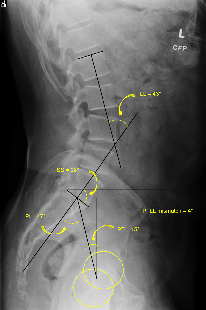
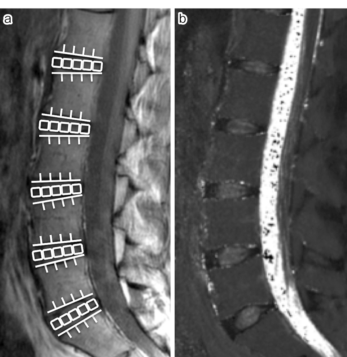
    Introduction: Although intervertebral disc degeneration (IVDD) and spinopelvic malalignment are likely key structural features of spinal degeneration and chronic low back pain (CLBP), the correlation analysis has not been fully conducted. This cross-sectional quantitative magnetic resonance imaging (MRI) T2 mapping study aimed to elucidate the association between IVDD and spinopelvic alignment in CLBP patients.

    Methods: The subjects included 45 CLBP patients (19 men and 26 women; mean age, 63.8 ± 2.0 years; range, 41-79 years). The T2 values of the anterior annulus fibrosus (AF), the nucleus pulposus (NP), and the posterior AF were evaluated using MRI T2 mapping. We compared the possible correlations of spinopelvic parameters with T2 values of anterior AF, NP, and posterior AF using Pearson's correlation coefficient analysis. T2 values in these regions were classified into upper (L1-L2 and L2-L3), middle (L3-L4), and lower (L4-L5 and L5-S1) disc levels, and we analyzed the correlations with spinopelvic parameters.

    Results: There were significant correlations of the anterior AF T2 values with lumbar lordosis (r = 0.51, p < 0.01), sacral slope (r = 0.43, p < 0.01), sagittal vertical axis (r = −0.40, p < 0.01), and pelvic tilt (r = −0.33, p < 0.01). In all lumbar levels, T2 values of anterior AF had significantly positive correlation with LL and significantly negative correlation with SVA. In lower disc level, T2 values of anterior AF had significantly positive correlation with SS and significantly negative correlation with PT. T2 values of NP and posterior AF had no significant correlations with spinopelvic parameters in all lumbar disc levels.

    Conclusions: In summary, this study indicated that the anterior AF degeneration is associated with hypolordosis of the lumbar spine, anterior translation of the body trunk, and posterior inclination of the pelvis in CLBP. Anterior AF degeneration in all lumbar disc levels was associated with hypolordosis of the lumbar spine and anterior translation of the body trunk. Anterior AF degeneration in lower disc level was associated with posterior inclination of the pelvis.

    Download PDF (466K)
  • Hideo Baba, Tsuyoshi Okudaira, Takayuki Yamaguchi, Shinichiro Hara, Hi ...
    2020 Volume 4 Issue 2 Pages 142-147
    Published: April 27, 2020
    Released on J-STAGE: April 27, 2020
    Advance online publication: October 20, 2019
    JOURNAL OPEN ACCESS

    Introduction: When surgery is performed for osteoporotic vertebral fractures, the extent to which kyphosis can be corrected by the intraoperative position of the body is often determined by preoperative radiography in the extension position. However, patients have difficulty adopting an adequate extension position due to the pain associated with their vertebral fracture. We place a pillow beneath the fractured vertebral body before surgery and take radiographs in the supine position to evaluate the extent to which the kyphosis can be corrected. This study aimed to examine the usefulness of this imaging method by comparing postoperative radiographs with preoperative radiographs taken with a pillow placed beneath the fractured vertebral body.

    Methods: Lateral preoperative radiographs were taken of the patients in seated flexion and extension positions and the supine position. Lateral radiographs (rollback) were also taken 5 min after placing a firm pillow 20 cm in diameter beneath the fractured vertebral body. The kyphotic angle was compared between preoperative lateral radiographs of patients in the flexion, extension, and supine positions, rollback, and postoperative lateral radiographs in the supine position.

    Results: The mean kyphotic angle was 33.3° in the flexion position, 28.3° in the extension position, 14.8° in the supine position, and 5.6° in rollback preoperatively and 6.4° postoperatively. The preoperative kyphotic angle differed from the postoperative kyphotic angle by ≥11° in 91% and 83% of participants in the flexion and extension positions, respectively; the difference was ≤ 5° in 30% and 61% of participants in the supine position and rollback, respectively. Differences in the postoperative angle were small in the order of rollback, supine position, extension position, and flexion position.

    Conclusions: Compared with radiographs taken in the flexion, extension, and supine positions, rollback showed little difference from postoperative radiographs, which showed almost the same angle as the intraoperative kyphotic angle.

    Download PDF (463K)
  • Aakash Agarwal, Amey Kelkar, Ashish Garg Agarwal, Daksh Jayaswal, Arvi ...
    2020 Volume 4 Issue 2 Pages 148-151
    Published: April 27, 2020
    Released on J-STAGE: April 27, 2020
    Advance online publication: October 20, 2019
    JOURNAL OPEN ACCESS

    Introduction: Recent literature identifies similar failure rates such as anchor pull-out and rod breakage, but a higher unplanned revision surgery with MAGEC rods than with traditional growth rods. Besides known failure modes such as rod fracture, infection, etc., failure to noninvasively distract the rods was cited as the main cause of such unplanned surgeries. The source of these data ranges from multicenter cohort studies to singular case series. These studies included explanted implants that had undergone failure in distraction mechanism, rod fracture, or infection, or had reached their maximum length. Nevertheless, in addition to identifying the overall mode of failure, it is equally important to identify the large-scale incidence of exclusive failures in comparison with standard instrumentation failure modes in spine surgery.

    Methods: The US Food and Drug Administration (FDA) Manufacturer and User Facility Device Experience (MAUDE) databases were searched for reports on MAGEC rods, and on standard instrumentation used for spinal fusion. The adverse events were recorded, tabulated, and analyzed.

    Results: A search of the US FDA MAUDE database yielded reports of 163 device-related adverse events. These included distraction mechanism failure (n=129), rod fracture (n=24), and minor voluntary reports of infection and tissue discoloration (n=10). For standard instrumentation usage in spine surgery, pedicle screw breakage post surgery (n=336), set screw damage during surgery (n=257), rod breakage post surgery (n=175), interbody cage breakage during surgery (n=118), and pedicle screw breakage during surgery (n=75) were identified as the top 5 failure modes.

    Conclusions: The study identified the distraction mechanism failure as the most common and growing complication associated with MAGEC rod usage in children with scoliosis, leading to unplanned invasive revision surgeries.

    Download PDF (145K)
  • Masahiro Inoue, Sumihisa Orita, Kazuhide Inage, Kazuki Fujimoto, Yasuh ...
    2020 Volume 4 Issue 2 Pages 152-158
    Published: April 27, 2020
    Released on J-STAGE: April 27, 2020
    Advance online publication: October 20, 2019
    JOURNAL OPEN ACCESS

    Introduction: There are several reports about invasive muscle injury during posterior spinal surgery. However, few reports have evaluated the association between the clinical symptoms and changes in the physical properties of the psoas major after oblique lateral interbody fusion (OLIF). Therefore, the current study aimed to investigate the relationship between the clinical symptoms and changes in the psoas major muscle before and after OLIF.

    Methods: Twenty-seven patients who underwent single-level OLIF following the diagnosis of degenerative lumbar disease were included in the study. The cross-sectional areas (CSAs) of the psoas major on the approaching and contralateral sides were measured in the axial computed tomography view of the surgical intervertebral space preoperatively and postoperatively at 1 week and 3, 6, and 12 months. The preoperative and postoperative changes in the CSAs were compared. Muscle degeneration was evaluated using axial magnetic resonance images at the same level as that in the CSA evaluation preoperatively and at 12 months postoperatively. Additionally, the relationship between these parameters and postoperative lower limb symptoms was investigated.

    Results: Significant swelling of the psoas major on the approach side was observed 1 week postoperatively (p < 0.05). No postoperative muscle degeneration was observed. Three cases of paresthesia in the front of the thigh were observed, but no association was found with changes in CSA in any of the cases.

    Conclusions: The OLIF approach caused swelling of the psoas major 1 week postoperatively with no more muscle degeneration in the mid-term. Although numbness of the lower limbs was found in some cases, no association was found with changes in CSA. Our study findings suggest that the OLIF approach causes temporary injury or swelling of the psoas major, but the long-term damage to the muscle is not significant.

    Download PDF (424K)
  • Takuma Koyama, Shurei Sugita, Takahiro Hozumi, Masanori Fujiwara, Kiyo ...
    2020 Volume 4 Issue 2 Pages 159-163
    Published: April 27, 2020
    Released on J-STAGE: April 27, 2020
    Advance online publication: November 01, 2019
    JOURNAL OPEN ACCESS

    Introduction: Cerebral spinal fluid leak from durotomy is a well-known risk with spinal surgeries. The aim of this study is to identify the incidence of unrecognized incidental durotomy during posterior surgery for spinal metastases and its risk factors.

    Methods: Participants comprised 75 patients who underwent posterior spine surgery for spinal metastases between January 2012 and December 2016. Cases with apparent durotomy noticed intraoperatively were excluded. Unrecognized durotomy was diagnosed as the presence of wide subcutaneous fluid retention on magnetic resonance imaging at least 3 months postoperatively. For comparison, 50 patients who underwent cervical laminoplasty due to cervical spondylotic myelopathy were examined using the same method. We also examined correlations between occurrence of durotomy and patient characteristics such as age, type of tumor, location of tumor (ventral or dorsal), extent of tumor, and history of radiotherapy before surgery.

    Results: Unrecognized durotomy occurred in 21 cases of spinal metastasis (26.7%) and in 1 case of cervical spondylotic myelopathy (2%), representing a significant difference between groups. Age, type of tumor, location of tumor, extent of tumor, and history of radiotherapy before surgery did not correlate significantly with occurrence of durotomy. No local trouble was observed in durotomy cases, except in one case with subcutaneous local infection.

    Conclusions: The incidence of unrecognized incidental durotomy is significantly higher during surgery for spinal metastases than that during surgery for degenerative disease.

    Download PDF (391K)
  • Hiroto Takenaka, Tatsunori Ikemoto, Junya Suzuki, Masayuki Inoue, Youn ...
    2020 Volume 4 Issue 2 Pages 164-170
    Published: April 27, 2020
    Released on J-STAGE: April 27, 2020
    Advance online publication: December 03, 2019
    JOURNAL OPEN ACCESS

    Introduction: The present study aimed to investigate the association between trunk muscle strength, lumbar spine bone mineral density (BMD), lumbar scoliosis angle (LSA), and appendicular skeletal muscle mass index (ASMI) and the severity locomotive syndrome (LS) using dual-energy X-ray absorptiometry (DXA) technology in elderly individuals.

    Methods: In this cross-sectional study, we enrolled 168 individuals aged >60 years. We measured their trunk muscle strength (flexion and extension) and BMD, LSA, and ASMI using DXA. We defined degenerative lumbar scoliosis (DLS) as LSA ≥ 10° by the Cobb method using the DXA image. The locomotor function was evaluated using the timed up-and-go (TUG) test and the 25-question Geriatric Locomotive Function Scale (GLFS-25) score. Normal locomotor function, LS-1, and LS-2 were defined as a GLFS-25 score of <7, ≥7 and <16, and ≥16, respectively. We compared the three groups, analyzing the associations between all variables and the locomotor function using univariate and multivariate analyses.

    Results: Although there was no significant difference in sex ratio, BMD, ASMI, and trunk-flexor strength, significant differences were observed in age (p < 0.01), the prevalence of DLS (p = 0.02), trunk-extensor strength (p < 0.01), and trunk-extensor/flexor strength ratio (p < 0.01) among the three groups. In multiple regression analyses, the significant risk factors of the TUG test were age (β = 0.26), body mass index (β = 0.36), LSA (β = 0.15), ASMI (β = −0.30), and trunk-extensor strength (β = −0.19), whereas the significant factor of the GLFS-25 score was trunk-extensor strength (β = −0.31).

    Conclusions: The results indicate that it is clinically important for LS to pay careful attention not only to BMD but also to lumbar scoliosis when DXA examination of the lumbar spine is routinely conducted. Moreover, it is essential to note that trunk-extensor strength is more important than trunk-flexor strength in maintaining locomotor function in elderly individuals.

    Download PDF (97K)
  • Paul R. Krafft, Brooks Osburn, Andrew C. Vivas, Gautam Rao, Puya Alikh ...
    2020 Volume 4 Issue 2 Pages 171-177
    Published: April 27, 2020
    Released on J-STAGE: April 27, 2020
    Advance online publication: December 20, 2019
    JOURNAL OPEN ACCESS

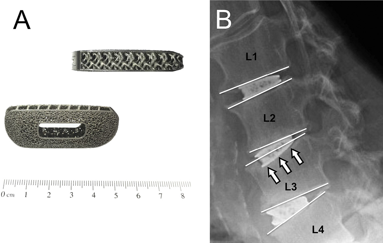
    Introduction: Implant subsidence is a potential complication of spinal interbody fusion and may negatively affect patients subjected to procedures relying on indirect decompression such as minimally invasive transpsoas lateral lumbar interbody fusion (LLIF). The porous architecture of a recently developed titanium intervertebral cage maximizes bone-to-implant contact and minimizes stress shielding in laboratory experiments; however, its subsidence rate in patients has not yet been evaluated. The goal of this current study was to evaluate implant subsidence in patients subjected to LLIF.

    Methods: Our institutional review board-approved single-center experience included 29 patients who underwent 30 minimally invasive LLIF from July 2017 to September 2018 utilizing the novel 3D-printed porous titanium implants. Radiographs, obtained during routine postoperative follow-up visits, were reviewed for signs of implant subsidence, defined as any appreciable compromise of the vertebral endplates.

    Results: Radiographic subsidence occurred in 2 cases (6.7%), involving 2 out of 59 porous titanium interbody cages (3.4%). Both cases of subsidence occurred in four-level stand-alone constructs. The patients remained asymptomatic and did not require surgical revision. Ten surgeries were stand-alone constructs, and 20 surgeries included supplemental posterior fixation.

    Conclusions: In our patient cohort, subsidence of the porous titanium intervertebral cage occurred in 6.7% of all cases and in 3.4% of all lumbar levels. This subsidence rate is lower compared to previously reported subsidence rates in patients subjected to LLIF using polyetheretherketone implants.

    Download PDF (341K)
TECHNICAL NOTE
  • Takaomi Kobayashi, Tadatsugu Morimoto, Kazumasa Maeda, Yu Toda, Hirohi ...
    2020 Volume 4 Issue 2 Pages 178-183
    Published: April 27, 2020
    Released on J-STAGE: April 27, 2020
    Advance online publication: January 29, 2020
    JOURNAL OPEN ACCESS

    Introduction: The transoral mandibular tongue-splitting approach is typically performed for the treatment of upper cervical tumor and instability but has not been performed for the treatment of upper cervical epidural abscess (UCEA). We report the first case of UCEA successfully treated with a transoral mandibular tongue-splitting approach.

    Technical Note: A 62-year-old man who had medical histories of tracheotomy with intubation and dermatopathy due to radiation therapy for the treatment of nasopharyngeal carcinoma presented with neck pain and limb weakness. The imaging examination revealed bone erosion of C2-C4 vertebrae and abscess at the level of C2-C4, supporting a diagnosis of UCEA. The transcervical approach could not be used for treatment; therefore, the transoral mandibular tongue-splitting approach was used successfully to perform decompression, debridement, and iliac bone grafting. Subsequently, we reviewed the literature pertaining to the use of the transoral mandibular tongue-splitting approach. The approach can be invasive and cause some complications. However, no fatal complications have been reported, and all patients demonstrated a favorable neurological outcome with reduced neurological deficits.

    Conclusions: This case and subsequent literature review suggest that the transoral mandibular tongue-splitting approach may be effective for the improvement of neurological outcomes without fatal complications in patients with UCEA. There may be an increasing number of patients with UCEA requiring the transoral mandibular tongue-splitting approach due to the increasing prevalence of immunocompromized status and the aging population.

    Download PDF (391K)
CLINICAL CORRESPONDENCE
feedback
Top