Spine Surgery and Related Research
Online ISSN : 2432-261X
ISSN-L : 2432-261X
Volume 7, Issue 5
Displaying 1-11 of 11 articles from this issue
ORIGINAL ARTICLE
  • Ryo Fukata, Takeo Furuya, Yuki Shiko, Yohei Kawasaki, Mayuko Kuwata, K ...
    2023 Volume 7 Issue 5 Pages 414-420
    Published: September 27, 2023
    Released on J-STAGE: September 27, 2023
    Advance online publication: March 13, 2023
    JOURNAL OPEN ACCESS

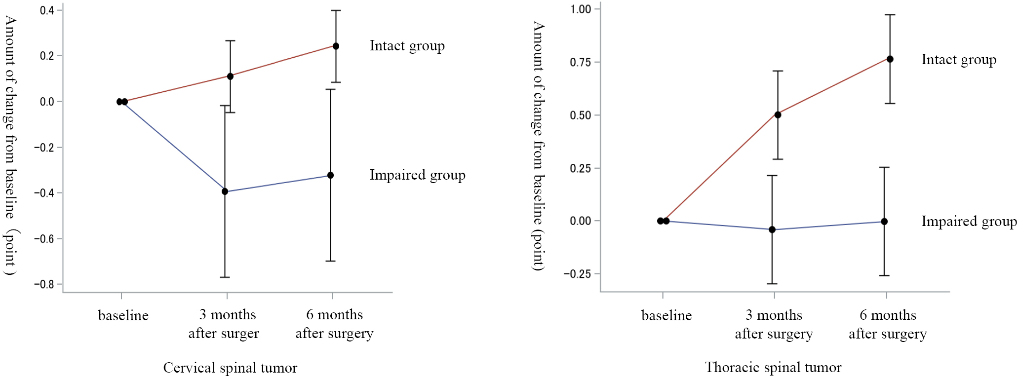
    Introduction: We investigated the effect of preoperative joint position sense in the big toe on the postoperative recovery of gait function after spinal tumor surgery.

    Methods: Seventy-three patients with spinal tumors who underwent surgery at our hospital between 2014 and 2019 and could be followed for at least 6 months after surgery were included. The patients were divided into the cervical spinal (41 cases) and thoracic spinal (32 cases) groups according to the localization of the tumor. These groups were further classified into an Impaired group (cervical spinal, 34 cases; thoracic spinal, 19 cases) and an Intact group (cervical spinal, 7 cases; thoracic spinal, 13 cases) according to the presence or absence of preoperative joint position sense in the big toe. The amount of change in ambulatory function from the preoperative period to 3 and 6 months postoperatively was compared between the Impaired and Intact groups within each tumor localization category.

    Results: Impaired preoperative joint position sense in the big toe in patients undergoing thoracic spinal tumor surgery delayed the recovery of gait function in the early postoperative period.

    Conclusions: In patients with thoracic spinal tumor surgery, the absence of preoperative joint position sense in the big toe delayed the recovery of postoperative gait function.

    Download PDF (299K)
  • Takaki Yoshiyama, Shunsuke Fujibayashi, Bungo Otsuki, Takayoshi Shimiz ...
    2023 Volume 7 Issue 5 Pages 421-427
    Published: September 27, 2023
    Released on J-STAGE: September 27, 2023
    Advance online publication: April 21, 2023
    JOURNAL OPEN ACCESS

    Introduction: Few articles have investigated patient satisfaction with laminoplasty in patients with cervical spondylotic myelopathy (CSM) alone, excluding other diseases, such as ossification of the posterior longitudinal ligament. In this study, we aimed to investigate patient satisfaction after double-door laminoplasty for CSM and determine the preoperative and postoperative factors that affect patient satisfaction.

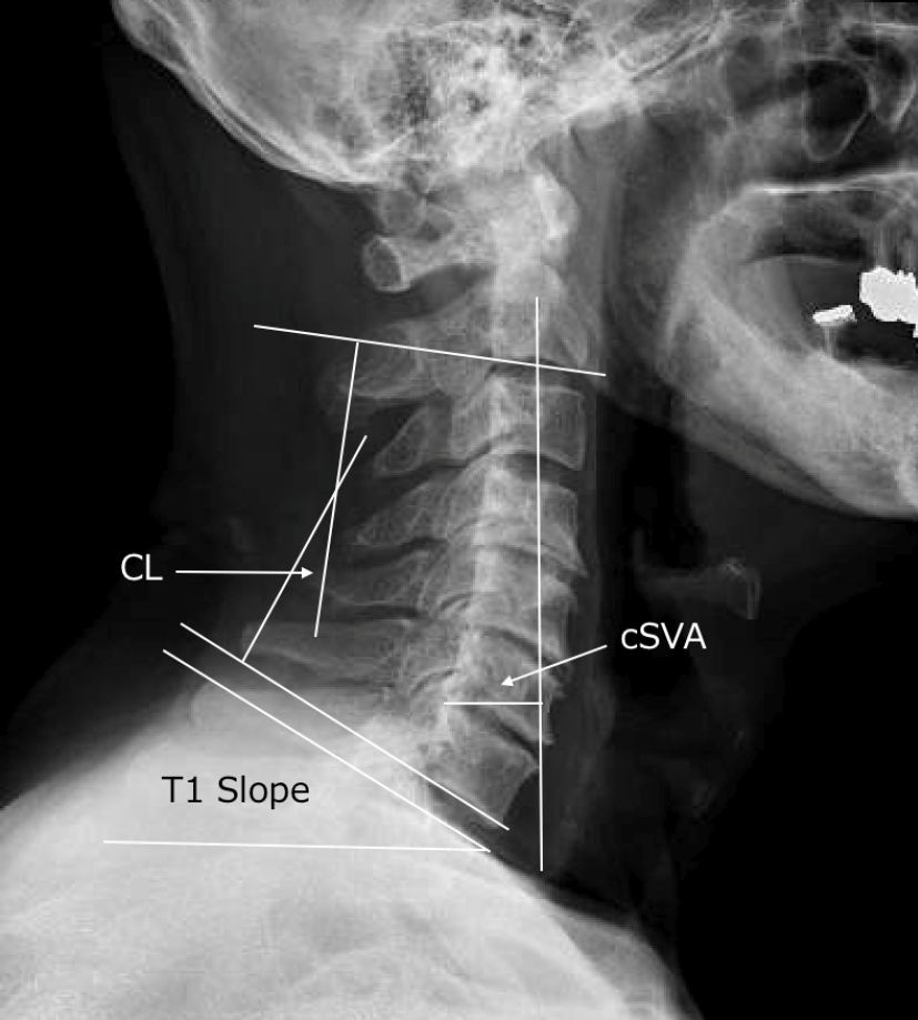
    Methods: We retrospectively reviewed cases of laminoplasty for CSM. We measured sagittal imaging parameters (cervical lordosis [CL], C2-C7 cervical sagittal vertical axis [cSVA], and T1 slope [T1S]), Japanese Orthopaedic Association (JOA) score, and patient-reported outcomes (PROs) such as the neck disability index (NDI) and visual analog scale (VAS) preoperatively, 3 months postoperatively, and 1 year postoperatively. In addition, a multiple regression analysis was performed to investigate factors affecting patient satisfaction.

    Results: Ninety patients were included in the analysis. After surgery, CL decreased significantly (p<0.01), whereas cSVA increased significantly (p<0.01). No significant differences were observed in the preoperative and postoperative T1S values (p=0.61). The JOA, NDI, and VAS scores significantly improved postoperatively (p<0.01). The median patient satisfaction was 85 (range, 12-100) at 1 year postoperatively and 80 (range, 25-100) at 3 months postoperatively. In the multiple regression analysis, lower-extremity sensory disorder in the JOA score at 1 year postoperatively (p<0.01) and VAS scores for neck pain preoperatively and 1 year postoperatively (p=0.01 and p<0.01, respectively) were determined as factors affecting patient satisfaction.

    Conclusions: Cervical laminoplasty is a useful and satisfactory surgical procedure to restore patient function. However, patients with severe preoperative and postoperative neck pain and those with severe postoperative sensory disorders of the lower extremities may be less satisfied with the procedure. It is important to keep these things in mind when treating patients.

    Download PDF (143K)
  • Fumitake Tezuka, Toshinori Sakai, Shiro Imagama, Hiroshi Takahashi, Ma ...
    2023 Volume 7 Issue 5 Pages 428-435
    Published: September 27, 2023
    Released on J-STAGE: September 27, 2023
    Advance online publication: April 21, 2023
    JOURNAL OPEN ACCESS

    Introduction: The number of patients on antithrombotic drugs for coronary heart disease or cerebrovascular disease has been increasing with the aging of society. We occasionally need to decide whether to continue or discontinue antithrombotic drugs before spine surgery. The purpose of this study is to understand the current perioperative management of antithrombotic drugs before elective spine surgery in Japan.

    Methods: In 2021, members of the Japanese Society for Spine Surgery and Related Research (JSSR) were asked to complete a web-based questionnaire survey that included items concerning the respondents' surgical experience, their policy regarding discontinuation or continuation of antithrombotic drugs, their reasons for decisions concerning the management of antithrombotic drugs, and their experience of perioperative complications related to the continuation or discontinuation of these drugs.

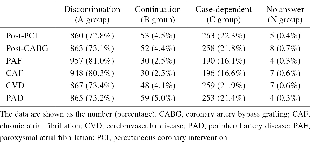
    Results: A total of 1,181 spine surgeons returned completed questionnaires, giving a response rate of 32.0%. JSSR board-certified spine surgeons comprised 75.1% of the respondents. Depending on the management policy regarding antithrombotic drugs for each comorbidity, approximately 73% of respondents discontinued these drugs before elective spine surgery, and about 80% also discontinued anticoagulants. Only 4%-5% of respondents reported continuing antiplatelet drugs, and 2.5% reported continuing anticoagulants. Among the respondents who discontinued antiplatelet drugs, 20.4% reported having encountered cerebral infarction and 3.7% reported encountering myocardial infarction; among those who discontinued anticoagulants, 13.6% reported encountering cerebral embolism and 5.4% reported encountering pulmonary embolism. However, among the respondents who continued antiplatelet drugs and those who continued anticoagulants, 26.3% and 27.2%, respectively, encountered an unexpected increase in intraoperative bleeding, and 10.3% and 8.7%, respectively, encountered postoperative spinal epidural hematoma requiring emergency surgery.

    Conclusions: Our findings indicate that, in principle, >70% of JSSR members discontinue antithrombotic drugs before elective spine surgery. However, those with a discontinuation policy have encountered thrombotic complications, while those with a continuation policy have encountered hemorrhagic complications.

    Download PDF (165K)
  • Ko Hashimoto, Yasuhisa Tanaka, Takumi Tsubakino, Takeshi Hoshikawa, Ch ...
    2023 Volume 7 Issue 5 Pages 436-442
    Published: September 27, 2023
    Released on J-STAGE: September 27, 2023
    Advance online publication: April 21, 2023
    JOURNAL OPEN ACCESS

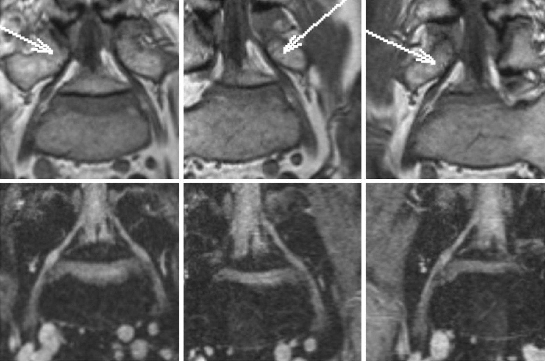
    Introduction: Imaging analysis of foraminal stenosis in the fifth lumbar (L5) nerve root remains to be a challenge because of the anatomical complexity of the lumbosacral transition. T2-weighted three-dimensional (3D) magnetic resonance images (MRI) have been dominantly used for diagnosis of lumbar foraminal stenosis, while the reliability of T1-weighted images (WI) has also been proven. In this study, we aim to compare the reliability and reproducibility of T1- and T2-weighted 3D MRI in diagnosing lumbar foraminal stenosis (LFS) of the L5 nerve root.

    Methods: In this study, 39 patients with unilateral L5 radiculopathy (20 had L4-L5 intracanal stenosis; 19 had L5-S foraminal stenosis) were enrolled, prospectively. T1- and T2-weighted 3D lumbar MRI were obtained from each patient. T1WI and T2WI were blinded and then separately reviewed twice by four examiners randomly. The examiners were instructed to answer the side of LFS or absence of LFS. The correct answer rate, sensitivity, specificity, and area under the curve were analyzed and compared between T1WI and T2WI. Also, intra- and interobserver agreements were calculated using kappa (κ) -statistics and compared in the same manner.

    Results: The average correct answer rate, sensitivity, specificity, and area under the curve of the T1WI/T2WI were 84.6%/80.1%, 82.9%/80.3%, 86.3%/81.3%, and 0.846/0.801, respectively. The intraobserver κ-values of the four examiners ranged from 0.692 to 0.916 (average: 0.762) and from 0.669 to 0.801 (average: 0.720) for T1WI and T2WI, respectively. The interobserver κ-values calculated in a round-robin manner (24 combinations in total) ranged from 0.544 to 0.790 (average: 0.657) and from 0.524 to 0.828 (average: 0.652), respectively.

    Conclusions: As per our findings, T1- and T2-weighted 3D MRI were determined to have nearly equivalent reliability and reproducibility in terms of diagnosing LFS of the L5 nerve root.

    Download PDF (637K)
  • Yusuke Murakami, Tadao Morino, Masayuki Hino, Hiroshi Misaki, Tomofumi ...
    2023 Volume 7 Issue 5 Pages 443-449
    Published: September 27, 2023
    Released on J-STAGE: September 27, 2023
    Advance online publication: April 21, 2023
    JOURNAL OPEN ACCESS

    Introduction: Some patients who have not been diagnosed with diffuse idiopathic skeletal hyperostosis (DISH) (patients in the preclinical stage of DISH [pre-DISH]) may develop DISH in the future. However, there are currently no clearly defined diagnostic criteria for pre-DISH. This study aims to define pre-DISH by analyzing the change in the ossification extent in each intervertebral space in the thoracic and lumbar spines over time using computed tomography (CT).

    Methods: Of the patients who underwent CT of the thoracic to pelvic region at least twice from 2009 to 2018, 188 who underwent CT at an interval of 5 years to 5 years and 2 months were enrolled. The prevalence of DISH during the first and second CT scans was investigated. The pre-DISH feature was defined, and the prevalence of pre-DISH on the first CT and the change after 5 years in patients with pre-DISH was investigated.

    Results: Of the 188 patients, 37 (19.7%) and 48 (25.5%) were diagnosed with DISH on the first and second CT scans, respectively. Pre-DISH was defined as the ossification characterized by the modified Mata score of three contiguous intervertebral spaces with a score of ≥2 points (222; 2 points, ligament ossification of half or more of the intervertebral disc height but incomplete fusion), and 52 patients were diagnosed with pre-DISH. Of the 52 patients with a score of ≥ (222), 11 (21.2%) were diagnosed with DISH 5 years later.

    Conclusions: Patients who have three contiguous intervertebral spaces with a modified Mata score of 2 or 3 points should be considered pre-DISH.

    Download PDF (672K)
  • Masanari Takami, Shunji Tsutsui, Motohiro Okada, Keiji Nagata, Hiroshi ...
    2023 Volume 7 Issue 5 Pages 450-457
    Published: September 27, 2023
    Released on J-STAGE: September 27, 2023
    Advance online publication: June 09, 2023
    JOURNAL OPEN ACCESS

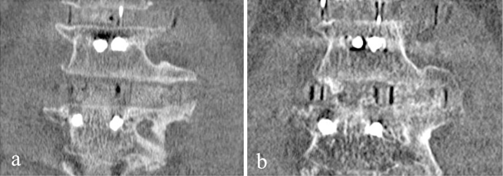
    Introduction: Despite the absence of bone grafting in the area outside the cage, lateral bridging callus outside cages (LBC) formation is often observed here following extreme lateral interbody fusion (XLIF) conversely to conventional methods of transforaminal lumbar interbody fusion and posterior lumbar interbody fusion. The LBC, which may increase stabilization and decrease nonunion rate in treated segments, has rarely been described. This study aimed to identify the incidence and associated factors of LBC following XLIF.

    Methods: We enrolled 136 consecutive patients [56 males, 80 females; mean age 69.6 (42-85) years] who underwent lumbar fusion surgery using XLIF, including L4/5 level with posterior fixation at a single institution between February 2013 and February 2018. One year postoperatively, the treated L4/5 segments were divided into the LBC formation and non-formation groups. Potential influential factors, such as age, sex, body mass index, bone density, height of cages, cage material (titanium or polyetheretherketone [PEEK]), presence or absence of diffuse idiopathic skeletal hyperostosis (DISH), and radiological parameters, were evaluated. Multivariate logistic regression analysis was performed for factors significantly different from the univariate analysis.

    Results: The incidence of LBC formation was 58.8%. Multivariate logistic regression analysis showed that the length of osteophytes [+1 mm; odds ratio, 1.29; 95% confidence interval, 1.17-1.45; p<0.0001] was significant LBC formation predictive factors. Receiver operating characteristic curve analysis demonstrated that the cut-off value for osteophyte length was 14 mm, the sensitivity was 58.8%, the specificity was 84.4%, and the area under the ROC curve for this model was 0.79.

    Conclusions: The incidence of LBC formation was 58.8% in L4/5 levels one year after the XLIF procedure. We demonstrated that the length of the osteophyte was significantly associated with LBC formation.

    Download PDF (550K)
CLINICAL CORRESPONDENCE
ERRATUM
feedback
Top