Spine Surgery and Related Research
Online ISSN : 2432-261X
ISSN-L : 2432-261X
Volume 6, Issue 1
Displaying 1-14 of 14 articles from this issue
REVIEW ARTICLE
  • Sihyong J. Kim, Ralph J. Mobbs, Pragadesh Natarajan, R. Dineth Fonseka ...
    2022 Volume 6 Issue 1 Pages 1-9
    Published: January 27, 2022
    Released on J-STAGE: January 27, 2022
    Advance online publication: June 11, 2021
    JOURNAL OPEN ACCESS

    Cortical bone trajectory (CBT) for posterior fixation with pedicle screws is considered a relatively new alternative trajectory that travels in the medio-lateral direction in the transverse plane and in the caudo-cephalad path in the sagittal plane. Various biomechanical studies have already validated its superior pullout strength and mechanical stability over the traditional trajectory of convergent pedicle screws. Due to the relatively medial starting point of this trajectory, the CBT also poses the clinical advantage of requiring a smaller surgical field of exposure, thus minimizing tissue and muscle injury while reducing operative time and intraoperative blood loss. The evolution of CBT through time has closely been linked to the unwavering philosophy of prioritizing patient outcomes, advancements in neuronavigational technology, and the mounting biomechanical, morphometric, and clinical evidence. In this historical review, we provide a unique perspective on how CBT surgical technique has developed through time, highlighting key milestones and attempting to explain its explosive rise in popularity.

    Download PDF (845K)
ORIGINAL ARTICLE
  • Koji Tamai, Akinobu Suzuki, Hiromitsu Toyoda, Hidetomi Terai, Masatosh ...
    2022 Volume 6 Issue 1 Pages 10-16
    Published: January 27, 2022
    Released on J-STAGE: January 27, 2022
    Advance online publication: June 11, 2021
    JOURNAL OPEN ACCESS

    Introduction: Mental well-being is essential for patient satisfaction. Therefore, a better understanding of the changes in the mental well-being of patients following spinal surgery can be useful to surgeons. We compared the 2-year postoperative change in the mental well-being of patients who underwent cervical and lumbar decompression surgery. Additionally, the predictive factors for improvement in mental well-being associated with both methods were evaluated.

    Methods: The patients who underwent spinal decompression surgery and were followed >2 years postoperatively were enrolled (lumbar cohort: n=111, cervical cohort: n=121). The 36-item Short-Form Health Survey (SF-36) mental component summary (MCS) was set as the mental well-being parameter, and the minimal clinically important difference (MCID) was defined as 4.0. After adjusting the cervical and lumbar cohorts using propensity scores, the improvements in the MCS were compared between the groups using a mixed-effect model. To identify predictors for improvements, the correlation between the MCS changes and preoperative clinical scores was evaluated. Subsequently, multivariate linear regression was applied, which included variables with p<0.10 in the former analysis as explanatory variables, and the change of MCS as the objective variable.

    Results: There were no significant differences in the MCS improvement between the adjusted cervical and lumbar cohorts; 47% and 49%, respectively, had MCS improvement score >MCIDs. However, predictors for the improvement were different between the two cohorts: SF-36 Social functioning in cervical surgery and lower back pain and SF-36 Role physical in lumbar surgery.

    Conclusions: Although there was no significant difference in the improvement in the mental well-being between patients who underwent either cervical or lumbar decompression surgery, less than half of the patients in both groups achieved a meaningful improvement. Preoperative back pain and personal activity were independent predictors in the lumbar cohort, while social functioning was the only predictor in the cervical cohort.

    Level of evidence: III

    Download PDF (132K)
  • Hideyuki Arima, Tomohiko Hasegawa, Yu Yamato, Go Yoshida, Tomohiro Ban ...
    2022 Volume 6 Issue 1 Pages 17-25
    Published: January 27, 2022
    Released on J-STAGE: January 27, 2022
    Advance online publication: August 23, 2021
    JOURNAL OPEN ACCESS

    Introduction: Kyphotic deformity is common after spinal tumor resection surgery. An adequate field of view is needed to resect the spinal cord tumor, and, in some cases, the facet joint must be removed during laminectomy, and fixation may be performed simultaneously. In this study, we investigated the incidence of postoperative deformity after spinal tumor resection and the factors associated with postoperative deformity.

    Methods: We retrospectively analyzed patients who underwent thoracic spinal cord tumor resection at a single institution between 2010 and 2017 and were followed up for at least 24 months after surgery. Fifty percent or more of the facet joint was removed during the laminectomy, and fixation was performed simultaneously. Patients were divided into two groups, with and without kyphotic deformity. Patient demographic characteristics (age at surgery and gender), whether they underwent primary surgery or reoperation, tumor level, pathological type, and surgical method were compared. Multiple linear regression analysis was performed to identify independent predictors of kyphotic deformity.

    Results: Thirty-one patients were found to be eligible. Thirteen patients had intramedullary spinal cord tumors. Laminectomy was performed in 52% (N=16), laminoplasty in 6% (N=2), and laminectomy and/or laminoplasty combined with fusion in 42% (N=13) of the patients. During a mean follow-up period of 66.8 months, 12 (39%) patients had postoperative kyphosis deformities, of which one patient (3%, a 12-year-old girl who underwent combined postoperative radiation therapy) underwent kyphosis correction surgery three years after surgery. The number of laminectomies was independently associated with kyphotic deformity.

    Conclusions: Although kyphotic deformity after spinal tumor surgery was observed in about 39% of the patients, corrective surgery was rarely required due to the progression of the deformity. The high number of laminectomies is a risk factor for postoperative kyphosis, and prophylactic fixation may be considered in cases of multiple laminectomies.

    Download PDF (374K)
  • Kazuo Nakanishi, Yasukazu Hijikata, Kazuya Uchino, Seiya Watanabe, Kos ...
    2022 Volume 6 Issue 1 Pages 26-30
    Published: January 27, 2022
    Released on J-STAGE: January 27, 2022
    Advance online publication: June 11, 2021
    JOURNAL OPEN ACCESS

    Introduction: There is a growing momentum for the collaboration between multiple disciplines for the prevention and treatment of skeletal-related events (SREs) in patients with metastatic spinal tumors. However, the effectiveness of multidisciplinary approaches remains unclear. Hence, we conducted an exploratory study to examine the impact of liaison treatment for metastatic spinal tumor (LMST) on the prevention of SREs among patients with a metastatic spinal tumor.

    Methods: This study was an exploratory interrupted time series conducted in a single medical center. Overall, 1,043 patients with a metastatic spinal tumor diagnosed between January 2011 and December 2020 were included. The LMST was implemented in January 2014. The LMST team consisted of the orthopedic surgery, thoracic surgery, breast and thyroid surgery, clinical oncology, urology, and radiology departments. Monthly joint conferences were held for patients with spinal instability, and the incidence of SRE was measured at 6-month intervals.

    Results: Throughout the study period, we identified 66 SRE incidences. After the implementation of the LMST, a level change of −5.2% (95% confidence interval [CI]: −11.7 to 1.3, p = 0.11) was observed. Subsequently, a post-implementation trend change of −0.3% (95% CI: −2.0 to 1.5, p = 0.75) beyond the baseline was noted.

    Conclusions: We suggest both immediate and gradual effects of the introduction of the LMST on deterring the development of SREs. Our results support the global trend of introducing a multidisciplinary approach for the treatment of metastatic spinal tumors.

    Download PDF (99K)
  • Masahiro Inoue, Takeshi Sainoh, Atsushi Kojima, Masatsune Yamagata, Ta ...
    2022 Volume 6 Issue 1 Pages 31-37
    Published: January 27, 2022
    Released on J-STAGE: January 27, 2022
    Advance online publication: June 11, 2021
    JOURNAL OPEN ACCESS

    Introduction: Condoliase is a newly approved drug that improves symptoms associated with lumbar disk herniation (LDH) by intradiscal administration. This study aimed to evaluate the mid-term outcomes of condoliase injection, examine the adverse events, including cases that required surgery after condoliase administration, and verify cases in which condoliase could be effective.

    Methods: We enrolled patients with LDH who were treated conservatively for at least six weeks and received condoliase. We assessed the visual analog scale (VAS) score, Japanese Orthopaedic Association Back Pain Evaluation Questionnaire, Oswestry Disability Index, disk height, and disk degeneration for up to 6 months, and we examined the complications. Furthermore, a 50% or more improvement in leg pain VAS score was considered effective. Factors related to symptom improvement were investigated by determining whether lower limb pain improved in six months.

    Results: In total, 84 patients were recruited (52 men, 32 women; mean age, 44.2 ± 17.1 [16-86 years]). The duration of illness was 6.7 ± 6.8 (1.5-30) months. All patient-based outcomes significantly improved at 4 weeks after the administration compared with pretreatment. The intervertebral disc height decreased significantly at four weeks after condoliase administration compared with that before administration. Progression of intervertebral disc degeneration occurred in 50% of the patients. Eleven patients underwent herniotomy due to poor treatment effects. Moreover, treatment in 77.4% of the patients was considered effective. A logistic regression analysis revealed that L5/S1 disk administration (p = 0.029; odds ratio, 5.94; 95% confidence interval, 1.20-29.45) were significantly associated with clinical effectiveness.

    Conclusions: Condoliase disk administration improved pain and quality of life over time. Condoliase disk administration was more effective in L5/S1 intervertebral administration.

    Download PDF (657K)
  • Yujiro Kagami, Hiroaki Nakashima, Kotaro Satake, Kenyu Ito, Satoshi Ta ...
    2022 Volume 6 Issue 1 Pages 38-44
    Published: January 27, 2022
    Released on J-STAGE: January 27, 2022
    Advance online publication: June 11, 2021
    JOURNAL OPEN ACCESS

    Introduction: This study aimed to investigate the ureteral running position from the viewpoint of the spine, and to identify the spinal level and left-right difference in the ureter at a dangerous location of ureteral injury during surgery.

    Methods: This retrospective study included 100 consecutive patients (39 males and 61 females; average age, 70.4 years). Preoperative contrast-enhanced computerized tomography (CT) scans obtained in the supine position for patients who underwent lateral lumbar interbody fusion (LLIF) were analyzed. The ureter location was divided into four regions on the axial CT images based on the lumbar disk levels as follows: A (ventral-medial), B (ventral-lateral), C (dorsal-medial), and D (dorsal-lateral). The C region surrounded by the vertebral body and the psoas muscle was assumed to have the highest probability of ureteral injury. We examined the characteristics of the ureteral position at each disc level.

    Results: In the upper lumbar spine, the ureter was outside the lateral dorsoventral axis from the contact point of the psoas muscle, while in the lower lumbar spine, it was inside the axis. The ureters located in the C region increased significantly in the lower lumbar disk levels (L1-L2 and L2-L3: 0%; L3-L4: 5.5%; L4-L5: 14.8%; L5-S: 31.5%). Comparing the left and right sides, especially at L4-L5, the ureter in the C region was observed in 21% of all ureters on the left side and in 9% on the right side. With respect to gender differences, the ureters present in the C region were significantly more common in women at lumbar disk levels L3-L4, L4-L5, and L5-S.

    Conclusions: The ureters in the C region were common on the left side and at lower lumbar disk levels. To avoid ureteral injury, it is necessary to confirm the location of the ureter by using preoperative images and performing LLIF carefully.

    Download PDF (452K)
  • Hirofumi Bekki, Takeshi Arizono, Akihiko Inokuchi, Ryuta Imamura, Taka ...
    2022 Volume 6 Issue 1 Pages 45-50
    Published: January 27, 2022
    Released on J-STAGE: January 27, 2022
    Advance online publication: June 11, 2021
    JOURNAL OPEN ACCESS

    Introduction: Due to the narrow portal of entry, microendoscopic laminectomy (MEL) is associated with a risk of postoperative spinal epidural hematoma (POSEH). This risk might be higher when performing multiple-level (m-) MEL. The purpose of this study is to clarify the incidence rate of POSEH following single-level (s-) and m-MEL by each interlaminar level and identify the risk factors for POSEH following m-MEL.

    Methods: A total of 379 patients underwent MEL of the lumbar spine (s-MEL, n=141; m-MEL, n=238). We determined the incidence of POSEH following s-MEL and m-MEL by each interlaminar level. For m-MEL, we clarified the correlation between POSEH and possible risk factors, such as operative findings, the sequence of operated interlaminar levels, and the preoperative cross-sectional dural area (CSA) on magnetic resonance imaging.

    Results: The incidence rate at L2/3 was significantly higher than that at L3/4 and L4/5. Patients who underwent L2/3 decompression at the end of the procedure showed a higher incidence of POSEH at the L2/3 level. Preoperative spinal stenosis was associated with POSEH at the L2/3 level, and CSA of 56 mm2 was a predictive factor for POSEH. Logistic regression analysis revealed that both were significant risk factors.

    Conclusions: In patients undergoing m-MEL, the incidence of POSEH is highest at the L2/3 level, and treatment of the L2/3 level at the end of the procedure and the presence of spinal stenosis are risk factors for POSEH.

    Download PDF (493K)
  • Shun Okuwaki, Toru Funayama, Akira Ikumi, Satoshi Matsuura, Haruo Kawa ...
    2022 Volume 6 Issue 1 Pages 51-57
    Published: January 27, 2022
    Released on J-STAGE: January 27, 2022
    Advance online publication: June 11, 2021
    JOURNAL OPEN ACCESS

    Introduction: Vertebral instability (VI) in osteoporotic vertebral fractures (OVFs) varies from mild to severe. The relationship between the VI of OVFs and independent factors, such as bone mineral density (BMD) and lumbar muscle volume, is unclear. This study aimed to investigate whether BMD and the cross-sectional area (CSA) of lumbar muscles are related to VI in OVFs.

    Methods: On the basis of the thoracolumbar lateral radiographs of 95 acute OVFs in postmenopausal women (mean age 80.6 years; range: 64-103 years), supine and standing vertebral collapse rates (CRsp and CRst, respectively) were determined. Subsequently, VI was defined as follows: VI=CRst−CRsp. Using axial T2-weighted magnetic resonance imaging (MRI), CSA of the psoas major, erector spinae, and multifidus muscles at the L3/4 intervertebral disc level were measured. The BMD of the lumbar spine and proximal femur (total hip) was measured for all participants using dual-energy X-ray absorptiometry. The patients were classified into group 1 (VI <20%) and group 2 (VI ≥20%).

    Results: We observed a negative correlation between VI and CSA of the erector spinae muscle (r=−0.3962, P<0.0001). No significant correlations were observed between VI and BMD. The CSA of the erector spinae muscle in group 2 was significantly lower than that in group 1 (P=0.0002). No significant difference in the BMD or the CSA of the psoas major or multifidus muscles was observed between the two groups. A multivariable analysis of factors of VI was performed. Both age (odds ratio [OR], 1.099; 95% confidence interval [CI], 1.015-1.189; P=0.020) and the CSA of the erector spinae (OR, 0.996; 95% CI, 0.993-0.999; P=0.020) were significant predictors of high VI.

    Conclusions: Although the severity of OVFs was related to the CSA of the erector spinae muscle, it was not associated with BMD.

    Download PDF (593K)
  • Hideki Shigematsu, Masato Tanaka, Sachiko Kawasaki, Keisuke Masuda, Yu ...
    2022 Volume 6 Issue 1 Pages 58-62
    Published: January 27, 2022
    Released on J-STAGE: January 27, 2022
    Advance online publication: June 11, 2021
    JOURNAL OPEN ACCESS

    Introduction: The loco-check is a simple tool for evaluating locomotive syndrome (LS), and a previous report suggested that it can be used to identify patients with stage 2 LS. The purpose of this study was to investigate the improvement in LS stage after surgery based on the loco-check in elderly patients with lumbar spinal stenosis (LSS) and to clarify the characteristics associated with improvement to non-stage 2 LS.

    Methods: We reviewed 40 elderly patients with LSS who underwent surgery at our institution. We compared the pre- and postoperative Japanese Orthopaedic Association score, loco-check, Oswestry Disability Index, EuroQoL-5 dimension utility values, and the EuroQoL-visual analog scale. We divided patients according to the presence or absence of stage 2 LS after surgery and compared their preoperative clinical findings and assessment measures.

    Results: Ninety percent of all patients had been preoperatively diagnosed with stage 2 LS according to the loco-check. After surgery, patients showed a decreased number of affirmative answers on the loco-check, according to which only 65% were postoperatively diagnosed with stage 2 LS. The receiver operating characteristic curve analysis identified less than four affirmative answers on the loco-check before LSS as predictive of improvement to non-stage 2 LS.

    Conclusions: Surgical treatment for elderly patients with LSS could improve LS. In patients with less than four affirmative answers on the loco-check preoperatively, improvement to non-stage 2 LS status may be possible.

    Download PDF (124K)
  • Hiroki Oba, Jun Takahashi, Yosuke Shibata, Tetsuro Ohba, Tomohiko Hase ...
    2022 Volume 6 Issue 1 Pages 63-70
    Published: January 27, 2022
    Released on J-STAGE: January 27, 2022
    Advance online publication: June 30, 2021
    JOURNAL OPEN ACCESS

    Introduction: Assessments of early postoperative bony union after posterior lumbar interbody fusion via computed tomography (CT) have revealed cases in which interbody fixation by bony union resulted in nonfusion due to bone absorption. The apparent bone union state reverted to a nonunion state several months later, exhibiting a so-called "fake union" phenomenon. Additionally, few reports have evaluated the effect of teriparatide on bony union. The present study aimed to evaluate the frequency of change in assessment from fusion to nonfusion in the postoperative follow-up of lumbar interbody fusion, compare the late postoperative bony union rates in groups with or without early postoperative fusion, and examine the effect of postoperative teriparatide in those groups.

    Methods: Sixty-nine subjects enrolled from multiple hospitals were prospectively evaluated following single-level lumbar interbody fusion. The patients were randomly allocated into treatment with or without weekly postoperative teriparatide. The subjects were then classified as having bony union or nonfusion at 2 months postoperatively, and fusion rates at 6 months were compared. For the evaluation of bony union, blinded radiological examinations were performed via CT. Additional comparisons were made according to teriparatide use.

    Results: The rate of nonunion at 6 months postoperatively in patients with fusion at 2 months postoperatively was 27.8%. Among subjects with bony union at 2 months postoperatively, the fusion rate at 6 months in those who received teriparatide was 93.3% (p=0.027) versus 57.1% in those who did not.

    Conclusions: The rate of nonunion at 6 months postoperatively in patients exhibiting union at 2 months after surgery was 27.8%. Postoperative weekly teriparatide treatment significantly reduced the rate of fake union.

    Download PDF (549K)
  • Daisuke Kurosawa, Eiichi Murakami, Hiroaki Koga, Hiroshi Ozawa
    2022 Volume 6 Issue 1 Pages 71-78
    Published: January 27, 2022
    Released on J-STAGE: January 27, 2022
    Advance online publication: June 11, 2021
    JOURNAL OPEN ACCESS

    Introduction: Sacroiliac joint (SIJ) arthrodesis using a minimally invasive technique, particularly the triangular implant system, is performed in patients with SIJ dysfunction in the United States and Europe. We report three-year clinical outcomes of the first minimally invasive SIJ arthrodesis procedures using the implants performed in Japan.

    Methods: Five patients (one man and four women; age: 56.4±16.9 years) with SIJ pain who underwent SIJ arthrodesis using a triangular implant system in 2017 were included. In addition to operation time and blood loss, pain intensity (visual analog scale [VAS]) and functional impairment (Oswestry disability index [ODI]) were assessed preoperatively and at a 36-month follow-up. Implant loosening and osseous bridging across the joint were evaluated using computed tomography images, and patients' satisfaction with the surgery was also assessed at 12 and 36 months.

    Results: The surgical time was 67.7±13.1 minutes, and blood loss was 7.4±6.9 mL. The mean VAS value improved significantly from 88.0±8.4 mm to 33.6±31.9 mm at 3 months and was maintained at 46.4±30.9 mm at 36 months (P<0.05). The mean ODI improved significantly from 76.4%±3.8% to 46.2%±21.9% at 6 months postoperatively (P<0.05) but had no significant improvements thereafter: 46.94±23.7% (12 months) and 66.4±8.6% (36 months). Three of five patients presented with at least one implant loosening on the sacrum side. No patient had osseous bridging across the joint. A total of 80% (4/5) of patients reported satisfaction with the surgery at 12 months and 60% (3/5) at 36 months.

    Conclusions: The mean VAS value and ODI significantly improved until 6 months after the surgery. However, the mean ODI was reaggravated at 36 months after the surgery. Osseous bridging across the joint was not observed in all patients. We should carefully keep an eye on further long-term results to evaluate the implant.

    Download PDF (1167K)
TECHNICAL NOTE
  • Motohiro Okada, Yukihiro Nakagawa, Munehito Yoshida, Hiroshi Yamada
    2022 Volume 6 Issue 1 Pages 79-85
    Published: January 27, 2022
    Released on J-STAGE: January 27, 2022
    Advance online publication: August 23, 2021
    JOURNAL OPEN ACCESS

    Introduction: Thoracic myelopathy due to ossification of the posterior longitudinal ligament (T-OPLL) is an indication for surgical treatment because the symptom is usually progressive. The surgery for T-OPLL is technically challenging for several reasons. Various operational procedures were developed for dealing with T-OPLL. The anterior decompression through a single posterior approach is a procedure to achieve the complete decompression via the direct resection of the ossified lesion, especially for the beak-type OPLL. Previous reports showed better postoperative outcomes using this method than using other procedures. However, the difficulty and risk of complications are also reported because of the blinded resection of the lesion positioning ventrally to the dura mater.

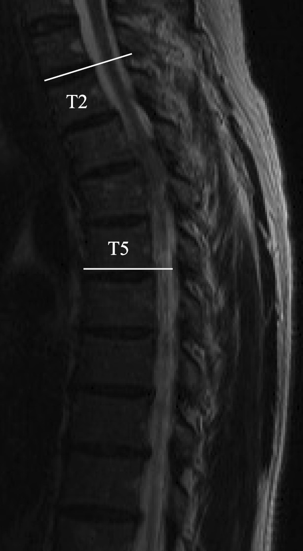
    Technical Note: We describe a novel method using an anterior decompression through a single posterior approach using an ultrasonic bone scalpel. The following procedure is for a case of beak-type OPLL at the T5-6 level. The posterior elements at T2-9 were exposed after a median skin incision was created above a spinous process. First, pedicle screws were inserted bilaterally at T3-5 and T7-9. After the laminectomies and dekyphosis maneuver at T3-9, the spinal cord compression by OPLL was evaluated using intraoperative ultrasonography. After the slight medial facetectomy and pediclotomy at T5-6, the ultrasonic bone scalpel was inserted through the bilateral side of the spinal cord. The tip of the handpiece was angled to reach OPLL. The resection of OPLL was performed under intraoperative spinal cord monitoring. The intraoperative ultrasonography revealed the normal pulsation of the spinal cord and the space between the vertebral body and dura mater after completing the resection of OPLL. Posterolateral fusion was completed with local bone and harvested iliac crest.

    Conclusions: The anterior decompression through a single posterior approach using an ultrasonic bone scalpel is a safe and effective treatment of thoracic OPLL.

    Download PDF (742K)
CLINICAL CORRESPONDENCE
feedback
Top